. 2016 Nov 14:6:37136.
doi: 10.1038/srep37136.

Periodic F-actin structures shape the neck of dendritic spines

Affiliations

Periodic F-actin structures shape the neck of dendritic spines

Julia Bär et al. Sci Rep. .

Abstract

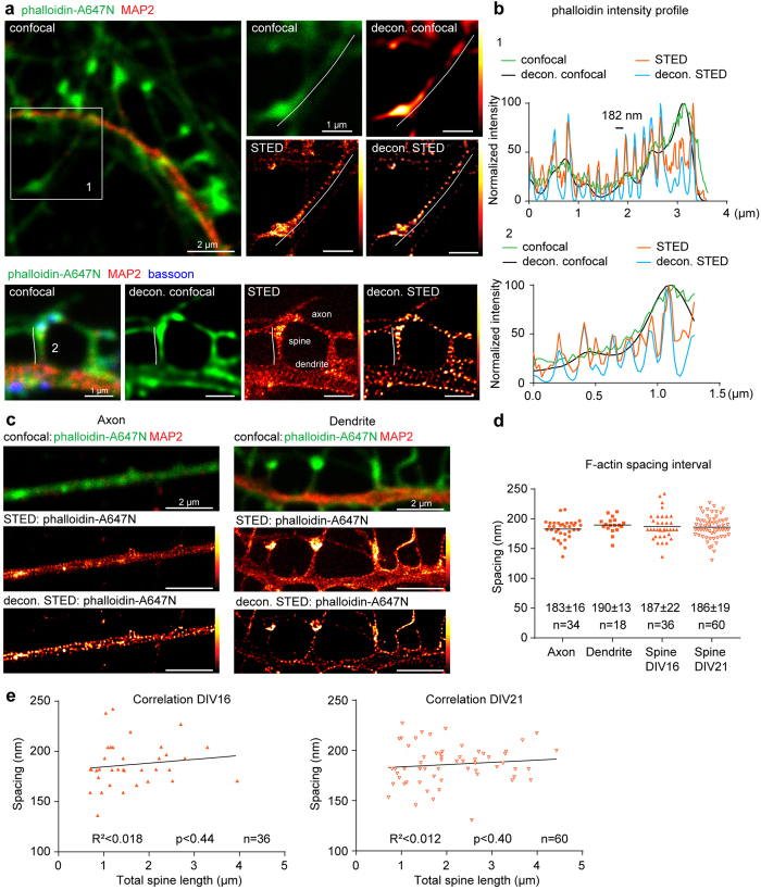
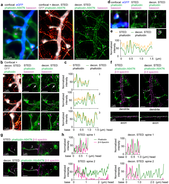
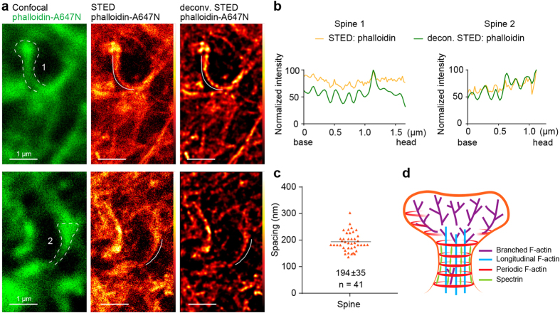
Most of the excitatory synapses on principal neurons of the forebrain are located on specialized structures called dendritic spines. Their morphology, comprising a spine head connected to the dendritic branch via a thin neck, provides biochemical and electrical compartmentalization during signal transmission. Spine shape is defined and tightly controlled by the organization of the actin cytoskeleton. Alterations in synaptic strength correlate with changes in the morphological appearance of the spine head and neck. Therefore, it is important to get a better understanding of the nanoscale organization of the actin cytoskeleton in dendritic spines. A periodic organization of the actin/spectrin lattice was recently discovered in axons and a small fraction of dendrites using super-resolution microscopy. Here we use a small probe phalloidin-Atto647N, to label F-actin in mature hippocampal primary neurons and in living hippocampal slices. STED nanoscopy reveals that in contrast to β-II spectrin antibody labelling, phalloidin-Atto647N stains periodic actin structures in all dendrites and the neck of nearly all dendritic spines, including filopodia-like spines. These findings extend the current view on F-actin organization in dendritic spines and may provide new avenues for understanding the structural changes in the spine neck during induction of synaptic plasticity, active organelle transport or tethering.


Figures

Figure 1
Figure 1. Periodic F-actin lattice is present in nearly all necks of dendritic spines.
(a) Top: Confocal image of a primary hippocampal neuron (DIV21) stained with anti-MAP2 antibodies and phalloidin-A647N with corresponding raw/deconvolved confocal and STED images of F-actin in higher magnification. Bottom: Confocal image of a mushroom-like spine from primary hippocampal cultures stained for actin (phalloidin), MAP2 and the presynaptic marker bassoon with corresponding deconvolved confocal, STED and raw STED image for phalloidin. Lines for intensity profile measurement (b) are indicated. (b) Normalized line profiles of phalloidin-Atto647N intensity along the spine necks indicated in (a) for raw and deconvolved confocal and STED images. (c) Representative confocal, raw and deconvolved STED images of periodic actin structures in axon (left) and dendrite (right). (d) Quantification of cortical F-actin periodicity in axons, dendrites and spines at DIV16 and DIV21 shows no difference in spacing interval. Indicated are mean ± standard deviation (Brown-Forsythe test for equal variances p = 0.20, 1-Way-ANOVA p = 62). Numbers of analyzed spines (n), dendritic or axonal segments (maximum 2 per neurite). from 3 independent experiments. (e) Periodic actin spacing does not correlate with spine length. The number of analyzed spines (n), Linear correlation (black line), coefficient of determination (R2) and p value (for slope being non-zero) are indicated.
Figure 2
Figure 2. Periodic structures are found in mushroom-like spines of primary hippocampal neurons and can be better identified by phalloidin-A647N than β-II spectrin staining.
(a) Confocal image of a DIV16 hippocampal neuron filled with eGFP (blue) and stained for phalloidin (green) and bassoon (magenta) with corresponding deconvolved STED images. Note that phalloidin fills the complete dendrite and spines, and that periodic actin structures are found in all spines. (b) Periodic actin structures in mushroom-like spines of hippocampal primary cultures (DIV16) filled with eGFP and stained for phalloidin and bassoon (left), including corresponding raw and deconvolved STED images for phalloidin and bassoon. (c) Normalized intensity profiles of spines indicated in (b). (d) Periodic actin patterns can extend into the spine head. Confocal image of an eGFP-filled spine stained for phalloidin and bassoon at the presynaptic site, and corresponding raw and deconvoled STED image of phalloidin and bassoon. Note the localization of bassoon in the center of the phalloidin staining. (e) Normalized intensity profile of actin structures in the spine head shown in (d) along the line indicated in the inlet. (f) 2-Color raw and deconvolved STED images of β-II spectrin (magenta) and phalloidin-A647N (green) show a periodic F-actin/spectrin organization in axons and dendrites. *indicates spines with β-II spectrin entering a spine base. (g) Phalloidin-A647N (green) is better suited to visualize periodic cytoskeleton organization than β-II spectrin stainings (magenta). Spine 1 represents an example where the cortical cytoskeletal lattice is organized by alternating phalloidin-A647N and β-II spectrin labelling. In case of spine 2, β-II spectrin is present at the beginning of the spine neck but then disappears, whereas F-actin periodicity is observed throughout the entire neck. *Indicates a spine lacking β-II spectrin stainings completely. Corresponding confocal images and MAP2 staining are in the Fig. S3. (h) Normalized intensity profiles of raw and deconvolved STED indicated in (g).
Figure 3
Figure 3. Phalloidin-A647N labels the periodic actin lattice in dendritic spines in acute hippocampal slices.
(a) Representative confocal images of 2 spines from hippocampal slices with phalloidin-Atto647N (green, left panel) and corresponding raw and deconvolved STED image (middle/right). (b) Normalized intensity profiles of phalloidin-A647N of the spines indicated in (a). More examples are in the Fig. S4. (c) Quantification of the spacing of periodic actin structures in spine necks of hippocampal slices. Mean ± standard deviation is indicated. (n) Number of analysed spines from 5 slices in 2 independent experiments. (d) Model of the cytoskeleton organization in dendritic spines.

References

    1. Tonnesen J., Katona G., Rozsa B. & Nagerl U. V. Spine neck plasticity regulates compartmentalization of synapses. Nat. Neurosci. 17, 678–685 (2014). - PubMed
    1. Adrian M., Kusters R., Wierenga C. J., Storm C., Hoogenraad C. C. & Kapitein L. C. Barriers in the brain: resolving dendritic spine morphology and compartmentalization. Front Neuroanat 8, 142 (2014). - PMC - PubMed
    1. Hering H. & Sheng M. Dendritic spines: structure, dynamics and regulation. Nat. Rev. Neurosci. 2, 880–888 (2001). - PubMed
    1. Bourne J. N. & Harris K. M. Balancing structure and function at hippocampal dendritic spines. Annu. Rev. Neurosci. 31, 47–67 (2008). - PMC - PubMed
    1. Sala C. & Segal M. Dendritic spines: the locus of structural and functional plasticity. Physiological reviews 94, 141–188 (2014). - PubMed

Publication types

LinkOut - more resources