. 2016 Dec 1;126(12):4674-4689.
doi: 10.1172/JCI86950. Epub 2016 Nov 14.

Perivascular macrophages mediate the neurovascular and cognitive dysfunction associated with hypertension

Perivascular macrophages mediate the neurovascular and cognitive dysfunction associated with hypertension

Giuseppe Faraco et al. J Clin Invest. .

Abstract

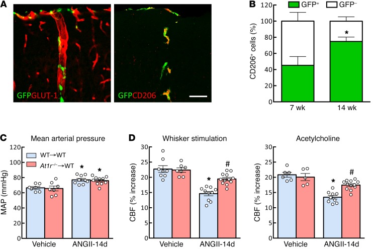
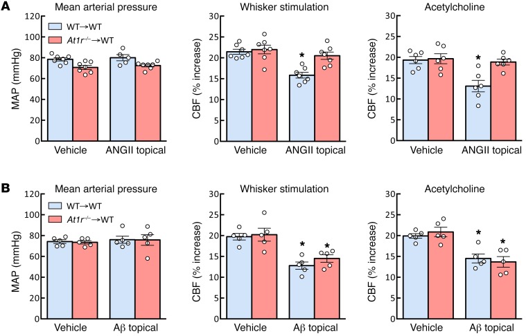
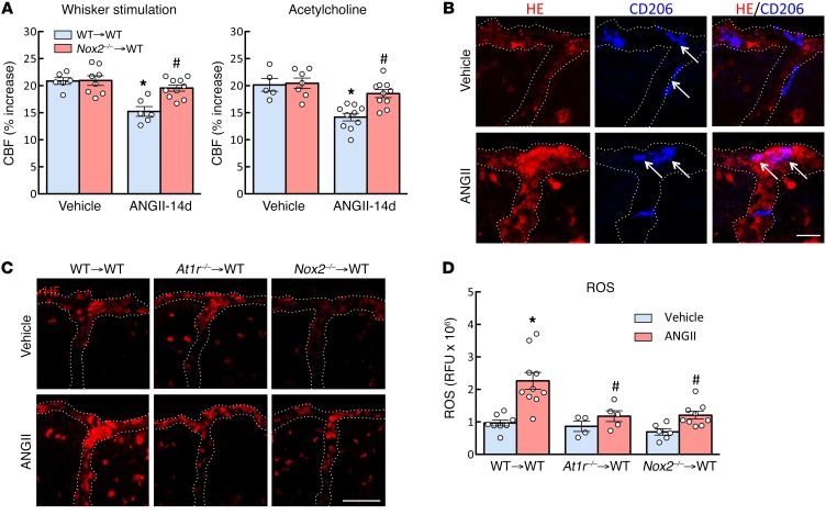
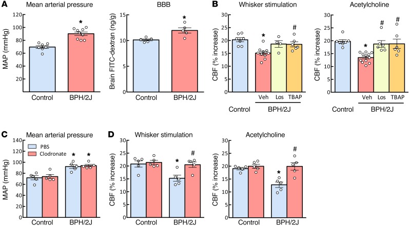
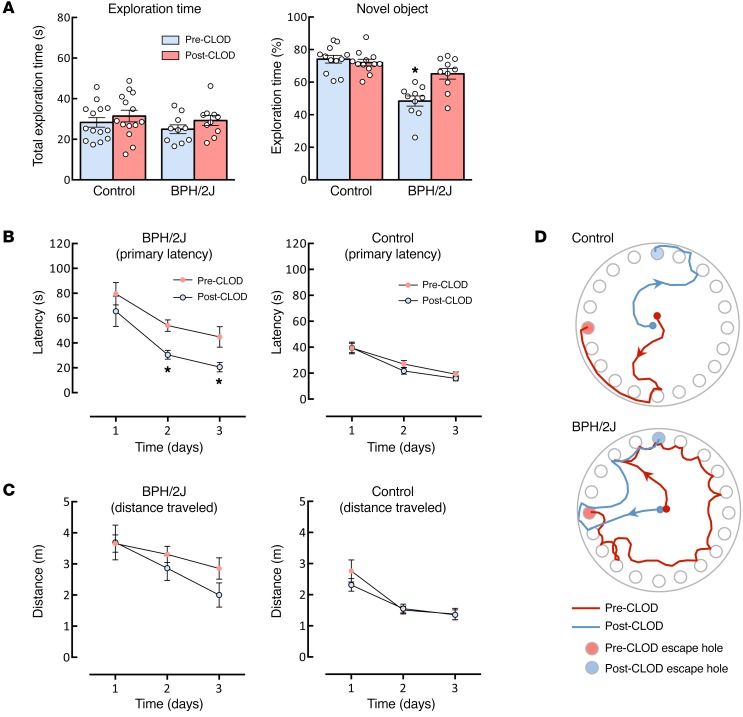
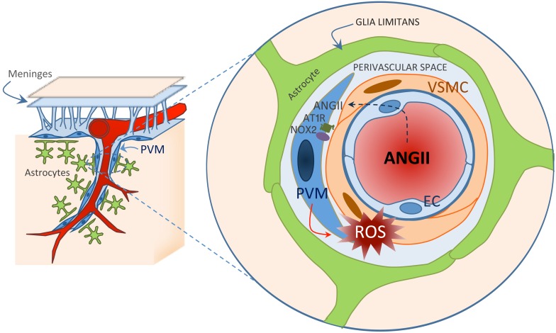
Hypertension is a leading risk factor for dementia, but the mechanisms underlying its damaging effects on the brain are poorly understood. Due to a lack of energy reserves, the brain relies on continuous delivery of blood flow to its active regions in accordance with their dynamic metabolic needs. Hypertension disrupts these vital regulatory mechanisms, leading to the neuronal dysfunction and damage underlying cognitive impairment. Elucidating the cellular bases of these impairments is essential for developing new therapies. Perivascular macrophages (PVMs) represent a distinct population of resident brain macrophages that serves key homeostatic roles but also has the potential to generate large amounts of reactive oxygen species (ROS). Here, we report that PVMs are critical in driving the alterations in neurovascular regulation and attendant cognitive impairment in mouse models of hypertension. This effect was mediated by an increase in blood-brain barrier permeability that allowed angiotensin II to enter the perivascular space and activate angiotensin type 1 receptors in PVMs, leading to production of ROS through the superoxide-producing enzyme NOX2. These findings unveil a pathogenic role of PVMs in the neurovascular and cognitive dysfunction associated with hypertension and identify these cells as a putative therapeutic target for diseases associated with cerebrovascular oxidative stress.


Conflict of interest statement

The authors have declared that no conflict of interest exists.

Figures

Figure 1
Figure 1. PVM identification.
(A) Electron micrograph of a neocortical arteriole (diameter 10 μm). A PVM containing lysosomes (electron dense) is seen at the top right of the vessel (scale bar: 1 μm). PVMs can be identified as perivascular cells (AD), positive for CD206 (BD), able to phagocytose i.c.v.-injected FITC-dextran (BD), weakly positive for the microglia marker IBA1 (C), and expressing the endothelial lymphatic vessel marker LYVE1 (D). Scale bars: 25 μm.
Figure 2
Figure 2. Slow pressor ANGII disrupts the BBB, leading to ANGII entry into the perivascular space and PVMs.
(A) Slow pressor ANGII gradually increases systolic blood pressure measured by tail-cuff plethysmography. *P < 0.05 vs. vehicle (Veh); n = 12–17 (2-way ANOVA and Bonferroni’s test). (B) ANGII plasma levels are increased after 2 weeks of ANGII administration. *P < 0.05 vs. Veh; n = 8–9 (Student’s t test). (C) Slow pressor hypertension but not acute i.v. ANGII administration increases BBB permeability to FITC-dextran (MW 3 kDa). *P < 0.05 vs. Veh; n = 5–7 (Student’s t test). (D) Confocal microscopy showing immunofluorescence labeling of biotinylated ANGII around cerebral blood vessels and in association with PVMs in mice treated with ANGII for 14 days but not in saline-treated mice (scale bar: 25 μm). (E) Orthogonal views (XY, XZ, YZ) illustrating colocalization of biotin-ANGII (green) and CD206 (blue). (F) Biotin-ANGII is associated with PVMs in mice treated with ANGII for 14 days but not in saline-treated mice or in mice acutely infused with ANGII. *P < 0.05 vs. Veh and acute ANGII; n = 3–4 per group; 86 ± 9 PVMs per animal (1-way ANOVA and Tukey’s test). (G) Electron micrographs of cortical arterioles showing immunoperoxidase labeling of biotinylated ANGII tracking along tight junctions (arrows) and reaching perivascular space (arrowheads) in mice treated with ANGII for 14 days (right) but not in saline-treated mice (left). Scale bar: 100 nm. PVM, perivascular macrophage; EC, endothelial cell; VSMC, vascular smooth muscle cells; BM, basement membrane.
Figure 3
Figure 3. PVM depletion by CLOD restores neurovascular function in ANGII slow pressor hypertension.
(A and B) Intracerebroventricular CLOD administration induces depletion of cells expressing the PVM marker CD206 in the somatosensory cortex assessed by histology. The vasculature is visualized by double label with the endothelial marker GLUT-1. The pial surface is at the top of the figure, and penetrating vessels can be seen diving into the neocortex. Scale bars: 50 μm. *P < 0.05 vs. PBS-Veh and PBS-ANGII; n = 5–10 per group (2-way ANOVA and Bonferroni’s test). (C) CLOD administration does not affect the increases in mean arterial pressure (MAP) induced by ANGII. *P < 0.05 vs. PBS-Veh and CLOD-Veh; n = 5–8 per group (2-way ANOVA and Bonferroni’s test). (D) CLOD does not alter the increase in CBF induced by whisker stimulation or cortical administration of ACh, but counteracts the attenuation of both responses induced by ANGII. *P < 0.05 vs. PBS-Veh and CLOD-Veh; #P < 0.05 vs. PBS-ANGII; n = 5–7 per group (2-way ANOVA and Bonferroni’s test).
Figure 4
Figure 4. PVM depletion counteracts the neurovascular dysfunction induced by topical neocortical application but not by acute i.v. ANGII administration.
(A) CLOD has no effect on the neurovascular dysfunction induced by acute i.v. administration of ANGII. *P < 0.05 vs. PBS-Veh and CLOD-Veh; n = 6 per group (2-way repeated-measures ANOVA and Bonferroni’s test). (B) CLOD-mediated PVM depletion rescues the neurovascular dysfunction induced by neocortical application of ANGII to the subarachnoid space (ANGII topical). *P < 0.05 vs. PBS-Veh and CLOD-Veh; n = 6–7 per group (2-way repeated-measures ANOVA and Bonferroni’s test).
Figure 5
Figure 5. PVM At1r deletion counteracts the harmful cerebrovascular effects of ANGII slow pressor hypertension.
(A) Fourteen weeks after transplant of GFP+ bone marrow in WT mice, GFP+ and CD206+ PVM surrounds GLUT-1+ vessels. Scale bar: 50 μm. (B) By 14 weeks after the transplant, approximately 80% of CD206+ cells in the somatosensory cortex are replaced by GFP+ bone marrow cells. *P < 0.05 vs. 7 weeks group [χ2(1) = 7.78]; n = 5 per group. (C) ANGII increases mean arterial pressure equally in WT mice with transplanted WT bone marrow (WT→WT) or At1r–/– marrow (At1r–/– WT). *P < 0.05 vs. WT→WT–Veh and At1r–/–→WT–Veh; n = 7–10 per group (2-way ANOVA and Bonferroni’s test). (D) At1r deletion in PVMs does not affect the CBF responses to whisker stimulation or ACh, but counteracts the attenuation of both responses induced by ANGII. *P < 0.05 vs. WT→WT–Veh and At1r–/–→WT–Veh; #P < 0.05 vs. WT→WT–ANGII; n = 7–12 per group (2-way ANOVA and Bonferroni’s test).
Figure 6
Figure 6. At1r deletion in PVMs rescues the neurovascular dysfunction induced by topical neocortical application of ANGII but not amyloid-β.
(A) At1r–/–→WT chimeras are protected from the cerebrovascular effects induced by topical neocortical application of ANGII. *P < 0.05 vs. WT→WT–Veh and At1r–/–→WT–Veh; n = 5–7 per group (2-way repeated-measures ANOVA and Bonferroni’s test). (B) The attenuation of the CBF responses induced by topical application of amyloid-β (Aβ), an effect not dependent on AT1Rs, is not affected. This finding attests to the specificity of the effect of PVM At1r deletion on the cerebrovascular dysfunction induced by ANGII. *P < 0.05 vs. WT→WT–Veh and At1r–/–→WT–Veh (2-way repeated-measures ANOVA and Bonferroni’s test).
Figure 7
Figure 7. NOX2 in PVMs is required for the cerebrovascular effects of ANGII slow pressor hypertension.
(A) Nox2 deletion in PVMs does not affect baseline cerebrovascular responses, but counteracts the cerebrovascular dysfunction induced by ANGII (Nox2–/–→WT chimeras). *P < 0.05 vs. WT→WT–Veh and Nox2–/–→WT–Veh; #P < 0.05 vs. WT→WT–ANGII; n = 5–10 per group (2-way ANOVA and Bonferroni’s test). (B) ANGII markedly increases ROS production in CD206+ cells (PVMs) assessed by dihydroethidine (DHE) microfluorography. Scale bar: 25 μm. (C and D) Deletion of At1r or Nox2 in PVMs attenuates the neurovascular oxidative stress induced by slow pressor ANGII in the neocortex. Scale bar: 50 μm. *P < 0.05 vs. WT→WT–Veh, At1r–/–→WT–Veh, and Nox2–/–→WT–Veh; #P < 0.05 vs. WT→WT–ANGII; n = 4–10 per group (2-way ANOVA and Bonferroni’s test).
Figure 8
Figure 8. PVMs mediate cerebrovascular dysfunction in chronically hypertensive BPH/2J mice.
(A) Mean arterial pressure and BBB permeability to FITC-dextran are increased in BPH/2J mice. *P < 0.05 vs. control; #P < 0.05 vs. Veh; n = 5–9 (Student’s t test). (B) Antagonism of AT1Rs with losartan or ROS scavenging with MnTBAP counteracts the neurovascular dysfunction in BPH/2J mice. *P < 0.05 vs. control; n = 4–7 per group (1-way ANOVA and Tukey’s test). (C and D) CLOD has no effect on the mean arterial pressure but completely reverses the attenuation in CBF response to whisker stimulation and ACh in BPH/2J mice. *P < 0.05 vs. PBS-control and CLOD-control; #P < 0.05 vs. PBS–BPH/2J; n = 5 per group (2-way ANOVA and Bonferroni’s test).
Figure 9
Figure 9. PVMs mediate cognitive dysfunction in chronically hypertensive BPH/2J mice.
(A) PVM depletion by CLOD rescues the recognition memory deficits assessed by the novel object recognition test in BPH/2J mice. *P < 0.05 vs. pre-CLOD–control and post-CLOD–control; n = 10–12 per group (2-way repeated-measures ANOVA and Bonferroni’s test). (B) PVM depletion does not affect the spatial memory assessed by Barnes maze test in control mice but rescues the deficits observed in BPH/2J mice. *P < 0.05 vs. pre-CLOD–BPH/2J; n = 15–20 per group (2-way repeated-measures ANOVA and Bonferroni’s test). (C) CLOD tends to reduce the distance traveled in BPH/2J mice, but the effect does not reach statistical significance. P > 0.05 vs. pre-CLOD–BPH/2J; n = 15–20 per group (2-way repeated-measures ANOVA and Bonferroni’s test). (D) Representative tracks for control and BPH/2J mice on acquisition day 3 before and after vehicle or CLOD injection.
Figure 10
Figure 10. Potential mechanisms by which ANGII hypertension leads to neurovascular dysfunction underlying cognitive deficits.
The left side of the figure illustrates a pial arteriole giving off a branch penetrating into the brain parenchyma and surrounded by astrocytic end-feet forming the glia limitans and by PVMs. As illustrated in the enlargement on the right, circulating ANGII reaches the perivascular space through a breach of the BBB and acts on ANGII type 1 receptors (AT1R) on PVMs, resulting in the activation of NOX2 and ROS production. Oxidative stress, in turn, leads to neurovascular dysfunction. PVM, perivascular macrophage; EC, endothelial cell; VSMC, vascular smooth muscle cell.

Comment in

References

    1. Rahimi K, Emdin CA, MacMahon S. The epidemiology of blood pressure and its worldwide management. Circ Res. 2015;116(6):925–936. doi: 10.1161/CIRCRESAHA.116.304723. - DOI - PubMed
    1. Faraco G, Iadecola C. Hypertension: a harbinger of stroke and dementia. Hypertension. 2013;62(5):810–817. doi: 10.1161/HYPERTENSIONAHA.113.01063. - DOI - PMC - PubMed
    1. Gorelick PB, et al. Vascular contributions to cognitive impairment and dementia: a statement for healthcare professionals from the American Heart Association/American Stroke Association. Stroke. 2011;42(9):2672–2713. doi: 10.1161/STR.0b013e3182299496. - DOI - PMC - PubMed
    1. Iadecola C. The overlap between neurodegenerative and vascular factors in the pathogenesis of dementia. Acta Neuropathol. 2010;120(3):287–296. doi: 10.1007/s00401-010-0718-6. - DOI - PMC - PubMed
    1. Lackland DT, et al. Factors influencing the decline in stroke mortality: a statement from the American Heart Association/American Stroke Association. Stroke. 2014;45(1):315–353. doi: 10.1161/01.str.0000437068.30550.cf. - DOI - PMC - PubMed

Publication types

MeSH terms