. 2016 Dec;157(12):2722-2730.
doi: 10.1097/j.pain.0000000000000692.

Dural stimulation in rats causes brain-derived neurotrophic factor-dependent priming to subthreshold stimuli including a migraine trigger

Affiliations

Dural stimulation in rats causes brain-derived neurotrophic factor-dependent priming to subthreshold stimuli including a migraine trigger

Carolina C Burgos-Vega et al. Pain. 2016 Dec.

Abstract

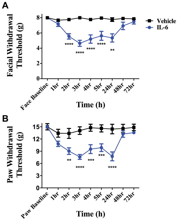
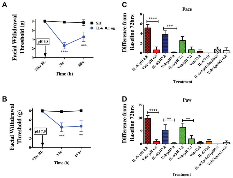
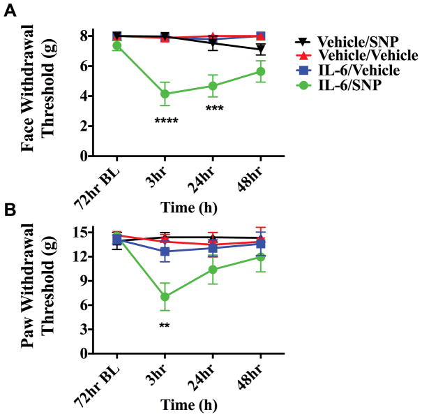
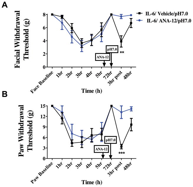
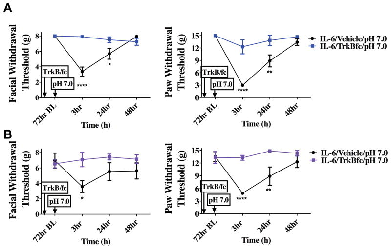
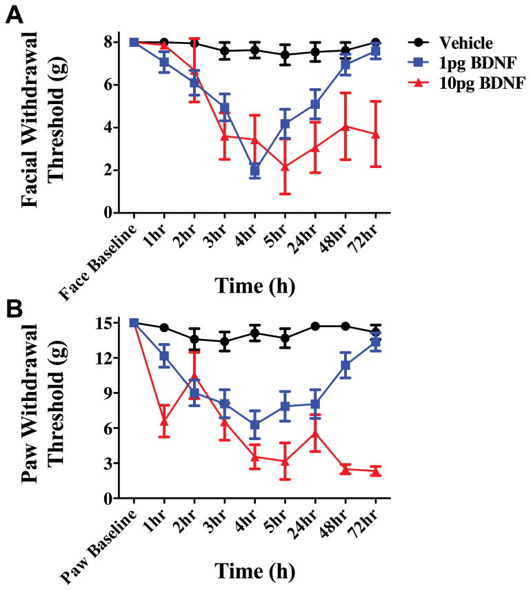
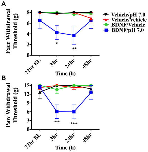
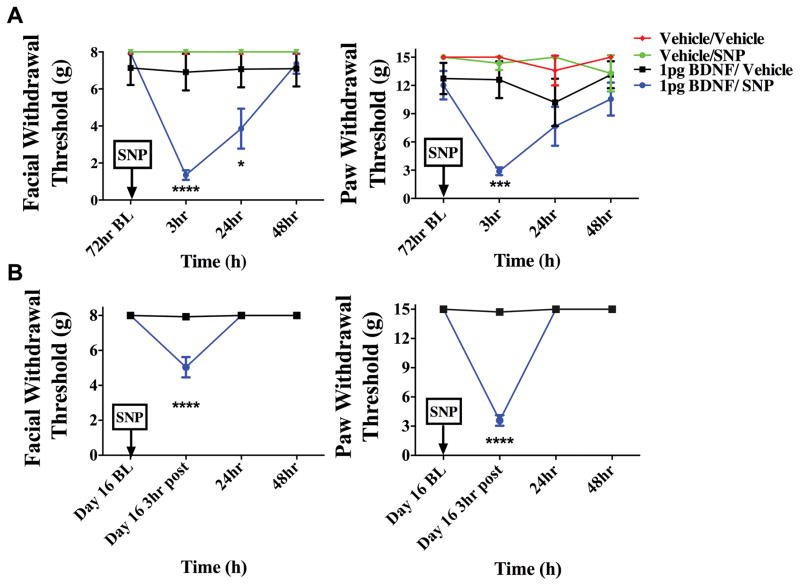
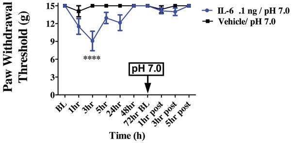
Migraine is one of the most common and most disabling disorders. Between attacks, migraine patients are otherwise normal but are sensitized to nonnoxious events known as triggers. The purpose of these studies was to investigate whether a headache-like event causes sensitization, or priming, to subsequent subthreshold events. Interleukin-6 (IL-6) was applied to the rat cranial dura mater which produced cutaneous facial and hind paw allodynia that lasted 24 hours. At 72 hours, IL-6-treated rats developed allodynia in response to dural stimulation with either a pH 6.8 or pH 7.0 solution and to a systemic nitric oxide (NO) donor, a well-known migraine trigger. Vehicle-treated rats did not respond to either pH stimulus or to the NO donor, demonstrating that IL-6 exposure primes rats to subthreshold stimuli. Inhibitors of brain-derived neurotrophic factor (BDNF) signaling given either systemically or intracisternally 24 hours after IL-6 eliminated responses to dural pH stimulation at 72 hours. Additionally, intracisternal administration of BDNF without previous dural stimulation produced allodynia and once resolved, animals were primed to dural pH 6.8/pH 7.0 and a systemic NO donor. Finally, hind paw IL-6 produced paw allodynia but not priming to paw injection of pH 7.0 at 72 hours demonstrating differences in priming depending on location. These data indicate that afferent input from the meninges produces BDNF-dependent priming of the dural nociceptive system. This primed state mimics the interictal period of migraine where attacks can be triggered by normally nonnoxious events and suggests that BDNF-dependent plasticity may contribute to migraine.


Conflict of interest statement

Potential Conflicts of Interest

The authors declare no conflicts of interest

Figures

Figure 1
Figure 1
Dural application of IL-6 produces headache-related behaviors. Withdrawal thresholds to tactile stimuli applied to the face (A) and hindpaws (B) were measured in animals prior to and after dural application of .1 ng IL-6 (n = 39) or Vehicle(pH 7.4) (n = 28). Administration of IL-6 produced significant allodynia that lasted 24 hours in both face and hindpaw. Two-factor analysis of variance (ANOVA) indicated a significant effect of both treatment and time of both the face and hindpaws. Significant differences among means for each group were determined by analysis of variance followed by Bonferroni post hoc test. (A) Facial: time F (8, 418) = 5.029, P < 0.0001, treatment F (1, 418) = 49.79,P < 0.0001; (B) Hindpaw: time F (8, 347) = 7.818, P < 0.0001, treatment F (1, 347) = 81.33, P < 0.0001.
Figure 2
Figure 2
Dural application of IL-6 primes animals to sub-threshold stimuli. Animals that received a prior application of IL-6 exhibit cutaneous allodynia when dural pH 6.8 (A, n=9 for all groups ) or pH 7.0 (B, n=10 for all groups) is applied after the resolution of allodynia at 72 hours. Withdrawal thresholds to tactile stimuli applied to the face (A & B) were measured in animals after application of dural pH 6.8 or pH 7.0 72 hours post IL-6. Significant differences among means for each group were determined by analysis of variance followed by Bonferroni post hoc test. Increasing the pH value to pH 7.2 did not produce significant facial allodynia in either IL-6 or vehicle-treated rats (C). ASIC3 blocker APETx2 was able to attenuate facial (C) and hindpaw (D) allodynia when administered with pH 6.8 at 72 hours post IL-6. Stimulation of the dura with Vehicle at 72 hours post IL-6 did not produce facial allodynia (C). Significant (**p < 0.01) differences among change of means at 72 hours for each group were determined using a t-test (C & D). (A) Facial: time F (2, 36) = 26.53, P <0.0001, treatment F (1, 87) = 0.0046,P =0.0001; (B) Facial: time F (2, 36) = 5.112, P =0.0111, treatment F (1, 36) = 19.70, P < 0.0001.
Figure 3
Figure 3
Dural application of IL-6 primes animals to systemic NO donors. Animals treated with meningeal IL-6 (n=8 IL-6/SNP 3mg/kg , n=7 IL-6/Vehicle) respond to systemic SNP; producing both facial (A) and hindpaw (B) allodynia at 72 hours post IL-6 while animals treated with Vehicle (n=5 for all groups)did not respond to SNP. Withdrawal thresholds to tactile stimuli applied to the face (A) and the hindpaw (B) after systemic SNP given 72 hours post IL-6. Significant differences among means for each group were determined by analysis of variance followed by Bonferroni post hoc test. Facial: time F (3, 88) = 1.578, P =0.2004, treatment F (3, 88) = 9.588,P <0.0001; Hind paw: time F (3, 80) = 1.172, P =0.3256 treatment F (3, 80) = 20.31, P < 0.0001.
Figure 4
Figure 4
Priming following dural IL-6 stimulation is dependent on BDNF signaling. Systemic administration of ANA-12(0.5 mg/kg) 24 hours following dural IL-6 application attenuated allodynia in the face (A) and the hindpaw (B) at 72 hours Animals given ANA-12 did not show significant responses to subsequent application of dural pH 7.0 vehicle (n=11) at 72 hours post IL-6 when compared with animals given dural Vehicle (n=7)(A &B). Significant differences among means for each group were determined with a one-way ANOVA followed by Bonferroni post hoc test. Facial: time F (8, 72) = 10.88, P < 0.0001, treatment F (1, 72) = .2654,P =0.6080; Hind paw: time F (8, 72) = 13.51, P < 0.0001, treatment F (1, 72) = 9.33, P = 0.0032.
Figure 5
Figure 5
Priming following dural IL-6 stimulation is dependent on BDNF signaling in the brainstem. TrkB-Fc was given i.c. 24 hours after dural application of IL-6. Only animals given i.c. vehicle (males n=12 A & females n=7 B) respond to dural pH 7.0 at 72 hours post IL6. Males treated with IL-6/i.c vehicle/ dural pH 7.0 showed significant facial and hindpaw responses at 3 hours and 24 hours. However, females treated with IL-6/i.c vehicle/dural pH 7.0 had significant facial responses at 3 hours and hindpaw responses at 3 hours and 24 hours. Both male(n=8 A) and female (n=7 B) animals treated with i.c. TrkB-Fc after IL-6 failed to display allodynia 3 hours post administration of pH 7.0 on to the dura. Significant differences among means for each group were determined one-way ANOVA followed by Bonferroni post hoc test. Males facial: time F (3, 72) = 11.74, P <0.0001, treatment F (1,72) = 10.29, P < 0.0001; Hind paw: time F (3, 64) = 1.944, P =0.134, treatment F (3, 64) = 13.21, P < 0.0001. Females Facial: time F (3, 64) = 20.92, P <0.0001, treatment F (3, 72) = 1.109, P =0.0013; Hind paw: time F (1, 72) = 6.152, P =0.0012, treatment F (3, 64) = 24.51, P < 0.0001
Figure 6
Figure 6
Intracisternal BDNF produces cutaneous allodynia. Administration of 1 or 10 picograms (pg) of BDNF i.c produces cutaneous facial (A) and hindpaw (B) allodynia. Allodynia induced by BDNF at 1 pg (n=20) resolved within 72 hours, whereas that induced by 10pg (n=6) of BDNF persisted beyond 72 hours. Vehicle (n=5) i.c. showed no significant results at any time point. Facial: time F (8, 252) = 5.85, P < 0.0001, treatment F (2,252) = 31.02, P < 0.0001; Hind paw: time F (3, 64) = 4.937 P < 0.0001, treatment F (3, 64) = 60.07, P < 0.0001
Figure 7
Figure 7
Intracisternal BDNF primes animals to sub-threshold dural pH stimulation. Animals treated with i.c. BDNF followed by pH 7.0(n=5 for all groups) application to the dura 72 hours later displayed facial allodynia (A &B) while BDNF-treated animals given i.c dural vehicle showed no response at this time point. Significance was determined by one-way ANOVA analysis if the differences of the means for each group, (****p < 0.0001, *p < 0.01) . Facial: time F (3, 64) = 0.7616, P <=0.5198, treatment F (3, 64) = 10.29, P < 0.0001; Hind paw: time F (3, 64) = 1.944, P =0.134, treatment F (3, 64) = 13.21, P < 0.0001
Figure 8
Figure 8
Priming following intracisternal BDNF lasts for at least several weeks. BDNF-treated animals developed facial allodynia when given i.p SNP at 72 hours (A) (BDNF/SNP n=10, BDNF/vehicle n=8, Vehicle/Vehicle n=5, Vehicle/SNP n=5) or day 16 (B) (n=8 for all groups) post BDNF treatment. A significant attenuation of withdrawal thresholds to tactile stimuli applied to the face were observed at both 3 hours and 24 hours, whereas hindpaw responses were only significant 3 hours after 72 hour SNP administration. However, on day 16 a significant attenuation of withdrawal thresholds was observed 3 hours after SNP administration for facial and hindpaw responses. Allodynia was not observed in i.c. BDNF-treated animals given vehicle at 72 hours or day 16(. Significant (****p < 0.0001) differences among means for each group were determined one-way ANOVA followed by Bonferroni post hoc test. (A) Facial: time F (3, 64) = 8.347, P =< 0.0001, treatment F (1, 64) = 12.30, P =0.0008; Hind paw: time F (3, 96) = 1.789, P =0.1544, treatment F (3, 96) = 11.55, P < 0.0001(B)Facial: time F (3, 56 = 26.59, P < 0.0001, treatment F (1,56) = 24.15, P < 0.0001; Hind paw: time F (8, 406) = 8.027, P < 0.0001, treatment F (1, 406) = 87.69, P < 0.0001
Figure 9
Figure 9
Hindpaw application of IL-6 fails to prime animals to subthreshold pH stimulation of the hindpaw. Animals received application of IL-6 in the hindpaw and show paw allodynia. Animals do not exhibit cutaneous allodynia when pH 7.0 is applied to the hindpaw (n=5 for all groups) after the resolution of IL-6 allodynia at 72 hours. Two-factor analysis of variance (ANOVA) indicated a significant effect of both treatment and time of the hindpaws. Significant differences among means for each group were determined by analysis of variance followed by Bonferroni post hoc test. Hind paw: time F (9, 90) = 4.11, P=0.0002, treatment F (1, 90) = 20.47, P < 0.0001.

References

    1. Andress-Rothrock D, King W, Rothrock J. An analysis of migraine triggers in a clinic-based population. Headache. 2010;50(8):1366–1370. - PubMed
    1. Ashkenazi A. Allodynia in cluster headache. Current pain and headache reports. 2010;14(2):140–144. - PubMed
    1. Autry AE, Monteggia LM. Brain-derived neurotrophic factor and neuropsychiatric disorders. Pharmacol Rev. 2012;64(2):238–258. - PMC - PubMed
    1. Bartsch T, Goadsby PJ. Increased responses in trigeminocervical nociceptive neurons to cervical input after stimulation of the dura mater. Brain. 2003;126(Pt 8):1801–1813. - PubMed
    1. Beggs S, Salter MW. The known knowns of microglia-neuronal signalling in neuropathic pain. Neurosci Lett. 2013;557(Pt A):37–42. - PubMed

Publication types

MeSH terms