. 2017 Jan 1;158(1):98-108.
doi: 10.1210/en.2016-1608.

Androgen Receptor in the Ovary Theca Cells Plays a Critical Role in Androgen-Induced Reproductive Dysfunction

Affiliations

Androgen Receptor in the Ovary Theca Cells Plays a Critical Role in Androgen-Induced Reproductive Dysfunction

Yaping Ma et al. Endocrinology. .

Abstract

Androgen and its receptor (AR) play a critical role in reproductive function under both physiological and pathophysiological conditions. Female AR global knockout mice are subfertile due to both neuroendocrine and ovarian defects. Female offspring from prenatally androgenized heterozygous AR pregnant mice showed rescued estrous cyclicity and fertility. Ar is expressed in granulosa cells, theca interstitial cells, and oocytes in the ovary. We created mice with theca-specific deletion of Ar (ThARKO) by crossing Cyp17-iCre mice that express Cre recombinase under cytochrome P450 17A1 (Cyp17) promoter with Arfl/fl mice. ThARKO mice exhibited no significant differences in pubertal onset or fertility compared with control littermates, and neither estrogen or testosterone levels were different between these groups. Therefore, Ar expression in theca cells likely does not influence fertility nor androgen levels in female mice. We then tested the role of AR in theca cells under hyperandrogenemic condition. After treatment with a pathophysiological level of dihydrotestosterone (DHT), control mice (control-DHT) showed acyclicity and infertility. However, estrous cycles and fertility were altered to a significantly less degree in ThARKO-DHT mice than in control-DHT mice. Messenger RNA (mRNA) levels of Lhcgr (luteinizing hormone receptor) and Timp1 (tissue inhibitor of metalloproteinase 1, and inhibitor of matrix metalloproteinase) were significantly lower in control-DHT ovary compared with control-no DHT ovaries, whereas mRNA levels of Fshr (follicle-stimulating hormone receptor) were significantly higher. Timp1 gene expression was comparable in the ThARKO-DHT and the control-no DHT ovary. We speculate that the preserved level of Timp1 in ThARKO-DHT mice contributes to retained reproductive function.


Figures

Figure 1.
Figure 1.
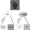
Animal models to study the reproductive role of AR in theca cells under physiological (normal androgen level) and pathophysiological (androgen excess) condition. WT, wild type.
Figure 2.
Figure 2.
AR expression in ovaries. (A) AR mRNA level, measured by qRT-PCR, was significantly reduced in the theca-interstitial cells (TI) of ThARKO compared with control littermates (Con). There were no significant differences in AR expression between control and ThARKO mice for granulosa cells, hypothalamus (Hypo), or pituitary. (B) Western blot was performed, and AR protein levels were quantified by densitometry in 3 independent experiments. The AR protein level was significantly reduced in the ovary of ThARKO compared with controls, but there was no change in the hypothalamus and pituitary. (C) Immunohistology staining of AR (5x and 20x objectives) in the ovaries showed AR expressed intensively in the granulosa cells in both ThARKO and control mice. AR was barely detectable in the theca cells of ThARKO ovary but was expressed widely in the theca-interstitial cells of control ovary (dark brown). Insets are the higher magnifications of the areas shown in the dashed boxes. Values are mean ± SEM; n = 4 to 7/group.
Figure 3.
Figure 3.
Female puberty and fertility. (A) ThARKO mice exhibited similar age of puberty onset, assessed by examination of vaginal opening and first estrus, as control mice. (B) Percentage of time spent in each of the stages (5 month old) was not significantly different between control and ThARKO mice. (C and D) There was no significant differences between ThARKO and controls in either (C) total numbers of litters per female or (D) numbers of pups per female. Values are mean ± SEM; n = 6 to 10/group. Con, control; D, diestrus; E, estrus; M, metestrus; NS, nonsignificant; P, proestrus.
Figure 4.
Figure 4.
Hormone levels. (A) Pituitary responses to GnRH stimulation were tested in the mornings of diestrus. There was no significant difference in LH secretion at either 10 or 20 minutes post GnRH stimulation between control and ThARKO mice. n = 8 to 10. (B) Androstenedione secretion with and without hCG. Ovary was collected at diestrus and cultured in McCoy5A medium for 3 hours. Medium was replaced with/without hCG. At 24 hours, medium was collected and androstenedione was measured. There was no significant difference in androstenedione secretion between control and ThARKO mice either without hCG or with hCG. For both groups of mice, androstenedione secretion was significantly increased by hCG addition compared with no hCG addition. n = 4/group. Values are mean ± SEM. Con, control; NS, nonsignificant.
Figure 5.
Figure 5.
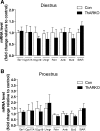
Expressions patterns of 8 genes necessary for steroidogenesis and ovary function at (A) diestrus and (B) proestrus. There were no significant differences in mRNA level between control (Con) and ThARKO mice for any gene at either estrous stage. Values are mean ± SEM; n = 5 to 11/group.
Figure 6.
Figure 6.
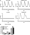
Estrous cycles. (A) Representative data for vaginal cytology from individual control (Con)-no DHT, ThARKO-no DHT, control-DHT, and ThARKO-DHT mice. (B) Percentage time spent in each stage of the estrous cycle was significantly differently among the 3 groups, analyzed by 1-way ANOVA followed by Tukey posttest. Percentage of time in proestrus (P) and estrus (E) was reduced, and time in metestrus (M)/diestrus (D) was increased in control-DHT compared with control-no DHT. The percentage time spent in each stage in ThARKO-DHT was in between that of control-no DHT and control-DHT. Bars with different letters represented values that are significantly different to those analyzed within the same cycle stage (P < 0.05). Values are mean ± SEM; n = 5 to 11.
Figure 7.
Figure 7.
Fertility with and without DHT. (A) Total number of litters and (B) pups per female were significantly reduced in control (Con)-DHT mice compared with control-no DHT mice during the 100 days of mating. ThARKO-DHT mice had a significantly improved fertility compared with control-DHT, although the fertility was still impaired compared with the control-no DHT mice. Bars with different letters represent significantly different values from each other with P < 0.05, analyzed by 1-way ANOVA followed by Tukey posttest. Values are mean ± SEM; n = 6 to 8/group.
Figure 8.
Figure 8.
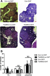
Ovary morphology and follicular numbers. (A) Histological sections of ovary from control and ThARKO mice with and without DHT 3 months after insertion of pellets (hematoxylin and eosin stain). (B) Numbers of follicles in each group. CL, antral follicles, and preantral follicles (primodial, primary, and secondary follicles) were examined in each group of ovaries. Control-DHT mice showed a significant reduction in numbers of CL compared with control-no DHT and ThARKO-DHT. In ThARKO-DHT mice, the number of CL was intermediate between control-no DHT and control-DHT mice. There were no significant differences in number of antral and preantral follicles among the 4 groups as analyzed by 1-way ANOVA followed by Tukey posttest. Bars with different letters represent significantly different values from each other with P < 0.05. Values are mean ± SEM; n = 5 to 21/group. Con, control; FC, follicle cyst; NS, nonsignificant.
Figure 9.
Figure 9.
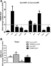
Gene expression patterns in ovary at dietrus. (A) Among the 10 genes related to steroid production and ovarian function examined by qRT-PCR, mRNA levels were significantly reduced in 6 (Lhcgr, Cyp17, Cyp19, StAR, Timp1, and Cdkn1b) and significantly increased in 2 (Fshr and Esr2) in control (Con)-DHT ovary compared with that of control-no DHT ovary 3 months after insertion of pellet. (B) Only Timp1 was recovered in ThARKO-DHT ovary compared with that of control-no DHT ovary. Bars with different letters represent significantly different values from each other with P < 0.05, analyzed by 1-way ANOVA followed by Tukey posttest. Values are mean ± SEM; n = 5 to 7/group.

References

    1. Walters KA. Role of androgens in normal and pathological ovarian function. Reproduction. 2015;149(4):R193–R218. - PubMed
    1. Hu YC, Wang PH, Yeh S, Wang RS, Xie C, Xu Q, Zhou X, Chao HT, Tsai MY, Chang C. Subfertility and defective folliculogenesis in female mice lacking androgen receptor. Proc Natl Acad Sci USA. 2004;101(31):11209–11214. - PMC - PubMed
    1. Shiina H, Matsumoto T, Sato T, Igarashi K, Miyamoto J, Takemasa S, Sakari M, Takada I, Nakamura T, Metzger D, Chambon P, Kanno J, Yoshikawa H, Kato S. Premature ovarian failure in androgen receptor-deficient mice. Proc Natl Acad Sci USA. 2006;103(1):224–229. - PMC - PubMed
    1. Walters KA, McTavish KJ, Seneviratne MG, Jimenez M, McMahon AC, Allan CM, Salamonsen LA, Handelsman DJ. Subfertile female androgen receptor knockout mice exhibit defects in neuroendocrine signaling, intraovarian function, and uterine development but not uterine function. Endocrinology. 2009;150(7):3274–3282. - PMC - PubMed
    1. Wu S, Chen Y, Fajobi T, DiVall SA, Chang C, Yeh S, Wolfe A. Conditional knockout of the androgen receptor in gonadotropes reveals crucial roles for androgen in gonadotropin synthesis and surge in female mice. Mol Endocrinol. 2014;28(10):1670–1681. - PMC - PubMed

Substances