Comparative Study
. 2016 Nov 14;4(1):119.
doi: 10.1186/s40478-016-0390-8.

Rubidium and potassium levels are altered in Alzheimer's disease brain and blood but not in cerebrospinal fluid

Affiliations
Comparative Study

Rubidium and potassium levels are altered in Alzheimer's disease brain and blood but not in cerebrospinal fluid

Blaine R Roberts et al. Acta Neuropathol Commun. .

Abstract

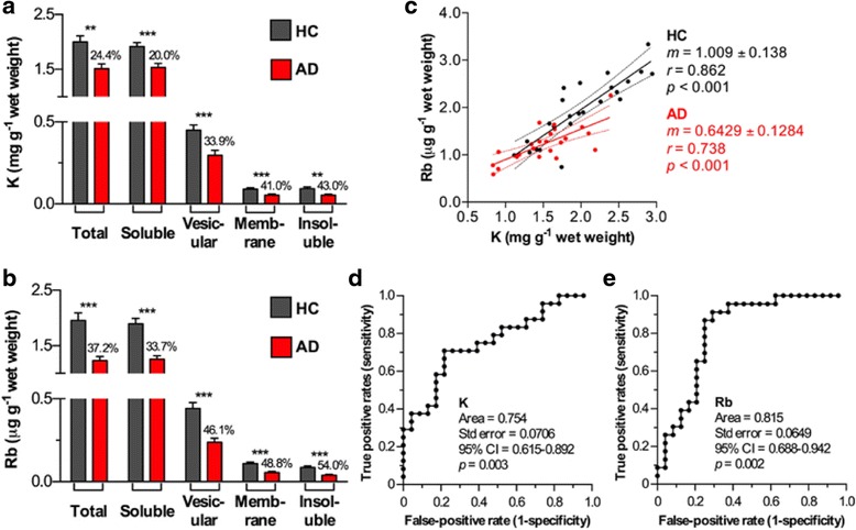
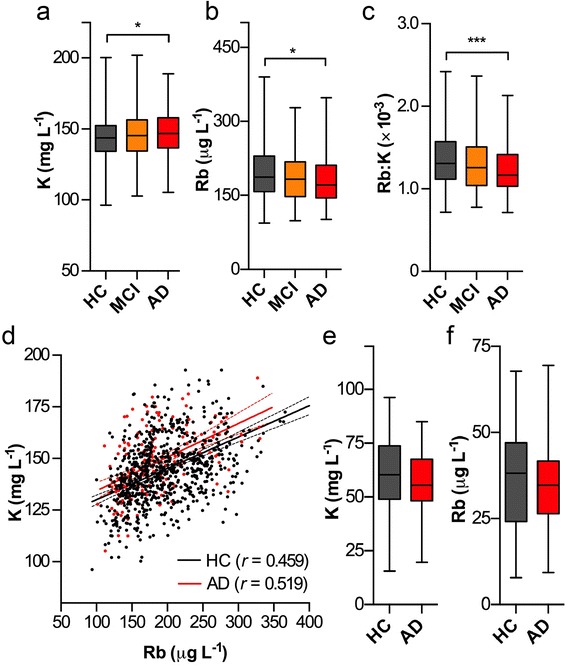
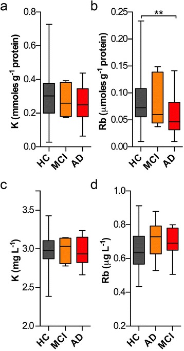
Loss of intracellular compartmentalization of potassium is a biochemical feature of Alzheimer's disease indicating a loss of membrane integrity and mitochondrial dysfunction. We examined potassium and rubidium (a biological proxy for potassium) in brain tissue, blood fractions and cerebrospinal fluid from Alzheimer's disease and healthy control subjects to investigate the diagnostic potential of these two metal ions. We found that both potassium and rubidium levels were significantly decreased across all intracellular compartments in the Alzheimer's disease brain. Serum from over 1000 participants in the Australian Imaging, Biomarkers and Lifestyle Flagship Study of Ageing (AIBL), showed minor changes according to disease state. Potassium and rubidium levels in erythrocytes and cerebrospinal fluid were not significantly different according to disease state, and rubidium was slightly decreased in Alzheimer's disease patients compared to healthy controls. Our data provides evidence that contrasts the hypothesized disruption of the blood-brain barrier in Alzheimer's disease, with the systemic decrease in cortical potassium and rubidium levels suggesting influx of ions from the blood is minimal and that the observed changes are more likely indicative of an internal energy crisis within the brain. These findings may be the basis for potential diagnostic imaging studies using radioactive potassium and rubidium tracers.


Figures

Fig. 1
Fig. 1
a, b K and Rb levels in fractionated brain homogenates were consistently decreased in AD brains (n = 30 per group; one-way ANOVA; ** p < 0.01; *** p < 0.001; percentage decrease in AD group compared to healthy controls shown). Error bars represent the standard error of the mean. c Both metals showed significant correlation regardless of clinical classification (p < 0.001), though the slope of the line of best fit was significantly decreased in the AD group (p < 0.05). d, e Both metals performed well at predicting AD via ROC curve analysis, with Rb slightly superior performance
Fig. 2
Fig. 2
K levels were significantly increased in AD serum (a; one-way ANOVA; * p < 0.05), whilst Rb levels were conversely decreased in AD (b; * p < 0.05); the latter mirroring our observation in brain homogenates. The ratio of Rb to K was significantly (*** p < 0.001) decreased in AD (c), though Rb and K levels were less well correlated in serum (d). In a subset (n = 30 per group) of erythrocytes from HC and AD groups, there was no significance difference in metal concentration (e, f). Boxes depict 25th and 75th percentiles and mean; error bars represent minimum to maximum values
Fig. 3
Fig. 3
K and Rb levels in platelets and CSF. a, b In platelets no significant changes were observed for K, though Rb was significantly decreased (p < 0.01; one-way ANOVA) in AD subjects compared to controls. c, d K and Rb levels in CSF remained unchanged. Boxes depict 25th and 75th percentiles and mean; error bars represent minimum to maximum values

References

    1. Mattson MP. Pathways towards and away from Alzheimer’s disease. Nature. 2004;430(7000):631–9. doi: 10.1038/nature02621. - DOI - PMC - PubMed
    1. Kamat PK, Kalani A, Rai S, Swarnkar S, Tota S, Nath C, Tyagi N. Mechanism of oxidative stress and synapse dysfunction in the pathogenesis of Alzheimer’s disease: understanding the Therapeutics strategies. Mol Neurobiol. 2014;53(1):648–61. doi: 10.1007/s12035-014-9053-6. - DOI - PMC - PubMed
    1. Butterfield D. Amyloid β-peptide (1-42)-induced oxidative stress and neurotoxicity: implications for neurodegeneration in Alzheimer’s disease brain. A review. Free Radic Res. 2002;36(12):1307–13. doi: 10.1080/1071576021000049890. - DOI - PubMed
    1. Crews L, Masliah E. Molecular mechanisms of neurodegeneration in Alzheimer’s disease. Hum Mol Genet. 2010;19(R1):R12–20. doi: 10.1093/hmg/ddq160. - DOI - PMC - PubMed
    1. Angelova PR, Abramov AY. Alpha-synuclein and beta-amyloid – different targets, same players: calcium, free radicals and mitochondria in the mechanism of neurodegeneration. Biochem Biophys Res Commun. 2016. - PubMed

Publication types