. 2016 Oct 6:(622):1-36.
doi: 10.3897/zookeys.622.9420. eCollection 2016.

Combined morphological and molecular data unveils relationships of Pseudobranchiomma (Sabellidae, Annelida) and reveals higher diversity of this intriguing group of fan worms in Australia, including potentially introduced species

Affiliations

Combined morphological and molecular data unveils relationships of Pseudobranchiomma (Sabellidae, Annelida) and reveals higher diversity of this intriguing group of fan worms in Australia, including potentially introduced species

María Capa et al. Zookeys. .

Abstract

Pseudobranchiomma (Sabellidae, Annelida) is a small and heterogeneous group of fan worms found in shallow marine environments and is generally associated with hard substrates. The delineation and composition of this genus is problematic since it has been defined only by plesiomorphic characters that are widely distributed among other sabellids. In this study we have combined morphological and molecular (mitochondrial and nuclear DNA sequences) data to evaluate species diversity in Australia and assess the phylogenetic relationships of these and other related sabellids. Unlike morphological data alone, molecular data and combined datasets suggest monophyly of Pseudobranchiomma. In this study, a new species of Pseudobranchiomma is described and three others are considered as potential unintentional introductions to Australian waters, one of them reported for the first time for the continent. Pseudobranchiomma pallidasp. n. bears 4-6 serrations along the radiolar flanges, lacks radiolar eyes and has uncini with three transverse rows of teeth over the main fang. In the new species the colour pattern as well is characteristic and species specific.

Keywords: dichotomous key; feather duster worms; invasive species; new species; sabellids; taxonomy; translocations.


Figures

Figure 1.
Figure 1.
Phylogenetic hypothesis of Pseudobranchiomma and related taxa. A Strict consensus of six most-parsimonious trees after analyses of morphological data (33 characters) and 26 members of Sabellidae rooted with Pseudopotamilla. Jack-knife support values are given (>50) B Strict consensus of three most parsimonious tree after implementation of implied weighting (constant of concavity k = 7) C Tree after maximum likelihood analyses of mitochondrial and nuclear dataset. Bootstrap values on nodes if >50; scale: average of nucleotide substitutions per site D Single most-parsimonious tree after analyses of the combined morphological and molecular datasets (12 taxa and 2239 characters); Jack-knife support values are given (>50).
Figure 2.
Figure 2.
Pseudobranchiomma cf. Pseudobranchiomma emersoni AM W.36365; photographs of preserved specimen; A Radiolar crown, dorsal view B Detached radiole C Detail of base of crown and single radiole D Anterior segments, ventral view (crown detached) E Same, lateral view F Same, dorsal view. vl, ventral lappet; vs, ventral sacs; white arrows, serrations of radiolar lateral flanges; black arrow, gap between ventral shields and thoracic tori.
Figure 3.
Figure 3.
Line drawings of chaetae and uncini of Pseudobranchiomma species in Australia; AF Pseudobranchiomma cf. Pseudobranchiomma emersoni GL Pseudobranchiomma cf. Pseudobranchiomma orientalis M–T Pseudobranchiomma pallida sp. n. UZ, A1 Pseudobranchiomma cf. Pseudobranchiomma schizogenica; A Thoracic uncinus B Abdominal uncinus C Superior thoracic chaeta D Inferior thoracic chaeta E, F Inferior abdominal chaetae G Thoracic uncinus H Abdominal uncinus I Superior thoracic chaeta J Inferior thoracic chaeta K, L Inferior abdominal chaetae M, N Thoracic uncini O Abdominal uncinus P, Q, R Inferior thoracic chaetae S Superior abdominal chaeta T Inferior abdominal chaeta U, V Thoracic uncini W Abdominal uncinus X, Y Superior thoracic chaetae Z, A1 Inferior thoracic chaetae. Scale bars: A–F = 2 µm; G–L = 4 µm; M–T = 2 µm; U–Z, A1 = 2 µm.
Figure 4.
Figure 4.
Pseudobranchiomma cf. Pseudobranchiomma orientalis AM W.32677, AM W.32679: Photographs. A Anterior end, ventral view B Detached radiole C Detail of lateral radiole, mid length D Detail of base lateral radiole E Detail of radiolar crown base with dorsal radiolar appendages F Detail of radiolar crown base with ventral sacs G Anterior thoracic parapodia H Posterior abdominal segments. dra, dorsal radiolar appendages; vs, ventral sac; black arrows, interramal eyespots.
Figure 5.
Figure 5.
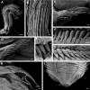
Pseudobranchiomma cf. Pseudobranchiomma orientalis AM W.37204: Scanning electron micrographs A Anterior end, lateroventral view B Detail of lateral flanges serrations C Notopodia, first thoracic segment D Notopodia, fifth thoracic segment E Uncini, second thoracic segment F Uncini, fifth thoracic segment G Posterior abdominal uncini H Neuropodia, mid abdominal segment I Posterior end, ventral view. Scale bars: A = 1 mm; B, I = 100 µm; C, D, G, H = 20 µm; E = 3 µm; F = 10 µm.
Figure 6.
Figure 6.
Pseudobranchiomma orientalis Type BMNH 85.12.1.393. Line drawings by Phyllis Knight-Jones. A–C Second dorsal radiole from different views D Holotype, divided in two, and partially covered by the tube, lateroventral view E Base of crown and anterior thoracic chaetigers, ventral view F Same, lateral view G Same, dorsal view H Thoracic parapodium I Abdominal parapodium. Scale bars: A–C = 1 mm; D = 2 mm; E–G = 1 mm; H–I = unknown.
Figure 7.
Figure 7.
Pseudobranchiomma pallida sp. n. AM W.36366: Photographs. A Whole specimen, ventral view B Whole specimen, lateral view C Lateral radioles D Detail of base of radiolar crown and anterior segments E Anterior thoracic segments, ventrolateral view F Anterior end, dorsal view. vl, ventral lappet; vs, ventral sac; white arrows, serrations of radiolar lateral flanges; black arrow, gap between ventral shields and thoracic tori.
Figure 8.
Figure 8.
Pseudobranchiomma cf. Pseudobranchiomma schizogenica. AM W.36368, AM W.36369. Photographs. A Anterior end, lateral view B Detail of base of crown and anterior segments, ventral view C Detail of lateral radiole D Anterior thoracic segments, lateral view E Anterior thoracic segments, dorsal view F Anterior thoracic segments, dorsal view. dra, dorsal radiolar appendages; vl, ventral lappet; vs, ventral sac; white arrows, serrations of radiolar lateral flanges.
Figure 9.
Figure 9.
Pseudobranchiomma cf. Pseudobranchiomma schizogenica. Scanning electron micrographs. A–F specimen from Queensland AM W.37205 G–L specimen from Western Australia AM W.37203. A Whole specimen, lateral view B Base of crown, dorsal view with dorsal lips and radiolar appendages C Notopodia, second thoracic segment D Uncini, third thoracic segment E Posterior abdominal neurochaetae F Uncini, posterior abdominal segment G Detail of lateral radiole with serrations in flanges H Notopodia, first thoracic segment I Uncini, fifth thoracic segment J Notopodia fourth thoracic segment K Neuropodia, mid abdominal chaetigers L Posterior abdominal uncini. Scale bars: A = 200 µm; B, G = 100 µm; C = 20 µm; D, F, I, L = 2 µm; E, H, J, K = 10 µm.
Figure 10.
Figure 10.
Pseudobranchiomma cf. Pseudobranchiomma schizogenica from Hawaii AM W.37206, AM W.37207: Scanning electron micrographs. A Detail of radiolar flanges serrations B Base of crown and anterior segments, ventral view C Lateral radioles D Anterior segments, dorsal view E Notopodia, first thoracic segment F Notopodia, second thoracic segment G Uncini, third thoracic segment H Neuropodia, posterior abdominal segment I Posterior abdominal uncini J Posterior end, ventral view (pygidium regenerating) K Posterior end, dorsal view. vl, ventral lappets; vs, ventral sac. Scale bars: A = 20 µm; B, C, J, K = 100 µm; D = 200 µm; E, H = 10 µm; F = 20 µm; G, I = 2 µm.
Figure 11.
Figure 11.
Pseudobranchiomma grandis from New Zealand. A Whole live animal B Section of radioles, showing paired radiolar eyes and serrations of lateral flanges. Photos by Rod Asher.

References

    1. Arias A, Giangrande A, Gambi MC, Anadón N. (2013) Biology and new records of the invasive species Branchiomma bairdi (Annelida: Sabellidae) in the Mediterranean Sea. Mediterranean Marine Science 14: 162–171. doi: 10.12681/mms.363 - DOI
    1. Baird W. (1865) Description of several new species and varieties of tubicolous Annelides = tribe Limivora of Grube, in the collection of the British Museum. Part 1. Journal of the Linnean Society of London 8: 10–22.
    1. Bush KJ. (1905) Tubicolous annelids of the tribes Sabellides and Serpulides from the Pacific Ocean. Harriman Alaska Expedition 12: 169–346.
    1. Capa M. (2008) The genera Bispira Krøyer, 1856 and Stylomma Knight-Jones, 1997 (Polychaeta, Sabellidae): systematic revision, relationships with close related taxa and new species from Australia. Hydrobiologia 596: 301–327. doi: 10.1007/s10750-007-9105-2 - DOI
    1. Capa M, Bybee DR, Bybee SM. (2010) Establishing species and species boundaries in Sabellastarte Krøyer, 1856 (Annelida: Sabellidae): an integrative approach. Organisms, Diversity and Evolution 10: 351–371. doi: 10.1007/s13127-010-0033-z - DOI

LinkOut - more resources