. 2016 Nov 11:7:22.
doi: 10.1186/s13100-016-0077-5. eCollection 2016.

Somatic retrotransposition is infrequent in glioblastomas

Affiliations

Somatic retrotransposition is infrequent in glioblastomas

Pragathi Achanta et al. Mob DNA. .

Abstract

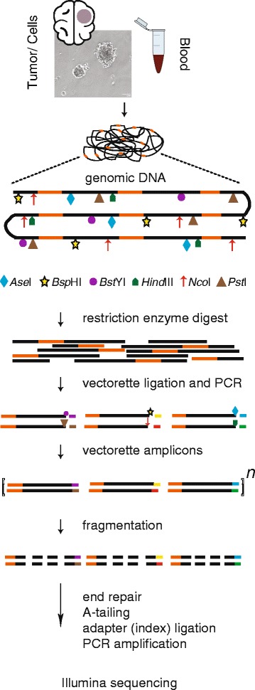
Background: Gliomas are the most common primary brain tumors in adults. We sought to understand the roles of endogenous transposable elements in these malignancies by identifying evidence of somatic retrotransposition in glioblastomas (GBM). We performed transposon insertion profiling of the active subfamily of Long INterspersed Element-1 (LINE-1) elements by deep sequencing (TIPseq) on genomic DNA of low passage oncosphere cell lines derived from 7 primary GBM biopsies, 3 secondary GBM tissue samples, and matched normal intravenous blood samples from the same individuals.

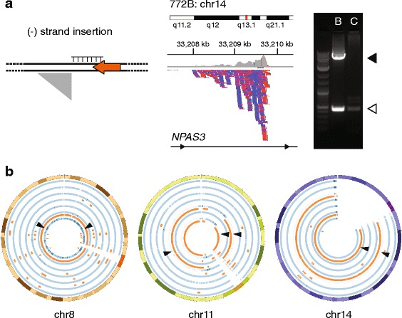
Results: We found and PCR validated one somatically acquired tumor-specific insertion in a case of secondary GBM. No LINE-1 insertions present in primary GBM oncosphere cultures were missing from corresponding blood samples. However, several copies of the element (11) were found in genomic DNA from blood and not in the oncosphere cultures. SNP 6.0 microarray analysis revealed deletions or loss of heterozygosity in the tumor genomes over the intervals corresponding to these LINE-1 insertions.

Conclusions: These findings indicate that LINE-1 retrotransposon can act as an infrequent insertional mutagen in secondary GBM, but that retrotransposition is uncommon in these central nervous system tumors as compared to other neoplasias.

Keywords: Cancer; Glioblastoma; LINE-1; Retrotransposition.


Figures

Fig. 1
Fig. 1
Transposon Insertion Profiling by sequencing (TIPseq) workflow. High molecular weight genomic DNA was extracted from primary and secondary glioblastoma (GBM) tumors, oncosphere cultures expanded from primary GBM, and matched blood samples from the same patients. Genomic DNA was then digested in six parallel reactions each using one of a panel of restriction enzymes. In the diagram, LINE-1 insertions are depicted as orange segments of the genomic DNA; restriction enzyme cuts sites are illustrated with different symbols. Vectorette oligonucleotides designed to match each restriction enzyme sticky end were ligated to the DNA fragments, and the 3′ ends of LINE-1 sequences and downstream DNA were amplified. Genomic DNA fragments without binding sites for the LINE-1 amplification primer are not enriched in this PCR. Amplified DNA was then randomly sheared and prepared for Illumina sequencing
Fig. 2
Fig. 2
TIPseq in primary glioblastoma GBM oncosphere lines and corresponding blood samples. a. TIPseq data. (Leftmost panel) The schematic depicts a minus (-) strand L1 as a leftward facing orange arrow. The LINE-1 sequence ends with a 3′ polyA tail, shown as a homopolymer of thymine (T) on the complementary strand. The gray right triangle illustrates the shape of sequencing reads piling up (vertically, downward) when mapped against in the reference genome (i.e., with genome coordinates depicted on the horizontal axis). (Central panel) TIPseq read alignments corresponding to an insertion detected in blood and not the patient’s oncosphere cell line. The insertion is in an intron of the NPAS3 gene (14q13.1). Read depth is illustrated on the top (gray) and individual reads are represented as blue and red bars denoting orientation. (Rightmost panel) An agarose gel electrophoresis of a validation PCR. The open arrowhead (lower) marks the pre-insertion allele and the solid arrowhead (upper) marks the amplicon spanning the LINE-1 insertion. The insertion is detected in the blood (B) sample for this patient and not the corresponding tumor cells (C). The LINE-1 is 5.4 kb. b. Copy number and loss of heterozygosity (LOH) studies on the oncosphere cell lines. Results for chromosomes 8, 11 and 14 are shown in Circos plots. The seven samples are each depicted as two circular tracks of data. The blue track indicates copy number; medium blue is diploid, darker blue shows amplifications, and lighter blue shows deletions. The orange track highlights regions with LOH. (Leftmost circle) Two insertions on chromosome 8 are marked with arrowheads at 25 and 120 MB; both were identified in a blood sample and not the corresponding patient’s oncosphere cell line, which showed a copy neutral LOH of the entire chromosome (847). (Central circle) Three LINE-1 insertions on chromosome 11 found in blood only are marked; two are the same insertion at 33.6 MB found in two different patient samples (847, 922). Both tumor cell lines had deletions with copy number decreases and LOH at this site. One of these cases (847) also had loss of material near 94.3 MB associated with deletion of a second LINE-1. (Rightmost circle) Two LINE-1 are marked with arrowheads at 30.2 and 33.2 MB on acrocentric chromosome 14. These were found in genomic DNA from blood, but were lost owing to LOH in the corresponding oncosphere lines (897, 772)
Fig. 3
Fig. 3
Expression and somatic retrotransposition of LINE-1 in secondary GBM. a. and b. LINE-1 ORF1p immunohistochemistry. a. Most primary GBMs and low grade gliomas do not have detectable LINE-1 ORF1p in this assay. Nuclei are counterstained in blue. b. About 74 % of secondary GBM cases are weakly, focally immunoreactive for LINE-1 ORF1p. (Brown). c-f. Identification of a somatically acquired LINE-1 insertion. c. The schematic depicts a plus (+) strand L1 as a rightward facing orange arrow with its 5′ inversion as a leftward facing blue arrow. The genomic LINE-1 sequence ends with a 3′ polyA tail. The gray right triangle illustrates the sequencing reads piling up (vertically, downward) when mapped against in the reference genome on the horizontal axis. d. TIPseq read alignments corresponding to an insertion detected in a secondary GBM tumor sample and not the patient’s blood DNA. The insertion is an intergenic LINE-1 on chromosome 17q22. Read depth is illustrated on the top (gray) and individual reads are stacked downward as blue and red bars denoting orientation. The greatest depth is immediately adjacent to the LINE-1 and extends 3′ of the element to create the triangular shape. e. An agarose gel electrophoresis of a validation PCR. The open arrowhead (lower) marks the pre-insertion allele and the solid arrowhead (upper) marks the amplicon that spans the LINE-1 insertion. The insertion is detected in the tumor (T) sample for this patient and not the corresponding blood cells (B). The LINE-1 is 5′ truncated at 1.8 kb. f. The annotated Sanger sequence for the LINE-1 insertion is shown in colored text: flanking unique genomic DNA (black), target site duplication (red), LINE-1 5′ inversion (blue), and LINE-1 3′ sequence and polyA tail (orange). Lowercase letters denote lower quality basecalls. These were confirmed by manually examining the trace file

References

    1. Cancer Genome Atlas Research N Comprehensive genomic characterization defines human glioblastoma genes and core pathways. Nature. 2008;455(7216):1061–1068. doi: 10.1038/nature07385. - DOI - PMC - PubMed
    1. Parsons DW, Jones S, Zhang X, Lin JC, Leary RJ, Angenendt P, Mankoo P, Carter H, Siu IM, Gallia GL, et al. An integrated genomic analysis of human glioblastoma multiforme. Science. 2008;321(5897):1807–1812. doi: 10.1126/science.1164382. - DOI - PMC - PubMed
    1. Burns KH, Boeke JD. Human transposon tectonics. Cell. 2012;149(4):740–752. doi: 10.1016/j.cell.2012.04.019. - DOI - PMC - PubMed
    1. Huang CR, Schneider AM, Lu Y, Niranjan T, Shen P, Robinson MA, Steranka JP, Valle D, Civin CI, Wang T, et al. Mobile interspersed repeats are major structural variants in the human genome. Cell. 2010;141(7):1171–1182. doi: 10.1016/j.cell.2010.05.026. - DOI - PMC - PubMed
    1. Beck CR, Collier P, Macfarlane C, Malig M, Kidd JM, Eichler EE, Badge RM, Moran JV. LINE-1 retrotransposition activity in human genomes. Cell. 2010;141(7):1159–1170. doi: 10.1016/j.cell.2010.05.021. - DOI - PMC - PubMed

LinkOut - more resources