. 2016 Mar 16;1(2):e000024.
doi: 10.1136/esmoopen-2015-000024. eCollection 2016.

Descriptive statistical analysis of a real life cohort of 2419 patients with brain metastases of solid cancers

Affiliations

Descriptive statistical analysis of a real life cohort of 2419 patients with brain metastases of solid cancers

Anna S Berghoff et al. ESMO Open. .

Abstract

Aim: We provide a descriptive statistical analysis of baseline characteristics and the clinical course of a large real-life cohort of brain metastases (BM) patients.

Methods: We performed a retrospective chart review for patients treated for BM of solid cancers at the Medical University of Vienna between 1990 and 2011.

Results: We identified a total of 2419 BM patients (50.5% male, 49.5% female, median age 59 years). The primary tumour was lung cancer in 43.2%, breast cancer in 15.7%, melanoma in 16.4%, renal cell carcinoma in 9.1%, colorectal cancer in 9.3% and unknown in 1.4% of cases. Rare tumour types associated with BM included genitourinary cancers (4.1%), sarcomas (0.7%). gastro-oesophageal cancer (0.6%) and head and neck cancers (0.2%). 48.7% of patients presented with a singular BM, 27.7% with 2-3 and 23.5% with >3 BM. Time from primary tumour to BM diagnosis was shortest in lung cancer (median 11 months; range 1-162) and longest in breast cancer (median 44 months; 1-443; p<0.001). Multiple BM were most frequent in breast cancer (30.6%) and least frequent in colorectal cancer (8.5%; p<0.001). Patients with breast cancer had the longest median overall survival times (8 months), followed by patients with lung cancer (7 months), renal cell carcinoma (7 months), melanoma (5 months) and colorectal cancer (4 months; p<0.001; log rank test). Recursive partitioning analysis and graded prognostic assessment scores showed significant correlation with overall survival (both p<0.001, log rank test). Evaluation of the disease status in the past 2 months prior to patient death showed intracranial progression in 35.9%, extracranial progression in 27.5% and combined extracranial and intracranial progression in 36.6% of patients.

Conclusions: Our data highlight the heterogeneity in presentation and clinical course of BM patients in the everyday clinical setting and may be useful for rational planning of clinical studies.

Keywords: brain metastases; descriptive analysis; disease status in end of life period; prognosis.


Conflict of interest statement

Conflicts of Interest: None declared.

Figures
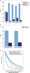
Figure 1
Figure 1
(A) Frequency of synchronous diagnosis of primary tumour and BM according to primary tumour type (B) Frequency of patients with asymptomatic BM at first diagnosis of primary tumour (C) Time from diagnosis to BM according to primary tumour type.
Figure 2
Figure 2
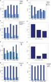
Clinical characteristics at diagnosis of brain metastases. (A) Extracranial involvement at diagnosis of BM according to primary tumour type (B) Frequency of progressive extracranial disease at diagnosis of BM according to primary tumour type (C) Frequency of brain only metastatic behaviour according to primary tumour type (D) Number of BM at first diagnosis of BM (E) Number of BM according to primary tumour type (F) Localisation of BM at diagnosis (G) Frequency of epileptic seizures at diagnosis of BM according to primary tumour type (H) Occurrence of additional leptomeningeal carcinomatosis after diagnosis of BM according to primary tumour type.
Figure 3
Figure 3
Recurrence pattern after initial treatment of brain metastases and cause of death. (A) Frequency of extracranial progression after initial treatment of BM according to primary tumour type (B) Frequency of intracranial progression after initial treatment of BM according to primary tumour type (C) Disease status in the end of life period (D) Disease status in the end of life period according to primary tumour type.
Figure 4
Figure 4
Frequency patterns of brain metastases over time. (A) Frequency of BM diagnosed between 1994 and 2010 (B) Frequency of BM according to primary tumour between 1994 and 2010 (C) Frequency of lung cancer BM in female and male patients between 1994 and 2010 (D) Overall survival from diagnosis of BM according to year of diagnosis.

References

    1. Caissie A, Nguyen J, Chen E et al. . Quality of life in patients with brain metastases using the EORTC QLQ-BN20+2 and QLQ-C15-PAL. Int J Radiat Oncol Biol Phys 2012;83:1238–45. 10.1016/j.ijrobp.2011.09.025 - DOI - PubMed
    1. Gavrilovic IT, Posner JB. Brain metastases: epidemiology and pathophysiology. J Neurooncol 2005;75:5–14. 10.1007/s11060-004-8093-6 - DOI - PubMed
    1. Barnholtz-Sloan JS, Sloan AE, Davis FG et al. . Incidence proportions of brain metastases in patients diagnosed (1973 to 2001) in the Metropolitan Detroit Cancer Surveillance System. J Clin Oncol 2004;22:2865–72. 10.1200/JCO.2004.12.149 - DOI - PubMed
    1. Bartsch R, Berghoff AS, Preusser M. Optimal management of brain metastases from breast cancer. Issues and considerations. CNS Drugs 2013;27:121–34. 10.1007/s40263-012-0024-z - DOI - PubMed
    1. Eichler AF, Chung E, Kodack DP et al. . The biology of brain metastases-translation to new therapies. Nat Rev Clin Oncol 2011;8:344–56. 10.1038/nrclinonc.2011.58 - DOI - PMC - PubMed

LinkOut - more resources