T2 Fluid-Attenuated Inversion Recovery Imaging of Uveal Melanomas and Other Ocular Pathology
- PMID: 27843906
- PMCID: PMC5091204
- DOI: 10.1159/000447265
T2 Fluid-Attenuated Inversion Recovery Imaging of Uveal Melanomas and Other Ocular Pathology
Abstract
Background/aims: This study describes patterns of intraocular lesions on T2 fluid-attenuated inversion recovery (FLAIR) imaging, exploring a prospective role of FLAIR imaging sequence in diagnosis and treatment.
Methods: A retrospective study of orbital magnetic resonance imaging (MRI) studies from the years 2000 to 2015 was performed. MRI sequences included: pre-contrast T1-weighted, T2-weighted, T2 FLAIR, and postcontrast T1 and T2 imaging gadolinium, which were evaluated by a neuroradiologist. Two cases of melanoma were correlated to their pathology.
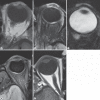
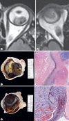
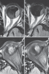
Results: Twenty-four patients with intraocular pathology were evaluated. All lesions, regardless of pigmentation, revealed previously described melanotic patterns on T1- and T2-weighted images; 80% of 10 melanomas localized were hyperintense on T2 FLAIR, which better delineated lesion margins. All of the four inflammatory pathologies on T2 FLAIR were hyperintense, as were 80% of the amelanotic neoplasms. Pathology of two large uveal melanomas paralleled the findings seen on T2 FLAIR.
Conclusions: T2 FLAIR appears beneficial in the demarcation of pigmented ocular lesions and may aid in determining protein content or previous treatment. Data also promote previous assertions that blood flow impacts intensity of lesions on T2 FLAIR. Further research is warranted.
Keywords: Choroidal hemangioma; Magnetic resonance imaging; Melanoma; Posttreatment changes; T2 fluid-attenuated inversion recovery; Uveal melanoma.
Figures


References
-
- Baek HJ, Lee SJ, Cho KH, Choo HJ, Lee SM, Lee YH, Suh KJ, Moon TY, Cha JG, Yi JH, Kim MH, Jung SJ, Choi JH. Subungual tumors: clinicopathologic correlation with US and MR imaging findings. Radiographics. 2010;30:1621–1636. - PubMed
-
- Bakri SJ, Sculley L, Singh AD. Imaging techniques for uveal melanoma. Int Ophthalmol Clin. 2006;46:1–13. - PubMed
-
- Gomori JM, Grossman RI, Shields JA, Augsburger JJ, Joseph PM, DeSimeone D. Choroidal melanomas: correlation of NMR spectroscopy and MR imaging. Radiology. 1986;158:443–445. - PubMed
-
- Mafee MF, Peyman GA, Grisolano JE, Fletcher ME, Spigos DG, Wehrli FW, Rasouli F, Capek V. Malignant uveal melanoma and simulating lesions: MR imaging evaluation. Radiology. 1986;160:773–780. - PubMed
LinkOut - more resources
Full Text Sources
Other Literature Sources

