

Trichoscopy of Focal Alopecia in Children - New Trichoscopic Findings: Hair Bulbs Arranged Radially along Hair-Bearing Margins in Aplasia Cutis Congenita
- PMID: 27843914
- PMCID: PMC5096236
- DOI: 10.1159/000445721
Trichoscopy of Focal Alopecia in Children - New Trichoscopic Findings: Hair Bulbs Arranged Radially along Hair-Bearing Margins in Aplasia Cutis Congenita
Abstract
Purpose: To establish whether trichoscopy can be useful in the differential diagnosis of patchy alopecia in children.
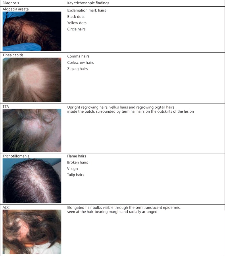
Procedures: The study was a retrospective analysis (2012-2015) and included 68 patients under 6 years of age. The inclusion criteria were age and the presence of 1-3 alopecia patches. A total of 124 alopecia patches were examined with the use of a videodermoscope: 102 alopecia areata, 8 tinea capitis, 6 trichotillomania, 3 temporal triangular alopecia and 5 aplasia cutis congenita.
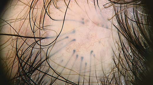
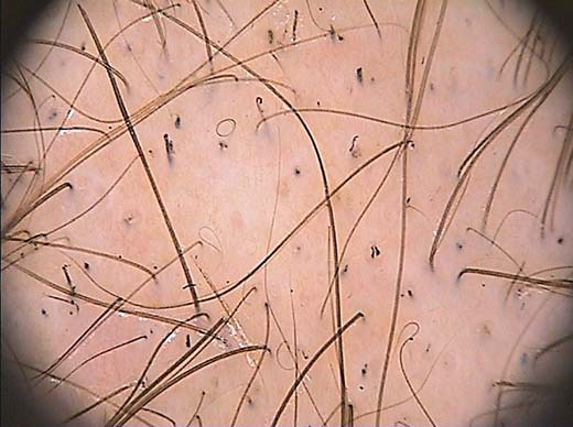
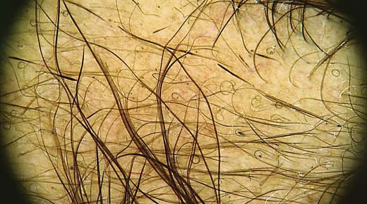
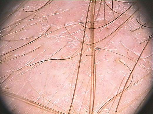
Results: In all aplasia cutis congenita lesions, trichoscopy revealed elongated hair bulbs visible through the semitranslucent epidermis, seen at the hair-bearing margin and radially arranged. Hair regrowth [upright regrowing hairs (44%), circular hairs (23%) and vellus hairs (20%)] was observed in the majority of alopecia areata patches. For triangular alopecia, upright regrowing hairs (100%; 3/3), vellus hairs (100%; 3/3) and circle hairs (33%; 1/3) were seen inside the alopecia patch.
Conclusion: Trichoscopy is a useful technique for the differential diagnosis of patchy alopecia in children. A novel finding in this study indicates that radially arranged hair bulbs visible through the translucent epidermis are characteristic of nonbullous type aplasia cutis congenita.
Keywords: Alopecia areata; Alopecia trichoscopy; Aplasia cutis congenital; Dermoscopy.
Figures
Similar articles
-
Clinical Significance of Trichoscopy in Common Causes of Hair Loss in Children: Analysis of 134 Cases.Int J Trichology. 2018 Jul-Aug;10(4):154-161. doi: 10.4103/ijt.ijt_101_17. Int J Trichology. 2018. PMID: 30386074 Free PMC article.
-
Trichoscopy of alopecia areata in children. A retrospective comparative analysis of 50 children and 50 adults.Pediatr Dermatol. 2019 Sep;36(5):640-645. doi: 10.1111/pde.13912. Epub 2019 Jul 11. Pediatr Dermatol. 2019. PMID: 31294493
-
Alopecia areata predictive score: A new trichoscopy-based tool to predict treatment outcome in patients with patchy alopecia areata.J Cosmet Dermatol. 2020 Mar;19(3):746-751. doi: 10.1111/jocd.13064. Epub 2019 Jul 13. J Cosmet Dermatol. 2020. PMID: 31301100
-
Hair shafts in trichoscopy: clues for diagnosis of hair and scalp diseases.Dermatol Clin. 2013 Oct;31(4):695-708, x. doi: 10.1016/j.det.2013.06.007. Dermatol Clin. 2013. PMID: 24075554 Review.
-
[Translated article] Trichoscopy in Alopecia Areata.Actas Dermosifiliogr. 2023 Jan;114(1):T25-T32. doi: 10.1016/j.ad.2022.08.027. Epub 2022 Nov 8. Actas Dermosifiliogr. 2023. PMID: 36368582 Review. English, Spanish.
Cited by
-
The Trichoscopic "Golf Club Set" Sign for Bullous Aplasia Cutis Congenita.Skin Appendage Disord. 2018 Oct;4(4):320-322. doi: 10.1159/000486463. Epub 2018 Feb 7. Skin Appendage Disord. 2018. PMID: 30410906 Free PMC article.
-
Dermoscopy of Aplasia Cutis Congenita: A Case Report and Review of the Literature.Dermatol Pract Concept. 2021 Jan 29;11(1):e2021154. doi: 10.5826/dpc.1101a154. eCollection 2021 Jan. Dermatol Pract Concept. 2021. PMID: 33614222 Free PMC article. No abstract available.
-
Trichoscopy in Alopecia Areata and Trichotillomania in Skin of Colour: A Comparative Study.Indian J Dermatol. 2023 Jan-Feb;68(1):78-84. doi: 10.4103/ijd.ijd_587_22. Indian J Dermatol. 2023. PMID: 37151271 Free PMC article.
-
1-Year Hospital-Based Observational Study of Trichoscopy Findings and Disease Activity in Alopecia Areata.Indian Dermatol Online J. 2020 Sep 19;11(6):965-969. doi: 10.4103/idoj.IDOJ_19_20. eCollection 2020 Nov-Dec. Indian Dermatol Online J. 2020. PMID: 33344348 Free PMC article.
-
Trichoscopic Evaluation of Focal Non-Cicatricial Alopecia in Egyptian Children.Dermatol Pract Concept. 2024 Oct 30;14(4):e2024238. doi: 10.5826/dpc.1404a238. Dermatol Pract Concept. 2024. PMID: 39652957 Free PMC article.
References
-
- Lencastre A, Tosti A. Role of trichoscopy in children's scalp and hair disorders. Pediatr Dermatol. 2013;30:674–682. - PubMed
-
- Rudnicka L, Olszewska M, Rakowska A, Kowalska-Oledzka E, Slowinska M. Trichoscopy: a new method for diagnosing hair loss. J Drugs Dermatol. 2008;7:651–654. - PubMed
-
- Inui S, Nakajima T, Itami S. Coudability hairs: a revisited sign of alopecia areata assessed by trichoscopy. Clin Exp Dermatol. 2010;35:361–365. - PubMed
-
- Rakowska A, Slowinska M, Olszewska M, Rudnicka L. New trichoscopy findings in trichotillomania: flame hairs, V-sign, hook hairs, hair powder, tulip hairs. Acta Derm Venereol. 2014;94:303–306. - PubMed
LinkOut - more resources
Full Text Sources
Other Literature Sources
