. 2016 Sep 28;3(11):844-853.
doi: 10.1002/acn3.344. eCollection 2016 Nov.

Archetypal NOTCH3 mutations frequent in public exome: implications for CADASIL

Affiliations

Archetypal NOTCH3 mutations frequent in public exome: implications for CADASIL

Julie W Rutten et al. Ann Clin Transl Neurol. .

Abstract

Objective: To determine the frequency of distinctive EGFr cysteine altering NOTCH3 mutations in the 60,706 exomes of the exome aggregation consortium (ExAC) database.

Methods: ExAC was queried for mutations distinctive for cerebral autosomal dominant arteriopathy with subcortical infarcts and leukoencephalopathy (CADASIL), namely mutations leading to a cysteine amino acid change in one of the 34 EGFr domains of NOTCH3. The genotype-phenotype correlation predicted by the ExAC data was tested in an independent cohort of Dutch CADASIL patients using quantified MRI lesions. The Dutch CADASIL registry was probed for paucisymptomatic individuals older than 70 years.

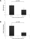
Results: We identified 206 EGFr cysteine altering NOTCH3 mutations in ExAC, with a total prevalence of 3.4/1000. More than half of the distinct mutations have been previously reported in CADASIL patients. Despite the clear overlap, the mutation distribution in ExAC differs from that in reported CADASIL patients, as mutations in ExAC are predominantly located outside of EGFr domains 1-6. In an independent Dutch CADASIL cohort, we found that patients with a mutation in EGFr domains 7-34 have a significantly lower MRI lesion load than patients with a mutation in EGFr domains 1-6.

Interpretation: The frequency of EGFr cysteine altering NOTCH3 mutations is 100-fold higher than expected based on estimates of CADASIL prevalence. This challenges the current CADASIL disease paradigm, and suggests that certain mutations may more frequently cause a much milder phenotype, which may even go clinically unrecognized. Our data suggest that individuals with a mutation located in EGFr domains 1-6 are predisposed to the more severe "classical" CADASIL phenotype, whereas individuals with a mutation outside of EGFr domains 1-6 can remain paucisymptomatic well into their eighth decade.


Figures

Figure 1
Figure 1
High frequency of EGFr cysteine altering NOTCH3 mutations in all populations in the ExAC database. Frequencies range from 0.4/1000 individuals in the African/African American population to 11.7/1000 in the South Asian population, with an overall frequency of 3.4/1000.*: Mutation previously described in cerebral autosomal dominant arteriopathy with subcortical infarcts and leukoencephalopathy (CADASIL) patients; †: Mutation reported three times in a homozygous state; (): Allele counts
Figure 2
Figure 2
Distribution of distinct EGFr cysteine altering NOTCH3 mutations in ExAC compared to those reported in cerebral autosomal dominant arteriopathy with subcortical infarcts and leukoencephalopathy (CADASIL) patients. Schematic representation of the NOTCH3 protein with 34 epidermal growth factor‐like repeat domains (EGFr) and the NOTCH3 EGFr encoding exons (exon 2–24). Exons 25–33 (and part of exon 24) encode the transmembrane and intracellular domains of NOTCH3; these have never been found to harbor CADASIL‐causing mutations. In the Dutch CADASIL registry and in CADASIL patients worldwide, most mutations are located in exon 4, which largely accounts for the predominance of mutations in EGFr domains 1–6. In ExAC, this predominance is not observed, as mutations are most frequent in exons 11–12 and 22, encoding EGFr domains 14–16 and 29–31, respectively. Pink bars above the EGFr domains represent the distribution of mutations across EGFr domains, where the height of the pink bar reflects the percentage of mutations in the respective EGFr domain. Exon colors reflect the number of mutations in each exon: gray indicates exons without mutations; blue indicates exons with mutations. The darker the color blue of the exon, the higher the number of distinct mutations.
Figure 3
Figure 3
Correlation between EGFr location of the NOTCH3 mutation and MRI lesion load in cerebral autosomal dominant arteriopathy with subcortical infarcts and leukoencephalopathy (CADASIL) patients. MRI quantification data from patients with a mutation in EGFr domains 1–6 (n = 32) compared to patients with a mutation in EGFr domains 7–34 (n = 8). (A) White matter hyperintensity volume (expressed as the % of total brain parenchymal volume), was significantly higher in patients with mutations in EGFr domains 1–6 than in patients with mutations in EGFr domains 7–34 (8.0% vs. 4.3%, P = 0.01). (B) The number of lacunar infarcts showed a similar trend (14.5 vs. 4.8, P = 0.05). Bars represent mean ± SEM, corrected for age, sex, smoking and hypertension.
Figure 4
Figure 4
Brain MRI of elderly asymptomatic and paucisymptomatic individuals with a NOTCH3 mutation compared to MRI in classical cerebral autosomal dominant arteriopathy with subcortical infarcts and leukoencephalopathy (CADASIL). (A–C) Brain MRI images of a classical CADASIL phenotype in the 6th decade. (A–B) FLAIR images showing extensive confluent symmetric white matter hyperintensities and multiple lacunar infarcts (arrows indicate random samples). (C) T2*‐weighted MRI showing multiple microbleeds (open arrows indicate random samples). (D–F) Brain MRI images of a female, diagnosed with CADASIL after predictive DNA testing at 78 years of age, with only very mild clinical symptoms. (D, E) FLAIR images showing symmetric white matter hyperintensities, but no lacunar infarcts. (F) T2*‐ weighted MRI showing some small microbleeds (open arrows). (G‐I) Dual echo images of a female who was still clinically asymptomatic at 73 years of age. MRI images were made at 58 years of age, showing very mild symmetric white matter hyperintensities, no lacunar infarcts and no microbleeds.

References

    1. Joutel A, Corpechot C, Ducros A, et al. Notch3 mutations in CADASIL, a hereditary adult‐onset condition causing stroke and dementia. Nature 1996;383:707–710. - PubMed
    1. Chabriat H, Joutel A, Dichgans M, et al. Cadasil. Lancet Neurol 2009;8:643–653. - PubMed
    1. Moreton FC, Razvi SS, Davidson R, et al. Changing clinical patterns and increasing prevalence in CADASIL. Acta Neurol Scand 2014;130:197–203. - PubMed
    1. Liem MK, Lesnik Oberstein SA, Haan J, et al. MRI correlates of cognitive decline in CADASIL: a 7‐year follow‐up study. Neurology 2009;72:143–148. - PubMed
    1. Viswanathan A, Gschwendtner A, Guichard JP, et al. Lacunar lesions are independently associated with disability and cognitive impairment in CADASIL. Neurology 2007;69:172–179. - PubMed