

Impaired neurovascular coupling in aging and Alzheimer's disease: Contribution of astrocyte dysfunction and endothelial impairment to cognitive decline
- PMID: 27845201
- PMCID: PMC5429210
- DOI: 10.1016/j.exger.2016.11.004
Impaired neurovascular coupling in aging and Alzheimer's disease: Contribution of astrocyte dysfunction and endothelial impairment to cognitive decline
Abstract
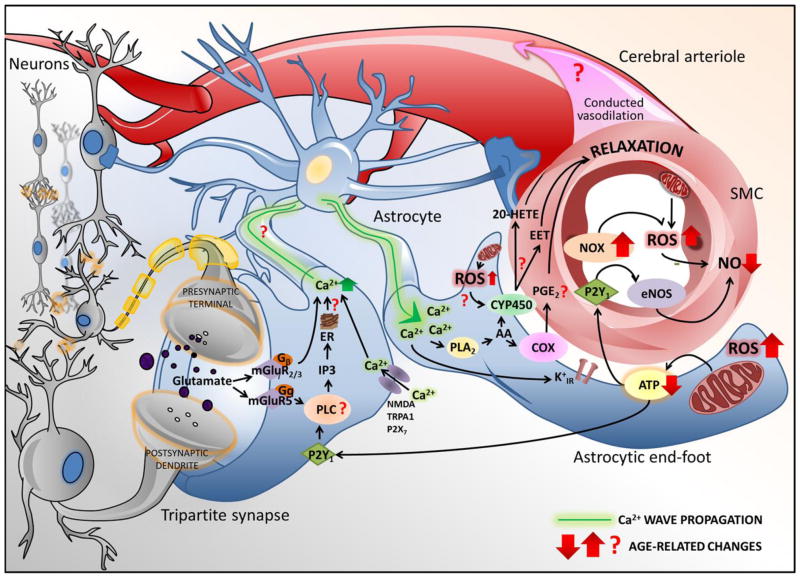
The importance of (micro)vascular contributions to cognitive impairment and dementia (VCID) in aging cannot be overemphasized, and the pathogenesis and prevention of age-related cerebromicrovascular pathologies are a subject of intensive research. In particular, aging impairs the increase in cerebral blood flow triggered by neural activation (termed neurovascular coupling or functional hyperemia), a critical mechanism that matches oxygen and nutrient delivery with the increased demands in active brain regions. From epidemiological, clinical and experimental studies the picture emerges of a complex functional impairment of cerebral microvessels and astrocytes, which likely contribute to neurovascular dysfunction and cognitive decline in aging and in age-related neurodegenerative diseases. This overview discusses age-related alterations in neurovascular coupling responses responsible for impaired functional hyperemia. The mechanisms and consequences of astrocyte dysfunction (including potential alteration of astrocytic endfeet calcium signaling, dysregulation of eicosanoid gliotransmitters and astrocyte energetics) and functional impairment of the microvascular endothelium are explored. Age-related mechanisms (cellular oxidative stress, senescence, circulating IGF-1 deficiency) impairing the function of cells of the neurovascular unit are discussed and the evidence for the causal role of neurovascular uncoupling in cognitive decline is critically examined.
Keywords: Cerebral circulation; Cerebrovascular; Functional hyperemia; Geroscience; Microcirculation; Neurovascular coupling; Senescence; VCI; VCID; Vascular aging.
Copyright © 2016 Elsevier Inc. All rights reserved.
Figures

References
-
- Amin-Hanjani S, Du X, Pandey DK, Thulborn KR, Charbel FT. Effect of age and vascular anatomy on blood flow in major cerebral vessels. J Cereb Blood Flow Metab. 2015;35:312–318. http://dx.doi.org/10.1038/jcbfm.2014.203. - DOI - PMC - PubMed
-
- Bailey-Downs LC, Mitschelen M, Sosnowska D, Toth P, Pinto JT, Ballabh P, Valcarcel-Ares MN, Farley J, Koller A, Henthorn JC, Bass C, Sonntag WE, Ungvari Z, Csiszar A. Liver-specific knockdown of IGF-1 decreases vascular oxidative stress resistance by impairing the Nrf2-dependent antioxidant response: A novel model of vascular aging. J Gerontol Biol Med Sci. 2012;67:313–329. - PMC - PubMed
-
- Balbi M, Ghosh M, Longden TA, Jativa Vega M, Gesierich B, Hellal F, Lourbopoulos A, Nelson MT, Plesnila N. Dysfunction of mouse cerebral arteries during early aging. J Cereb Blood Flow Metab. 2015;35:1445–1453. http://dx.doi.org/10.1038/jcbfm.2015.107. - DOI - PMC - PubMed
-
- Bhat R, Crowe EP, Bitto A, Moh M, Katsetos CD, Garcia FU, Johnson FB, Trojanowski JQ, Sell C, Torres C. Astrocyte senescence as a component of Alzheimer’s disease. PLoS One. 2012;7:e45069. http://dx.doi.org/10.1371/journal.pone.0045069. - DOI - PMC - PubMed
-
- Blanco VM, Stern JE, Filosa JA. Tone-dependent vascular responses to astrocyte-derived signals. Am J Physiol Heart Circ Physiol. 2008;294:H2855–2863. http://dx.doi.org/10.1152/ajpheart.91451.2007. - DOI - PMC - PubMed
Publication types
MeSH terms
Substances
Grants and funding
LinkOut - more resources
Full Text Sources
Other Literature Sources
Medical
Miscellaneous
