. 2016 Nov 15:6:36747.
doi: 10.1038/srep36747.

An iron detection system determines bacterial swarming initiation and biofilm formation

Affiliations

An iron detection system determines bacterial swarming initiation and biofilm formation

Chuan-Sheng Lin et al. Sci Rep. .

Abstract

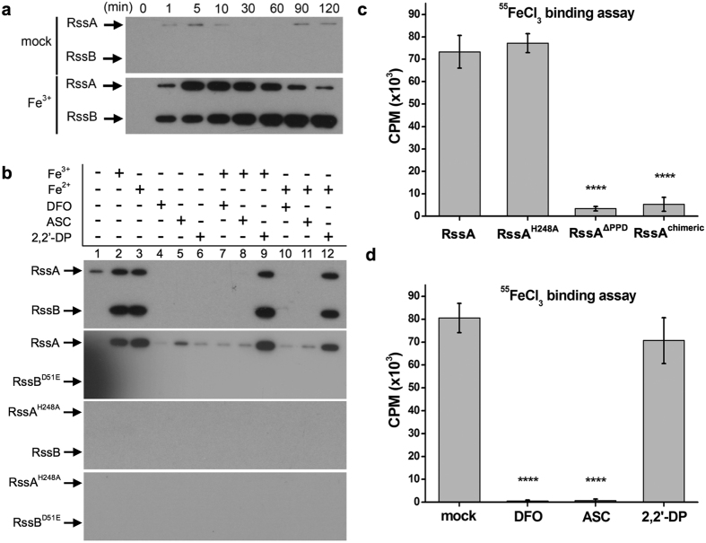
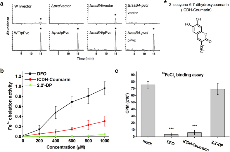
Iron availability affects swarming and biofilm formation in various bacterial species. However, how bacteria sense iron and coordinate swarming and biofilm formation remains unclear. Using Serratia marcescens as a model organism, we identify here a stage-specific iron-regulatory machinery comprising a two-component system (TCS) and the TCS-regulated iron chelator 2-isocyano-6,7-dihydroxycoumarin (ICDH-Coumarin) that directly senses and modulates environmental ferric iron (Fe3+) availability to determine swarming initiation and biofilm formation. We demonstrate that the two-component system RssA-RssB (RssAB) directly senses environmental ferric iron (Fe3+) and transcriptionally modulates biosynthesis of flagella and the iron chelator ICDH-Coumarin whose production requires the pvc cluster. Addition of Fe3+, or loss of ICDH-Coumarin due to pvc deletion results in prolonged RssAB signaling activation, leading to delayed swarming initiation and increased biofilm formation. We further show that ICDH-Coumarin is able to chelate Fe3+ to switch off RssAB signaling, triggering swarming initiation and biofilm reduction. Our findings reveal a novel cellular system that senses iron levels to regulate bacterial surface lifestyle.


Figures

Figure 1
Figure 1. Fe3+ controls swarming initiation and biofilm formation in S. marcescens through the TCS RssAB.
(a,b) Swarming pattern (a) and migration radius (b) of WT and ΔrssBA S. marcescens on LB swarming plates containing Fe3+ (100 μM) and/or DFO (0.3 mM). (c) Swarming migration radius of WT and ΔrssBA on DMM swarming plates containing Fe3+ at the indicated concentration (0–10 μM). The number in the lower right corner of each swarming plate image represents the duration of the lag phase in hours. Migration radius (mm) corresponds to mean ± SEM (n = 3). ND, not detected. (d) As mentioned in Methods, biofilm of WT and ΔrssBA S. marcescens in LB broth containing the indicated concentration of Fe3+ and/or DFO (0.3 mM) was quantified by monitoring absorbance at 630 nm. (e,f) In LB condition with or without Fe3+ (100 μM) and DFO (0.3 mM), swarming radius (e) and biofilm quantification (f) of ΔrssBA harboring the vector pACYC184 or the recombinant pACYC184 plasmid containing different constructs of RssB and RssA driven by their native promoters. RssBD51E and RssAH248A: constructs with point mutations at conserved phosphorylation sites; RssAΔPPD: RssA with deletion in periplamic domain (PPD, amino acids 32–163; RssAchimeric: a chimeric RssA whose periplasmic domain was replaced with the periplasmic domain of QseC. The results represent means ± SEM from three independent experiments (n = 3). Statistical analysis was performed using one-way ANOVA. For Fig. 1d, *, ** and *** represent P < 0.05, 0.01, 0.001, and 0.0001, respectively, compared to the untreated sample. # and ## represent P < 0.05 and 0.01, respectively, compared to the group treated with DFO but without Fe3+. For Fig. 1f, *P < 0.05; **P < 0.01 compared to LB group.
Figure 2
Figure 2. Fe3+ availability regulates RssAB signaling status during swarming and biofilm development.
(a) Representative image of cytolocalization of EGFP-tagged RssB. Cytoplasmic and membrane location of EGFP-RssB indicates ON (activated) and OFF (inactivated) RssAB signaling status, respectively. Scale bar, 3 μm. (b) WT S. marcescens harboring the pEGFP-RssBA::Sm plasmid encoding EGFP-RssB and RssA was used to evaluate the state of RssAB signaling during swarming (2–8 hr). Swarming assays were performed on LB plates containing 0.1% arabinose with or without Fe3+ (100 μM) and DFO (0.3 mM). Cellular localization of EGFP-RssB was monitored to quantify the percentage of activated and inactivated RssAB signaling. (c) Quantification of RssAB signaling status during swarming progression (1–4 hr) on DMM swarming plates supplemented with Fe3+ at the indicated concentration (0–10 μM). (d) Quantification of RssAB signaling status during biofilm development (12–48 hr) in LB condition with or without Fe3+ (100 μM) and DFO (0.3 mM). Percentage of cell type is shown as mean from three independent experiments performed in triplicate. At least 200 cells were counted for each assay condition.
Figure 3
Figure 3. RssA binds Fe3+ through its periplasmic domain and transphosphorylates RssB.
(a) His-tagged RssA and RssB reconstituted in liposomes with or without Fe3+ was supplemented with [γ32P]ATP (50 μCi), collected at the indicated time points, and examined by radiography imaging. (b) Liposomes containing His-tagged RssA, RssB, nonphosphorylatable RssA or RssB were harvested 30 min after addition of [γ32P]ATP and examined by radiography imaging. (c) His-tagged RssA, RssAH248A (His248 mutated to Ala), RssAΔPPD (RssA with deletion in periplasmic domain), and RssAchimeric (a chimeric RssA whose periplasmic domain was replaced with the periplasmic domain of QseC) were reconstituted in liposomes containing 500 nM 55FeCl3. After incubation, disrupted liposomes passed through NTA column to remove unbound 55FeCl3. Iron-bound membrane proteins were eluted and subjected to radioactivity analysis of liquid scintillation counting (counts per min, CPM). (d) His-tagged periplasmic domain of RssA was incubated with 55FeCl3 and mock (distilled water), DFO (0.3 mM), ASC (0.3 mM), or 2,2′-DP (0.3 mM). Radioactivity was determined in the periplasmic domain eluted from Ni2+-NTA column. Statistical analysis was performed using one-way ANOVA. For Fig. 1c, ****P-value < 0.0001, compared to RssA group. For Fig. 1d, *** and **** correspond to P-values < 0.001 and <0.0001, respectively, compared to mock group.
Figure 4
Figure 4. pvc cluster regulated by RssB is involved in regulating swarming and biofilm formation.
(a) Schematic map of the pvc cluster, RssB-P binding site, and pvc cluster deletion mutant. Red dash lines represent RssB-P binding sites (−349 to +38) of the pvcA promoter region. For construction of pvc cluster deletion mutants (Δpvc), genomic region between two asterisks (*) was replaced with Smr cassette. (b) EMSA was employed to confirm the interaction between phosphorylated RssB (RssB-P) and promoter region of pvcA (PpvcA). Digoxigenin (DIG)-labeled DNA fragments were incubated with purified GST, GST-RssBD51E or GST-RssB-P, followed by analysis by non-denaturing PAGE. Negative control (NC) was performed by incubating GST-RssB-P with the DNA sequence between M13F/M13R in the plasmid pBIISK. (c) During swarming progression (2–6 hr) with different iron conditions, relative expression of RssB downstream genes (flhDC and pvcA), normalized to 16S rRNA, in WT and ΔrssBA was respectively determined by qRT-PCR. (d,e) Swarming migration radius (d) and biofilm formation (e) of each strain of S. marcescens. Strain harboring pPvc encoding pvc cluster under pBAD promoter with 0.01% arabinose. The results shown represent means ± SEM from three independent experiments (n = 3). Statistical analysis was performed using one-way ANOVA. For Fig. 4c, * and ** corresponding to P-value < 0.05 and <0.01 in comparison with LB group of each strain, respectively. For Fig. 4e, * corresponding to P-value < 0.05 compared to WT.
Figure 5
Figure 5. ICDH-Coumarin produced from pvc gene cluster chelates Fe3+ and blocks iron binding to RssA.
(a) LC-MS chromatogram of ethyl acetate extracts from WT, Δpvc, ΔrssBA, and ΔrssBA-pvc bacteria harboring pBAD33 (vector) or pPvc encoding pvc cluster under DMM broth with arabinose (0.3%). *Indicates the peak and structure of ICDH-Coumarin as determined by NMR. (b) Fe3+ chelation activity of DFO, ICDH-Coumarin, and 2,2′-DP at the indicated concentration. (c) His-tagged periplasmic domain of RssA was incubated with 55FeCl3 and distilled water (mock), DFO (0.3 mM), ICDH-Coumarin (0.3 mM), or 2,2′-DP (0.3 mM). Radioactivity was determined from the eluted periplasmic domain. One-way ANOVA with *** corresponding to P-value < 0.001 compared to mock group.
Figure 6
Figure 6. ICDH-Coumarin induces swarming initiation and represses biofilm formation by modulating extracellular Fe3+ availability and RssAB signaling.
(a) Migration radius of WT, ΔrssBA, Δpvc, and ΔrssBA-pvc strains on LB swarming plates supplemented with ICDH-Coumarin (0–300 μM). (b) Quantified RssAB signaling of WT and Δpvc carrying pEGFP-RssBA::Gm during swarming progression (2–8 hr) on LB swarming plates containing arabinose (0.1%) and ICDH-Coumarin (0–300 μM) is shown. (c) Migration radius of WT and Δpvc on LB swarming plates supplemented with or without Fe3+ (100 μM) and ICDH-Coumarin (300 μM). (d) Biofilm of WT and Δpvc in LB condition supplemented with or without Fe3+ (100 μM) and ICDH-Coumarin (300 μM) was determined by monitoring absorbance at 630 nm. The results shown represent means ± SEM from three independent experiments (n = 3). One-way ANOVA with * and ** represent P values < 0.05 and <0.01 compared to LB group; # represents P-value < 0.05 compared to ICDH-Coumarin group.
Figure 7
Figure 7. Proposed model for the control of swarming and biofilm formation by the RssAB-ICDH-Coumarin-iron pathway.
(a) Low extracellular Fe3+ deactivates RssAB signaling and relieves transcription of the pvc cluster and flhDC, which encodes the master regulator of flagellum biosynthesis This in turn increases flagellum and production of ICDH-Coumarin which subsequently chelates Fe3+ to maintain low availability of free Fe3+. These processes induce swarming and repress biofilm formation. (b) High extracellular Fe3+ activates RssAB signaling, leading to phosphorylation of RssB, repression of pvc cluster and flhDC transcription, and reduced ICDH-Coumarin and flagellum production. High Fe3+ availability sustains activation of RssAB signaling. These processes lead to biofilm formation and inhibit swarming migration by restraining bacteria in the swarming lag phase.

References

    1. Andrews S. C., Robinson A. K. & Rodriguez-Quinones F. Bacterial iron homeostasis. FEMS Microbiol Rev 27, 215–237 (2003). - PubMed
    1. Touati D. Iron and oxidative stress in bacteria. Arch Biochem Biophys 373, 1–6 (2000). - PubMed
    1. Yeom J., Imlay J. A. & Park W. Iron homeostasis affects antibiotic-mediated cell death in Pseudomonas species. J Biol Chem 285, 22689–22695 (2010). - PMC - PubMed
    1. Marx J. J. Iron and infection: competition between host and microbes for a precious element. Best Pract Res Clin Haematol 15, 411–426 (2002). - PubMed
    1. Braun V. & Hantke K. Recent insights into iron import by bacteria. Curr Opin Chem Biol 15, 328–334 (2011). - PubMed

Publication types

MeSH terms