. 2016 Nov 15:6:37217.
doi: 10.1038/srep37217.

Biomechanics of subcellular structures by non-invasive Brillouin microscopy

Affiliations

Biomechanics of subcellular structures by non-invasive Brillouin microscopy

Giuseppe Antonacci et al. Sci Rep. .

Erratum in

Abstract

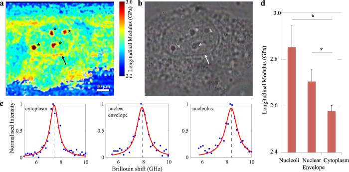
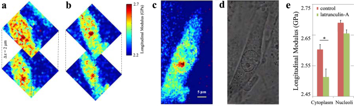
Cellular biomechanics play a pivotal role in the pathophysiology of several diseases. Unfortunately, current methods to measure biomechanical properties are invasive and mostly limited to the surface of a cell. As a result, the mechanical behaviour of subcellular structures and organelles remains poorly characterised. Here, we show three-dimensional biomechanical images of single cells obtained with non-invasive, non-destructive Brillouin microscopy with an unprecedented spatial resolution. Our results quantify the longitudinal elastic modulus of subcellular structures. In particular, we found the nucleoli to be stiffer than both the nuclear envelope (p < 0.0001) and the surrounding cytoplasm (p < 0.0001). Moreover, we demonstrate the mechanical response of cells to Latrunculin-A, a drug that reduces cell stiffness by preventing cytoskeletal assembly. Our technique can therefore generate valuable insights into cellular biomechanics and its role in pathophysiology.


Figures

Figure 1
Figure 1. Confocal Brillouin microscope and stiffness calibration.
Optical setup diagram of the confocal Brillouin microscope (a) and longitudinal modulus of polyacrylamide hydrogels of different stiffness (b). The stiffness of the polyacrylamide gels was varied by adjusting the amount of crosslinker from 0.05% to 1.2% w/v bisacrylamide. The hydrogels (n = 3 for each crosslink concentration) were measured using the Brillouin microscope to determine their longitudinal modulus. The longitudinal modulus appears to increase linearly with the hydrogel crosslink concentration (R2 > 0.99), indicating that Brillouin light scattering provides a meaningful measure of bulk stiffness.
Figure 2
Figure 2. High-resolution Brillouin image of a single cell in vitro.
A cross section through a single cultured human umbilical vein endothelial cell imaged using both Brillouin (a) and phase contrast (b) microscopy at 100x magnification. Cellular structures, such as the nuclear envelope (arrow) and nucleoli (*), are clearly recognisable in both image modalities. Representative Brillouin (Anti-Stokes) spectral peaks from cytoplasm, nuclear envelope and nucleoli (c). From the Brillouin shift and the associated longitudinal modulus, it is apparent that these structures exhibit different mechanical properties compared to the surrounding cytoplasm. In particular, the nuclear envelope and nucleoli exhibit higher longitudinal moduli (2.78 ± 0.05 GPa, p < 0.0001 and 3.12 ± 0.07 GPa, p < 0.0001 respectively) than the nucleus and cytoplasm that surrounds them (2.51 ± 0.04 GPa) (values reported as mean ± s.d.). A bar-plot represents the differences (mean ± SEM) between the longitudinal modulus as measured in the cytoplasm, nucleoli, and nuclear envelope of all experiments (*p < 0.001) (d).
Figure 3
Figure 3. Cellular stiffness in response to latrunculin-A.
Two cross sections along the z-axis through a cell before (a) and after (b) exposing the cell to latrunculin-A. A Brillouin image of the same cell taken at higher sampling resolution after drug exposure (c) and the associated phase-contrast image (d). Latrunculin is a toxin that prevents polymerisation of actin filaments and thereby decreases cell stiffness. Note that actin filaments are predominantly found in the cytoplasm and are absent from nucleoli. Indeed, cytoplasmic stiffness is decreased from 2.57 ± 0.05 to 2.46 ± 0.06 GPa (p < 0.0001), whereas nucleolar stiffness is not substantially affected by latrunculin-A exposure with a small decrease from 2.68 ± 0.07 GPa to 2.64 ± 0.06 GPa (p = 0.65) (values reported as mean ± s.d.). A bar-plot represents the change (mean ± SEM) in the longitudinal modulus of the cytoplasm and nucleoli in response to latrunculin-A of all experiments (*p < 0.001) (e). These data show that Brillouin microscopy is capable of measuring both spatial and temporal variations in stiffness in a physiologically relevant stiffness range that facilitates the study of cellular biomechanics.

References

    1. Cunningham K. S. & Gotlieb A. I., The role of shear stress in the pathogenesis of atherosclerosis, Laboratory investigation 85, 9–23 (2005). - PubMed
    1. Overby D. R. et al., Altered mechanobiology of Schlemm’s canal endothelial cells in glaucoma, PNAS 111, 13876–13881 (2014). - PMC - PubMed
    1. Li Q. S., Lee G. Y. H., Ong C. N. & Lim C. T. AFM indentation study of breast cancer cells. Biochemical and Biophysical Research Communications 374, 609–613 (2008). - PubMed
    1. Lee G. Y. & Lim C. T, Biomechanics approaches to studying human diseases. Trends in biotechnology 25, 111–118 (2007). - PubMed
    1. Kuznetsova T. G., Starodubtseva M. N., Yegorenkov N. I., Chizhik S. A. & Zhdanov R. I. Atomic force microscopy probing of cell elasticity. Micron 38, 824–833 (2007). - PubMed

Publication types

MeSH terms