. 2017 Mar 21;8(12):19137-19155.
doi: 10.18632/oncotarget.13268.

Hyaluronan activates Hyal-2/WWOX/Smad4 signaling and causes bubbling cell death when the signaling complex is overexpressed

Affiliations

Hyaluronan activates Hyal-2/WWOX/Smad4 signaling and causes bubbling cell death when the signaling complex is overexpressed

Li-Jin Hsu et al. Oncotarget. .

Abstract

Malignant cancer cells frequently secrete significant amounts of transforming growth factor beta (TGF-β), hyaluronan (HA) and hyaluronidases to facilitate metastasizing to target organs. In a non-canonical signaling, TGF-β binds membrane hyaluronidase Hyal-2 for recruiting tumor suppressors WWOX and Smad4, and the resulting Hyal-2/WWOX/Smad4 complex is accumulated in the nucleus to enhance SMAD-promoter dependent transcriptional activity. Yeast two-hybrid analysis showed that WWOX acts as a bridge to bind both Hyal-2 and Smad4. When WWOX-expressing cells were stimulated with high molecular weight HA, an increased formation of endogenous Hyal-2/WWOX/Smad4 complex occurred rapidly, followed by relocating to the nuclei in 20-40 min. In WWOX-deficient cells, HA failed to induce Smad2/3/4 relocation to the nucleus. To prove the signaling event, we designed a real time tri-molecular FRET analysis and revealed that HA induces the signaling pathway from ectopic Smad4 to WWOX and finally to p53, as well as from Smad4 to Hyal-2 and then to WWOX. An increased binding of the Smad4/Hyal-2/WWOX complex occurs with time in the nucleus that leads to bubbling cell death. In contrast, HA increases the binding of Smad4/WWOX/p53, which causes membrane blebbing but without cell death. In traumatic brain injury-induced neuronal death, the Hyal-2/WWOX complex was accumulated in the apoptotic nuclei of neurons in the rat brains in 24 hr post injury, as determined by immunoelectron microscopy. Together, HA activates the Hyal-2/WWOX/Smad4 signaling and causes bubbling cell death when the signaling complex is overexpressed.

Keywords: Hyal-2; Smad; WWOX; hyaluronan; hyaluronidase.


Conflict of interest statement

CONFLICTS OF INTEREST

The authors declare no conflicts of interest.

Figures
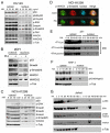
Figure 1
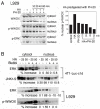
Figure 1. Hyaluronan induces nuclear accumulation of WWOX, Smads and others proteins
A. Prostate DU145 cells were treated with high-molecular-weight hyaluronan (HA; 105−106 kDa; 50 μg/ml; Sigma) for 1 hr. Nuclear translocation of full length WWOX, isoform WWOX2, p-FAK, p-Smad2/3, Smad4, p-ERK, and pSer46-p53 is shown (~100–300% increases). Time course analysis showed that in response to a low level of HA at 100 ng/ml, WWOX and Smad4 relocated to the nucleus in 40 min (Supplementary Figure S1). B. Breast MCF7 cells were treated with HA for 1 hr. Nuclear translocation of WWOX, WWOX2, p53, p-ERK and p-Smad2/3 was shown. C, D. p53-deficient NCI-H1299 cells were treated with medical grade HA for 1 hr. Nuclear translocation of WWOX, p-WWOX (Tyr33 phosphorylation) and Smads is shown by Western blotting and fluorescence microscopy (~100–300% increases in nuclear localization using 25 μg/ml HA). 25 μg/ml HA was used in (D). Note the colocalization of p-WWOX with p-Smad2/3 in the cytosol and their nuclear accumulation post HA treatment. Scale bar, 10 μm. E-G. Mouse breast cancer 4T1 cells (wild type and 2 cold-resistant variants), human monocytic THP-1 cells, and human leukemia Jurkat T cells were treated with HA (25 μg/ml) for indicated times. Nuclear accumulation of indicated proteins occurred in 10-20 min. c = medium control; α-Tub=α-Tubulin.
Figure 2
Figure 2. Wild type WWOX is necessary for HA induction of protein nuclear translocation
A, B. WWOX-deficient breast MDA-MB-231 and MDA-MB-435S were treated with high-molecular-weight HA (25 μg/ml) for 1 hr. Nuclear translocation of each indicated protein was retarded (<10% increase in nuclear localization compared to controls). C, D. Exposure of wild type Wwox+/+ MEF cells to HA (25 μg/ml) resulted in relocation of endogenous Smad4 into the nucleus in 5 min (also see Supplementary Information Figures S3 and S5). When knockout Wwox−/− MEF cells were stimulated with HA, Smad4 appeared to relocate into nucleus in 4 hr (also see Supplementary Figures S2-S4). The extent of Smad4 nuclear localization was quantified (n=4; 20 cells per count). Scale bar, 10 μm.
Figure 3
Figure 3. Blebbistatin blocks HA-mediated protein nuclear translocation
A. HA (100 μg/ml) was predigested with PH-20 (100 units/ml) for various indicated times. The digested HA could not induce nuclear translocation of WWOX, p-WWOX and p-ERK in L929 cells (representative data from 2 experiments). The extent of p-WWOX nuclear translocation is quantified (right panel). B. L929 and 4T1-Luc-c1d cells were pre-treated with blebbistatin (10 μM) for 1 hr, followed by exposure to HA (25 and 50 μg/ml) for determining the relocation of indicated proteins to the nucleus by Western blotting. Blebbistatin blocked HA-induced nuclear relocation of ERK, JNK and pY33-WWOX. p-WWOX = pY33-WWOX; Blebbi = blebbistatin.
Figure 4
Figure 4. HA increases the complex formation of Hyal-2, WWOX and Smads, followed by reduction
A. Indicated cell lines were treated with HA (50 μg/ml) for 30 min at 37°C. Cytosolic fractions were used for reducing SDS-PAGE and Western blotting. HCT116 cells were treated with androgen (100 ng/ml) for 30 min, which led to increased pY216-Hyal-2 expression. B. Immunostaining of non-permeabilized COS7 cells shows the expression of Hyal-2 on cell surface. Hyal-2 is clustered on the surface of non-permeabilized MDA-MB-231 cells. Scale bar, 5 μm. C. HA (25 μg/ml) rapidly induced translocation of Hyal-2 to the nucleus in the wild type Wwox+/+ MEF cells, followed by reduction. In the knockout Wwox−/− MEF cells, approximately 60% of endogenous Hyal-2 is present in the nucleus. HA reduces the nuclear localization. D. By immunoprecipitation using WWOX antibody, HA increased the WWOX/Hyal-2 complex in the cytosol, whereas the nuclear level of the complex was still low in THP-1 cells. In U937 cells, HA reduced the cytosolic WWOX/Hyal-2 complex in 30 min and showed the increased complex in the nucleus. E. HA (25 μg/ml) reduced the cytosolic Hyal-2/WWOX/Smads complex in SK-N-SH cells in 30 min, as determined by immunoprecipitation using Hyal-2 antibody. Immunoprecipitation by Smad3 antibody revealed the presence of the Hyal-2/WWOX/Smads complex in resting cells and HA decreased the complex. F. Jurkat T cells and L929R fibroblasts were treated with HA (25 μg/ml) for 30 min, followed by processing immunoprecipitation with WWOX antibody. HA reduced the complex formation of cytosolic WWOX/Hyal-2/ERK. G. HA reduced the complex formation of Hyal-2/WWOX/Smad4 in EGFP-expressing COS7 cells. Also, ectopic EGFP-dn-WW suppressed the complex formation. In the input, one-tenth amounts of the cell lysates were loaded in the SDS-PAGE. H. The Hyal-2/WWOX/Smad4 complex was not affected by hyaluronidase PH-20 treatment of MCF7 cells for 1 hr.
Figure 5
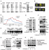
Figure 5. Tyr33 phosphorylation of WWOX is essential for interacting with Smad4 and Hyal-2, and Smad4 competitively blocks the binding of WWOX to membrane Hyal-2
A. Ras rescue-based protein/protein interaction in the cytoplasm by yeast two-hybrid was performed [16, 21, 37, 39]. In positive controls, binding of WWOX with p53 and MafB self-interaction are shown, as evidenced by the growth of yeast at 37°C using a selective agarose plate containing galactose. No yeast growth at 37°C was observed for the empty pSos/pMyr vectors in negative controls. The N-terminal first WW domain of WWOX (WWOXww) bound Smad4 and Hyal-2. Alteration of Tyr33 to Arg33 in the first WW domain, WWOXww(Y33R), abolished its interaction with Smad4 and Hyal-2. Dominant negative WWOX, dn-WWOX(K28T/D29V), also bound Smad4. The mitochondria-targeting area (amino acid 209-273) of the C-terminal SDR domain did not bind Smad4. No direct interaction was observed for Hyal-2 and Smad4. B, C. In this competitive binding assay, yeast cells were transfected with Hyal-2 (target; green) for anchoring onto cell membrane, in the presence of a constant amount of WWOX (bait; dark blue) and various amounts of Smad4 (competitor; red). % Colony growth = (survival colonies at 37°C) / (total colonies grown at 22°C) (see brackets). Smad4 competitively blocked the binding of WWOX to membrane Hyal-2, and thereby prevented yeast growth.
Figure 6
Figure 6. Induction of WWOX and Smads nuclear accumulation by agonist Hyal-2 antibodies and antisense mRNA
A. Compared to non-immune serum (cont) and medium control (med), exposure of NCI-H1299 cells to agonist antibodies against Hyal-2 (1:1000 dilution) for 30 min resulted in spontaneous nuclear translocation of WWOX and Smads (~75-200% increase in nuclear localization; see the nuclear protein levels without HA treatment; representative data from 2 experiments). These cells were further exposed to HA (100 μg/ml) for 30 min. B. Similarly, suppression of Hyal-2 expression by antisense mRNA (~70% reduction) spontaneously induced nuclear translocation of WWOX and Smads in TβRII-deficient colon HCT116 cells. Without Hyal-2, HA could not increase nuclear translocation of these proteins (~100-200% increase in nuclear localization; see the nuclear protein levels without HA treatment; representative data from 2 experiments). C. In resting cells, Hyal-2 binds WWOX in the cell membrane or in the lysosome. HA increases the formation of the Hyal-2/WWOX/Smads complex for relocating to the nucleus. Without WWOX, Hyal-2 may spontaneously accumulate in the nucleus.
Figure 7
Figure 7. Time-lapse FRET microscopy for HA-activated signaling pathways
A. DU145 cells were transiently transfected with ECFP-Smd4, EGFP-WWOX and DsRed-monomer-p53 expression constructs. HA (25 μg/m) induced energy transfer from ECFP-Smad4 to EGFP-WWOX and then to DsRed-monomer-p53 for the Smad4/WWOX/p53 signaling. Data are shown as FRETc (FRET concentration). B, C. Dominant negatives for WWOX and p53 abolished the signaling. D. HA also induced signaling for ECFP-Smd4, EGFP-Hyal-2(−sp) and DsRed-monomer-WWOX. Cytosolic EGFP-Hyal-2(−sp) is devoid of the GPI linkage. E, F. Both antisense mRNA targeting Hyal-2 and dn-WWOX abolished the HA-induced signaling. G. In negative controls, HA did not induce signaling in cells expressing ECFP, EGFP and DsRed. H. The Smad4/WWOX/p53-expressing cells started to undergo membrane blebbing post stimulation with HA for 1.5 hr (see the star in the left). No cell death occurred. In contrast, Smad4/Hyal-2/WWOX-expressing cells underwent bubbling cell death [51, 52], post HA stimulation for 5-16 hr (see the star for the generated bubble in the right). Also, see Supplementary Video legends and Videos S1–S9. I. Similarly, HA failed to induce the activation of the IκBα/ERK/WWOX signaling [42].
Figure 8
Figure 8. Immunoelectron microscopy analysis for HA induction of WWOX/Hyal-2 nuclear translocation
A. HCT116 cells were treated with HA for 20 min and then subjected to processing for immunoelectron microscopy using antibodies against WWOX and Hyal-2, plus indicated immunogold particles. Where indicated, the enlarged areas were from 40,000 to 80,000x magnifications. Shown in the column at right are digitally enlarged immunogold particles in the nuclei. B. Immunostaining for p-WWOX and Smad4 particles is shown.
Figure 9
Figure 9. Hyal-2, WWOX and Smad4 synergistically induce apoptosis
A. L929 cells were electroporated with Hyal-2 and/or Smad4 constructs, cultured 48 hr, and subjected to cell cycle analysis by FACS. Hyal-2 could not bind Smad4 (see Figure 5), and did not enhance the apoptotic function of Smad4 (% apoptosis = % subG1 phase of the cell cycle). B. Under similar conditions, L929 cells were transfected with Hyal-2, WWOX, and/or Smad4. Hyal-2 enhanced WWOX-mediated apoptosis. In combination, these 3 proteins increased cell death. C. In contrast, WWOXsi blocked Smad4-mediated apoptosis. In controls, scramble siRNA had no effect. WWOXsi, siRNA-targeting WWOX. All data are average from two experiments B, C. Unless otherwise indicated, 5 μg DNA constructs were used in electroporation. Data for both G1 and SubG1 phases are shown for all experiments.
Figure 10
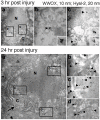
Figure 10. Nuclear accumulation of Hyal-2 and WWOX in apoptotic nuclei of cortical neurons during traumatic brain injury
Rats were stabbed with needles into their brains to induce traumatic brain injury. Post injury for 3 and 24 hr, rats were sacrificed. There were increased numbers of apoptotic neurons in the brain cortex, with the presence of the Hyal-2/WWOX complex in the apoptotic nuclei of neurons.

Similar articles

Cited by

References

    1. Stern R. Devising a pathway for hyaluronan catabolism: are we there yet? Glycobiology. 2003;13:105r–15r. - PubMed
    1. Sherman LS, Matsumoto S, Su W, Srivastava T, Back SA. Hyaluronan Synthesis, Catabolism, and Signaling in Neurodegenerative Diseases. Int J Cell Biol. 2015;2015:368584. Review. - PMC - PubMed
    1. Schmaus A, Bauer J, Sleeman JP. Sugars in the microenvironment: the sticky problem of HA turnover in tumors. Cancer Metastasis Rev. 2014;33:1059–79. - PubMed
    1. McAtee CO, Barycki JJ, Simpson MA. Emerging roles for hyaluronidase in cancer metastasis and therapy. Adv Cancer Res. 2014;123:1–34. - PMC - PubMed
    1. Frost GI, Mohapatra G, Wong TM, Csoka AB, Gray JW, Stern R. HYAL1(LUCA-1), a candidate tumor suppressor gene on chromosome 3p21.3, is inactivated in head and neck squamous cell carcinomas by aberrant splicing of pre-mRNA. Oncogene. 2000;19:870–7. - PubMed

MeSH terms

LinkOut - more resources