. 2017 Jan 3;8(1):863-882.
doi: 10.18632/oncotarget.13273.

TRIM28 multi-domain protein regulates cancer stem cell population in breast tumor development

Affiliations

TRIM28 multi-domain protein regulates cancer stem cell population in breast tumor development

Patrycja Czerwińska et al. Oncotarget. .

Abstract

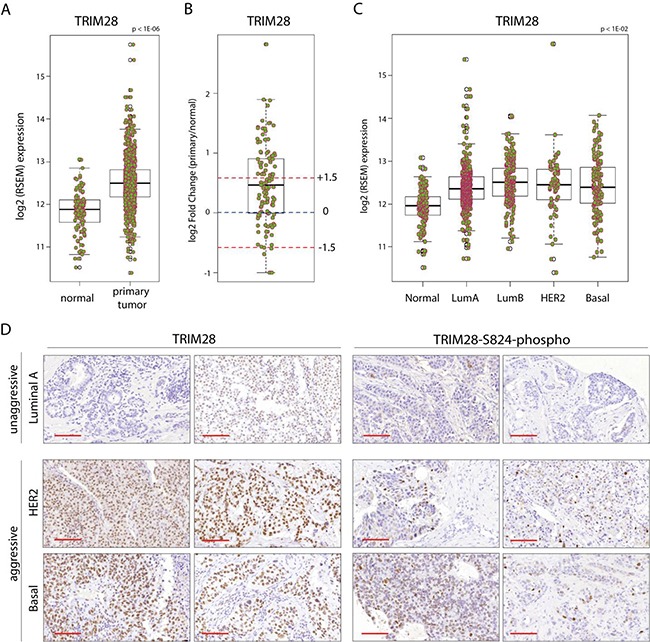
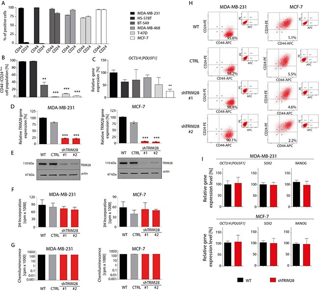
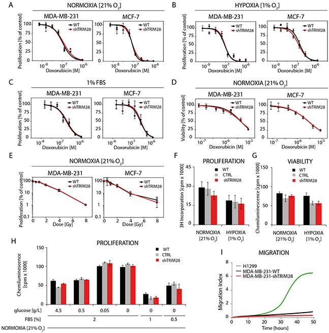
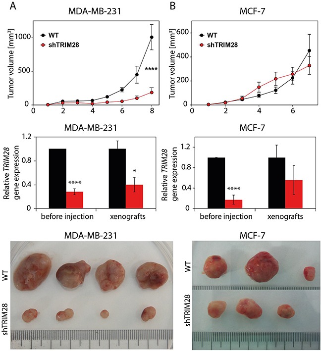
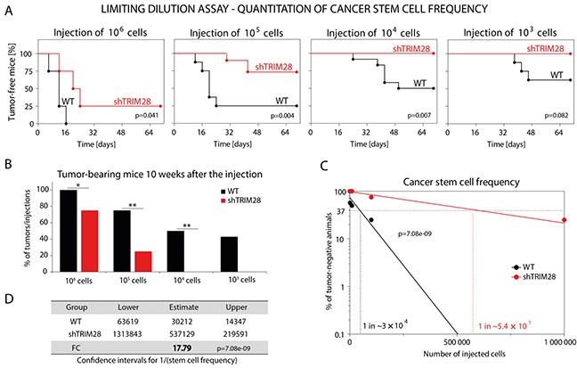
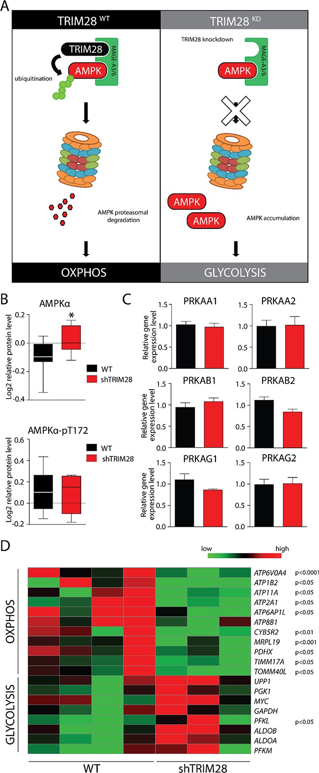
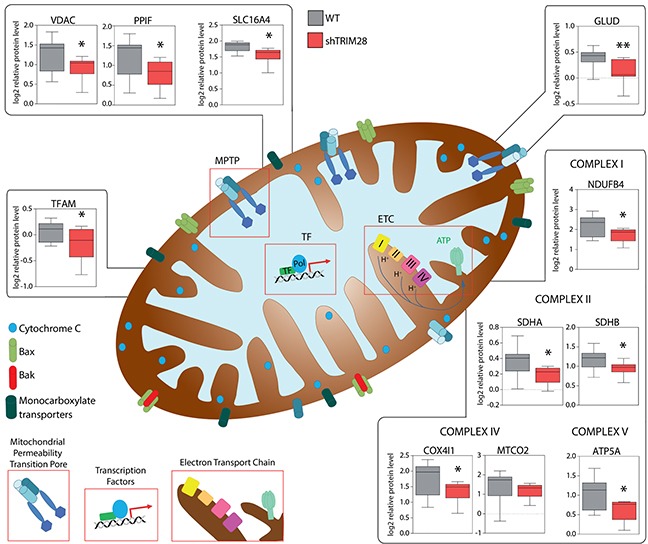
The expression of Tripartite motif-containing protein 28 (TRIM28)/Krüppel-associated box (KRAB)-associated protein 1 (KAP1), is elevated in at least 14 tumor types, including solid and hematopoietic tumors. High level of TRIM28 is associated with triple-negative subtype of breast cancer (TNBC), which shows higher aggressiveness and lower survival rates. Interestingly, TRIM28 is essential for maintaining the pluripotent phenotype in embryonic stem cells. Following on that finding, we evaluated the role of TRIM28 protein in the regulation of breast cancer stem cells (CSC) populations and tumorigenesis in vitro and in vivo. Downregulation of TRIM28 expression in xenografts led to deceased expression of pluripotency and mesenchymal markers, as well as inhibition of signaling pathways involved in the complex mechanism of CSC maintenance. Moreover, TRIM28 depletion reduced the ability of cancer cells to induce tumor growth when subcutaneously injected in limiting dilutions. Our data demonstrate that the downregulation of TRIM28 gene expression reduced the ability of CSCs to self-renew that resulted in significant reduction of tumor growth. Loss of function of TRIM28 leads to dysregulation of cell cycle, cellular response to stress, cancer cell metabolism, and inhibition of oxidative phosphorylation. All these mechanisms directly regulate maintenance of CSC population. Our original results revealed the role of the TRIM28 in regulating the CSC population in breast cancer. These findings may pave the way to novel and more effective therapies targeting cancer stem cells in breast tumors.

Keywords: KAP1; TRIM28; breast cancer stem cells; epigenetics; pluripotency.


Conflict of interest statement

CONFLICTS OF INTEREST

The authors disclose no potential conflicts of interest.

Figures

Figure 1
Figure 1. TRIM28 gene is overexpressed in breast cancer
A. Box plot presents the TRIM28 gene expression level in primary breast tumor and normal tissue from TCGA project. The black line presents median, box shows interquantile region and whiskers - the highest (max) and the lowest (min) value. Outliers are also depicted. B. Differential expression analysis of TCGA BRCA patient samples for which tumor and matched normal tissues are available. Fold changes +/− 1.5 fold change are depicted. The black line presents median, box shows interquantile region and whiskers - the highest (max) and the lowest (min) value. Outliers are also depicted. C. TRIM28 gene expression is distinct between different BRCA PAM50 intrinsic subtypes. D. IHC for total TRIM28 and phosphorylated TRIM28 protein (TRIM28-S824) in selected breast cancer samples confirmed higher levels of TRIM28/phospho-TRIM28 in more aggressive breast cancer subtypes (basal and HER2+). Scale = 100 μm.
Figure 2
Figure 2. TRIM28 knockdown does not affect breast cancer cell proliferation, cell viability and the percentage of CD44+/CD24−/low breast cancer stem cell population in vitro
A. 6 breast cancer cell lines (MDA-MB-231, HS-578T, BT-549, MDA-MB-468, T-47D and MCF-7) are characterized by different proportion of CD44-positive and CD24-positive cells as determined using FACS analysis. B. Identification of a CD44+/CD24−/low subpopulation in breast cancer cell lines by flow cytometry. Relatively to MDA-MB-231 breast cancer cell line, MCF-7 and T-47D luminal breast cancer cell line and MDA-MB-468 basal-like breast cancer cell line are poor in the population of CD44+/CD24−/low cancer stem cells. Error bars, SD; n = 3; ** p < 0.01; *** p < 0.001. C. The level of OCT3/4 pluripotency marker in selected breast cancer cell lines in vitro was determined using RT-qPCR analysis. Relatively to MDA-MB-231 breast cancer cell line, MCF-7 cells express OCT3/4 at the lowest level in vitro. Error bars, SD; n = 4; ** p < 0.01. D, E. MDA-MB-231 and MCF-7 cells stably infected with lentiviral vectors expressing TRIM28 shRNA (sh#1 or sh#2) or with empty vector (CTRL) were analyzed using RT-qPCR (A) and Western blot (B) for TRIM28 gene and protein levels. Error bars, SD; n = 3; *** p < 0.001. F, G. TRIM28 downregulation does not affect cell proliferation (F) and viability (G) in vitro as determined using an 3H-thymidine-incorporation assay and ATPlite™ Luminescence Assay, respectively. Error bars, SD; n = 4; p > 0.05. H. The comparison of CD44+/CD24−/low cells frequency in breast cancer cell lines upon reduction of TRIM28 level in vitro. TRIM28 knockdown does not affect the percentage of breast cancer stem cell population in vitro. I. TRIM28 downregulation does not affect the expression of pluripotency markers OCT3/4, SOX2 and NANOG in vitro in MDA-MB-231 (upper panel) and MCF-7 (bottom panel) breast cancer cell lines as determined using RT-qPCR. Error bars, SD; n = 4; p > 0.05.
Figure 3
Figure 3. TRIM28 knockdown does not affect breast cancer cell chemo- and radioresistance in vitro
A, B, C. The dose response curves show the relative proliferation in vitro (3H-thymidine incorporation assay) of TRIM28WT and TRIM28KD (sh#1) cells after doxorubicin treatment in normoxia (A), hypoxia (B) and low level of serum (C). Error bars, SD; n = 4; p > 0.05. D. The dose response curves presenting relative cell viability in vitro (ATPlite™ Luminescence Assay) of TRIM28WT and TRIM28KD (sh#1) cells after doxorubicin treatment in normoxia. Error bars, SD; n = 4; p > 0.05. E. Radiation dose response curves show the relative proliferation of TRIM28WT and TRIM28KD (sh#1) cells 80 hours after γ-irradiation. Proliferation was analyzed using an 3H-thymidine incorporation assay. Error bars, SD; n = 3; p > 0.05. F, G, H. The proliferation and viability of breast cancer cells in normoxia (F), hypoxia (G) or in low level of serum and/or glucose (H) is not affected in vitro by TRIM28 knockdown as determined using 3H-thymidine incorporation assay and ATPlite™ Luminescence Assay, respectively. Error bars, SD; n = 3; p > 0.05. I. Preliminary results from in vitro migration assay using xCELLigence® RTCA DP instrument revealed very low potential of MDA-MB-231 triple-negative breast cancer cells to migrate in vitro when attracted with 10% FBS containing medium. Lung cancer H1299 cell line was used as a positive control.
Figure 4
Figure 4. TRIM28 protein regulates tumor growth in vivo
Upper panel: Kinetics of tumor growth in a xenograft mouse model. TRIM28WT and TRIM28KD (sh#1) cells from the MDA-MB-231 A. and MCF-7 B. cell line were subcutaneously injected into athymic nude mice, and tumor size was measured weekly for 7-8 weeks. Error bars, SEM; **** p < 0.0001. Middle panel: TRIM28 gene expression was downregulated in TRIM28KD (sh#1) xenografts, as confirmed by RT-qPCR. Error bars, SD; * p < 0.05; **** p < 0.0001. Bottom panel: The image shows a representative group of MDA-MB-231 (A) and MCF-7 (B) tumors excised 7-8 weeks after injection.
Figure 5
Figure 5. TRIM28 knockdown leads to downregulation of pluripotency and mesenchymal markers and inhibition of stem cell-related pathways in MDA-MB-231 xenografts
A. Box plots presenting relative expression of selected pluripotency markers in TRIM28WT and TRIM28KD (sh#1) xenografts evaluated using the RT-qPCR analysis. Error bars, SEM; p > 0.05. B. Heatmap of selected pluripotency markers expressed in xenografts based on RNA sequencing. TRIM28 gene expression was efficiently downregulated in all of the TRIM28KD (sh#1) samples, as shown at the bottom of the heatmap of pluripotency markers. Statistical significance (p-value) is presented in the figure. Green = downregulation; Red = upregulation. C. IHC staining confirmed downregulation of OCT3/4 and SOX2 pluripotency markers in TRIM28KD (sh#1) MDA-MB-231 xenografts compared with TRIM28WT tumors. Scale = 50 μm. D. Protein coding transcripts differentially expressed upon TRIM28 knockdown. 1634 markers were significantly (FDR 5%) downregulated and 1384 markers were significantly upregulated in TRIM28KD tumors when compared to TRIM28WT xenografts. List of 3018 differentially expressed transcripts was further used for Gene Set Enrichment Analysis (GSEA). E. Summary of significantly changed (p < 0.05) gene sets after TRIM28 knockdown identified using GSEA analysis. Signaling pathways involved in the regulation of stem cell phenotype were inhibited in TRIM28KD (sh#1) xenografts. In contrast, gene sets that were upregulated in TRIM28KD (sh#1) xenografts included several pathways involved in protein metabolism and translational regulation. F. Box plots presenting relative expression of selected mesenchymal markers in TRIM28WT and TRIM28KD (sh#1) xenografts evaluated using the RT-qPCR analysis. Error bars, SEM; * p < 0.05. G. Heatmap of selected mesenchymal markers expressed in xenografts based on RNA sequencing. Statistical significance (p-value) is presented in the figure. Green = downregulation; Red = upregulation. H. IHC staining confirmed the downregulation of N-CADHERIN (mesenchymal marker) and the upregulation of E-CADHERIN (epithelial marker) in TRIM28KD (sh#1) MDA-MB-231 xenografts compared to TRIM28WT tumors. Scale = 50 μm. I. TRIM28-dependant downregulation of selected mesenchymal markers and inhibition of “cadherin switch” was further confirmed using RPPA analysis. Error bars, SD; * p < 0.05.
Figure 6
Figure 6. TRIM28 gene depletion reduces the number of cancer stem cells in MDA-MB-231 breast cancer xenografts
A. Limiting dilution assay was performed to estimate the hypothetical frequency of cancer stem cells in CD44+CD24−/low-enriched MDA-MB-231 breast cancer cell line upon TRIM28 knockdown. TRIM28WT and TRIM28KD (sh#1) MDA-MB-231 cancer cells were injected subcutaneously in serial dilutions (106, 105, 104 and 103 of cells per injection) into athymic nude mice and the ability of cancer cells to induce tumor growth in vivo was examined for 10 weeks. TRIM28 depletion (sh#1) reduced the ability of MDA-MB-231 breast cancer cells to induce tumor growth in vivo. B. Injection of TRIM28KD MDA-MB-231 (sh#1) cells resulted in a reduced number of xenografts compared with TRIM28WT cells 10 weeks after the engraftment. C, D. Hypothetical frequency of cancer stem cells in TRIM28WT and TRIM28KD (sh#1) MDA-MB-231 xenografts. TRIM28 knockdown significantly diminished the number of cancer stem cells in MDA-MB-231 xenografts (FC = 17.79, p = 7.08e-09). The calculation of the estimated stem cell frequency for each condition was performed using ELDA software (ref. 51).
Figure 7
Figure 7. AMPK accumulation upon TRIM28 knockdown mediates metabolic switch from OXPHOS to glycolysis in cancer cells
A. MAGE-A3/6-TRIM28 cancer-specific ubiquitin ligase targets AMPKα kinase for proteasomal degradation in TRIM28WT breast cancer cells. However, TRIM28 knockdown (right panel) should shut down AMPKα ubiquitination and proteasomal degradation, resulting in AMPKα accumulation-mediated metabolic switch. B. Level of AMPKα and phospho-AMPKα (T172) in TRIM28WT an TRIM28KD xenografts was determined by RPPA analysis. Error bars, SD; * p < 0.05. C. AMPKα protein is composed of 3 independent subunits (α, β and γ) and each of AMPKα subunits is encoded by at least two gene isoforms. As presented on bar graphs, TRIM28 downregulation does not affect the level of AMPK encoding genes as determined with RNA-Seq. Error bars, SD. D. Heatmap of selected metabolism-associated markers expressed in xenografts based on RNA sequencing. Statistical significance (p-value) is presented in the figure. Green = downregulation; Red = upregulation.
Figure 8
Figure 8. TRIM28-dependant inhibition of triple-negative breast tumor growth is mediated by metabolic changes/attenuation of oxidative phosphorylation in cancer cells
RPPA analysis revealed significant downregulation of many proteins engaged in mitochondrial electron transport chain (ETC), formation of mitochondrial permeability transition pore (MPTP) and regulation of mitochondrial transcription suggesting that TRIM28-dependant inhibition of triple-negative breast tumor growth is mediated by metabolic changes in the tumor cells.

Similar articles

Cited by

References

    1. Visvader JE, Lindeman GJ. Cancer stem cells: Current status and evolving complexities. Cell Stem Cell. 2012;10:717–28. - PubMed
    1. Czerwinska P, Kaminska B. Regulation of breast cancer stem cell features. Contemp Oncol. 2015;1A:7–15. - PMC - PubMed
    1. Muñoz P, Iliou MS, Esteller M. Epigenetic alterations involved in cancer stem cell reprogramming. Mol Oncol. 2012;6:620–36. - PMC - PubMed
    1. Mani SA, Guo W, Liao MJ, Eaton EN, Ayyanan A, Zhou AY, Brooks M, Reinhard F, Zhang CC, Shipitsin M, Campbell LL, Polyak K, Brisken C, et al. The epithelial-mesenchymal transition generates cells with properties of stem cells. Cell. 2008;133:704–15. - PMC - PubMed
    1. Li L, Neaves WB. Normal stem cells and cancer stem cells: The niche matters. Cancer Res. 2006;66:4553–7. - PubMed

MeSH terms

Substances