. 2017 Jan 2;13(1):114-132.
doi: 10.1080/15548627.2016.1252889. Epub 2016 Nov 15.

Ubiquitination of ERMES components by the E3 ligase Rsp5 is involved in mitophagy

Affiliations

Ubiquitination of ERMES components by the E3 ligase Rsp5 is involved in mitophagy

Naïma Belgareh-Touzé et al. Autophagy. .

Abstract

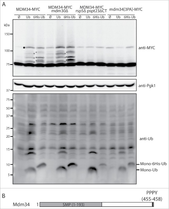
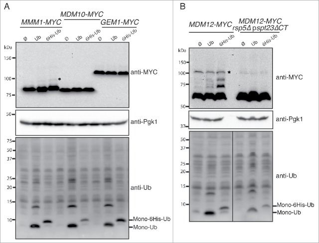
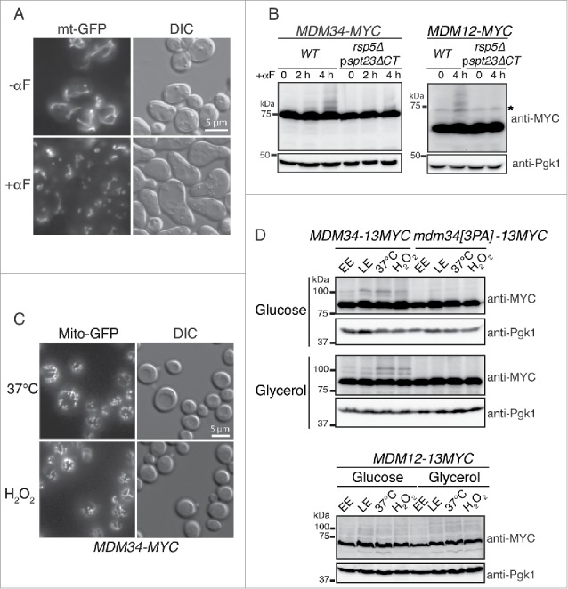
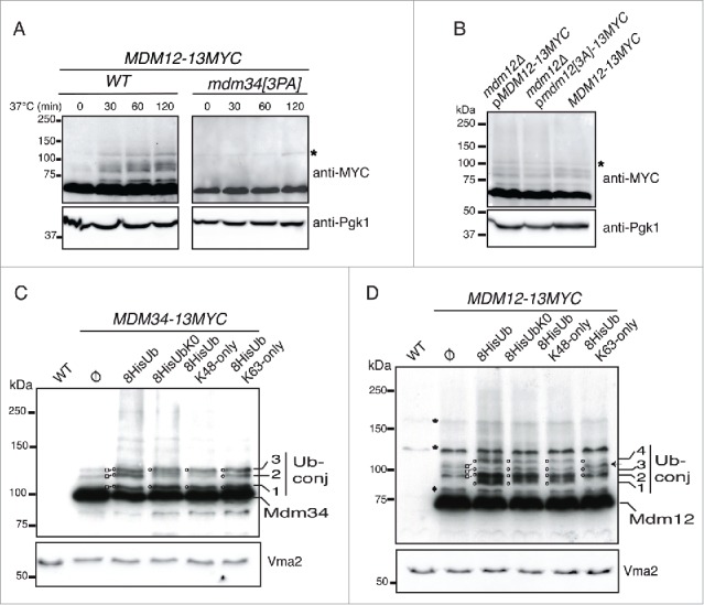
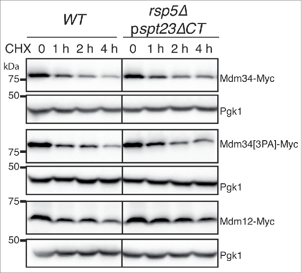
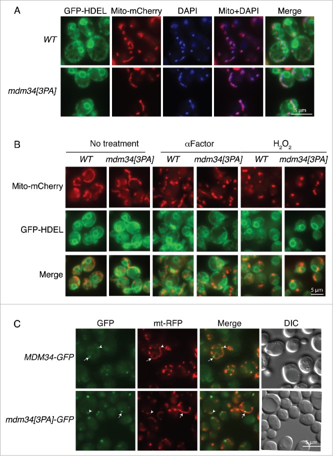
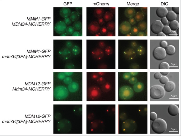
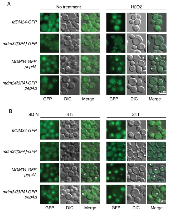
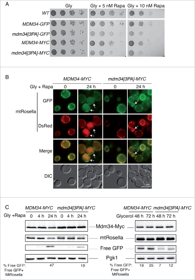
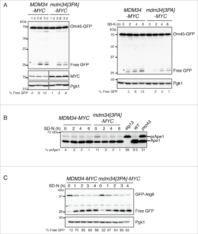
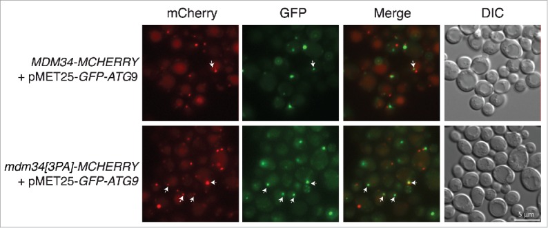
Mitochondria are dynamic organelles that undergo permanent fission and fusion events. These processes play an essential role in maintaining normal cellular function. In the yeast Saccharomyces cerevisiae, the endoplasmic reticulum-mitochondrial encounter structure (ERMES) is a marker of sites of mitochondrial division, but it is also involved in a plethora of other mitochondrial functions. However, it remains unclear how these different functions are regulated. We show here that Mdm34 and Mdm12, 2 components of ERMES, are ubiquitinated by the E3 ligase Rsp5. This ubiquitination is not involved in mitochondrial dynamics or in the distribution and turnover of ERMES. Nevertheless, the ubiquitination of Mdm34 and Mdm12 was required for efficient mitophagy. We thus report here the first identification of ubiquitinated substrates participating in yeast mitophagy.

Keywords: ER; ERMES; Mdm12; Mdm34; Rsp5; S. cerevisiae; mitochondria; mitophagy; ubiquitin.


Figures

Figure 1.
Figure 1.
Mdm34 is directly ubiquitinated by Rsp5. (A) MDM34-MYC, MDM34-MYC mdm30Δ, MDM34-MYC rsp5Δ pspt23ΔCT and mdm34[3PA]-MYC strains transformed with plasmids expressing ubiquitin (Ub), ubiquitin tagged with 6-histidines (6His-Ub) or with an empty plasmid (Ø) were grown at 30°C in YNB with glucose as the carbon source. Copper (CuSO4, 100 µM) was added to the cell cultures for 1 h to induce ubiquitin synthesis from the CUP1 promoter. Total protein extracts were prepared and analyzed by western immunoblotting with antibodies directed against MYC (anti-MYC), ubiquitin (anti-Ub), or phosphoglycerate kinase (anti-Pgk1) as a loading control. Monoubiquitin (Mono-Ub) and monoubiquitin tagged with 6 histidine residues (Mono-6His-Ub) are indicated. □, Mdm34-MYC conjugated with ubiquitin; ′, Mdm34-MYC conjugated with 6His-ubiquitin; *, Nonspecific band. (B) Schematic representation of Mdm34. The PPPY motif is shown in black and the SMP domains (synaptotagmin-like, mitochondrial and lipid-binding proteins) in gray.
Figure 2.
Figure 2.
Mdm12 is a new target of Rsp5 ubiquitination. (A, B) The MMM1-MYC, MDM10-MYC, GEM1-MYC (A) MDM12-MYC, and MDM12-MYC rsp5Δ pspt23ΔCT (B) strains bearing plasmids encoding ubiquitin (Ub), ubiquitin tagged with 6 histidines (6His-Ub) or an empty plasmid (Ø) were grown at 30°C in YNB supplemented with glucose. CuSO4 (100 µM) was added to the cell cultures for 1 h to induce ubiquitin synthesis from the CUP1 promoter. Total protein extracts were prepared and analyzed by western immunoblotting with anti-MYC (anti-MYC), anti-ubiquitin (anti-Ub) and anti-phosphoglycerate kinase (anti-Pgk1) antibodies. Pgk1 was used as a loading control. •,Glycosylated forms; *, nonspecific band.
Figure 3.
Figure 3.
Fission induction promotes the ubiquitination of Mdm34 and Mdm12. (A) Wild-type cells transformed with pYX142-mt-GFP and grown to early exponential growth phase were incubated in the presence (+αF) or absence (−αF) of 50 µg/mL α factor. Live cells were examined with differential interference contrast (DIC) and for GFP fluorescence under a Zeiss microscope. (B) MDM34-MYC, MDM34-MYC rsp5Δ pspt23ΔCT, MDM12-MYC, MDM12-MYC rsp5Δ pspt23ΔCT cells were grown to early exponential growth phase in YPD. We added 50 µg/mL α factor (αF) to the medium and total protein extracts were prepared at the times indicated. Proteins were analyzed by western immunoblotting with anti-MYC and anti-phosphoglycerate kinase (anti-Pgk1) antibodies. Pgk1 was used as a loading control. *, nonspecific band. (C) The MDM34-MYC strain expressing Mito-GFP from a vector integrated into the chromosome was grown at 30°C in YPD to mid-exponential growth phase. It was then shifted to 37°C or stressed by incubation with 2 mM H2O2 for 2 h. Fluorescence microscopy confirmed the presence of fragmented mitochondria after treatment. (D) The MDM34-13MYC, mdm34[3PA]-13MYC and MDM12-13MYC strains were grown to early exponential phase (EE), late exponential phase (LE), heated at 37°C (37°C) or incubated at 30°C in the presence of 2 mM H2O2 (H2O2) for 2 h, in medium containing glucose (YPD) or glycerol (YPG). Total protein extracts were prepared and subjected to western immunoblotting with antibodies against MYC (anti-MYC) and phosphoglycerate kinase (anti-Pgk1) as a loading control.
Figure 4.
Figure 4.
Analysis of the ubiquitination pattern of Mdm34, Mdm12 and PY mutants. (A) MDM12-13MYC MDM34-FLAG and MDM12-13MYC/mdm34[3PA]-FLAG cells were grown to early exponential growth phase in YPD at 24°C. They were then shifted to 37°C and total protein extracts were prepared at the times indicated. The ubiquitination pattern of Mdm12-13MYC was analyzed by western immunoblotting with antibodies against MYC and Pgk1. Pgk1 was used as a loading control. *, nonspecific band. (B) mdm12Δ pMDM12-13MYC, mdm12Δ pmdm12[3PA]-13MYC and the MDM12-13MYC strains were grown to late exponential growth phase to induce the ubiquitination of Mdm12. Total protein extracts were prepared and the ubiquitination pattern of Mdm12-13MYC was analyzed by western immunoblotting with antibodies against MYC and Pgk1 as a loading control. *, nonspecific band. (C and D) A wild-type strain without tagged genes (WT), and the MDM12-13MYC and MDM34-13MYC strains transformed with plasmids expressing 8-His tagged wild-type ubiquitin (8His-Ub), Ub-K0 (8His-UbK0), Ub-K48only (8HisUb-K48only), Ub-K63only (8HisUb-K63only) or with an empty plasmid (Ø) were grown to early exponential growth phase at 24°C in YNB with glucose as the carbon source. Cells were shifted to 37°C to induce protein ubiquitination and copper (CuSO4, 100 µM) was added to the cell cultures for 1 h to induce ubiquitin synthesis from the CUP1 promoter. Total protein extracts were prepared and analyzed by western immunoblotting and autoradiography with antibodies directed against MYC (anti-MYC), ubiquitin (anti-Ub), or vacuolar membrane ATPase (anti-Vma2) as a loading control. Various slowly migrating bands are indicated by numbers. □, Mdm34-13MYC and Mdm12-13MYC ubiquitinated species; O, Mdm34-13MYC and Mdm12-13MYC 8His-ubiquitinated species; *, nonspecific bands; ♦, uncharacterized slowly migrating Mdm12-13MYC moiety;←, New band; Ub-conj, ubiquitin conjugated to Mdm34-13MYC and Mdm12-13MYC.
Figure 5.
Figure 5.
Analysis of the stability of the Mdm34, Mdm12 and mutant Mdm34[3PA]proteins. WT and rsp5Δ pspt23ΔCT strains bearing chromosomal genes encoding 9MYC-tagged MDM34, mdm34[3PA], or MDM12 were grown to mid-exponential growth phase in YPD at 24°C. The cells were then shifted to 37°C, and protein synthesis was blocked by adding cycloheximide (CHX, 200 µg/mL). Protein extracts were prepared at the times indicated and analyzed by western immunoblotting with an anti-MYC antibody. Pgk1 was used as a loading control.
Figure 6.
Figure 6.
Mutation of the PY motif of Mdm34 does not affect ER or mitochondrial morphology, mitochondrial fission or Mdm34 localization. (A) WT or mdm34[3PA] strains bearing GFP-HDEL (ER marker) and Mito-mCherry expressed from vectors integrated into the chromosome were grown to early exponential growth phase in YPD at 30°C. Mitochondrial DNA was stained by adding 1 µg/mL DAPI to the growth medium and incubating for a further 2 to 3 h. Cells were washed twice and processed for fluorescence microscopy. (B) The same cells as in (A) were either left untreated (No treatment) or incubated with 50 µg/mL α factor for 3 h or with 2 mM H2O2 for 2 h. The cells were then washed twice and processed for fluorescence microscopy. (C) MDM34-GFP and mdm34[3PA]-GFP strains transformed with pYX142-mt-RFP were grown in YPD at 30°C before microscopy. Arrows indicate GFP at mitochondrial fission sites and arrowheads indicate GFP at mitochondrial tips. Cell shape was assessed with DIC.
Figure 7.
Figure 7.
Mutation of the PY motif of Mdm34 does not affect the localization of Mdm12 and Mmm1. MDM34-MCHERRY and MDM34[3PA]-MCHERRY strains bearing chromosomal genes encoding MDM12-GFP and MMM1-GFP were grown to the exponential growth phase in glycerol-containing medium (YPG). Cells were washed twice and processed for fluorescence microscopy. Cell shape was assessed with DIC.
Figure 8.
Figure 8.
Analysis of Mdm34-GFP localization in conditions inducing ubiquitination and mitophagy. (A) MDM34-GFP, mdm34[3PA]-GFP, MDM34-GFP pep4Δ and mdm34[3PA]-GFP pep4Δ strains were grown to exponential growth phase in complete synthetic medium. The cells were divided into 2 sets. The first set was treated with 2 mM H2O2 for 2 h (A) and the second set was washed 3 times with water and the cells were resuspended in SD-N medium and cultured for 4 or 24 h (B). GFP fluorescence was analyzed with a GFP filter set and cell morphology was investigated with DIC. Fluorescent structures inside the vacuole are indicated by a star symbol (*).
Figure 9.
Figure 9.
Mutation of the PY motif of MDM34 affects mitophagy. (A) The WT, MDM34-GFP, mdm34[3PA]-GFP, MDM34-MYC and mdm34[3PA]-MYC strains were grown to mid-exponential growth phase in YPD, washed twice in water and subjected to serial 5-fold dilution. Cells were then spotted onto solid complete medium containing glycerol as a carbon source (Gly) and supplemented with the indicated concentrations of rapamycin (Rapa). The plates were then incubated at 30°C. (B and C) The MDM34-MYC and mdm34[3PA]-MYC strains transformed with a plasmid encoding mitochondrion-targeted Rosella (mtRosella) were grown to very early exponential growth phase in YNB with glycerol as the carbon source and supplemented with the appropriate amino acids. After 24 h in glycerol-containing medium, the cultures were split in 2: one-half of the culture was treated with 200 nM rapamycin (Rapa) to induce mitophagy and the second-half of the culture was allowed to grow for 48 to 72 h. The cells of the first half of the culture were observed by fluorescence microscopy at time 0 (just before the addition of rapamycin) and after 24 h in the presence of rapamycin (A). Arrows indicate cells with mtRosella in the vacuole. (C) Total protein extracts were obtained from these cells after 0, 4 and 24 h of rapamycin treatment (C, left panel) and from the cells of the second culture after 48 and 72 h of culture in glycerol-containing medium (C, right panel). Protein extracts were analyzed by western immunobloting with antibodies against MYC, GFP (to detect full-length mtRosella and free GFP) and Pgk1, as a loading control. The ratio of free GFP to mtRosella was determined by densitometry with Image Lab 3.0.1 software (Bio-Rad). Fluorescence images and protein gel blots shown were representative images from 3 independent experiments.
Figure 10.
Figure 10.
Mutation of the PY motif of MDM34 affects mitophagy but not nonselective bulk autophagy. (A and B). The MDM34-MYC OM45-GFP and mdm34[3PA]-MYC OM45-GFP strains were grown to early exponential growth phase in YNB with glycerol as the carbon source and supplemented with the appropriate amino acids. After one d in glycerol-containing medium, the cultures were split in 2: one half of the culture was left untreated for 3 d (A left part) and the other was washed 3 times with water and resuspended in SD-N medium and incubated for 2, 4 and 6 h (A, right panel and B). Protein extracts from atg1Δ, pep4Δ and a wild-type (WT) strain cultured to exponential growth phase in YPG were also prepared (B). Total protein extracts were prepared and analyzed by western immunobloting with antibodies against GFP (to detect full-length OM45-GFP and free GFP), MYC, Ape1 and Pgk1, as a loading control. MDM34-MYC and mdm34[3PA]-MYC strains, transformed with pGFP-ATG8, were grown as in (B) and protein extracts were analyzed by western immunoblotting with anti-GFP and anti-Pgk1 antibodies. The percentage of Free GFP corresponds to the ratio free GFP: (free GFP + Om45-GFP) (A) or free GFP: (free GFP + GFP-Atg8) (C). The percentage of prApe1 corresponds to the ratio prApe1: prApe1+Ape1. *, nonspecific band.
Figure 11.
Figure 11.
Analysis of GFP-ATG9 localization in the WT and mutant MDM34[3PA] strains. MDM34-MCHERRY and MDM34[3PA]-MCHERRY strains transformed with pMET25-GFP-ATG9 were cultured in minimal medium containing glycerol and then transferred to SD-N medium for 1 h for the induction of starvation. Cells were then imaged for the assessment of mCherry (Red) and GFP (Green) fluorescence, and cell shape was visualized with DIC. The arrows indicate the colocalization of red and green signals.

References

    1. Westermann B. Mitochondrial fusion and fission in cell life and death. Nat Rev Mol Cell Biol 2010; 11:872-84; PMID:21102612; http://dx.doi.org/ 10.1038/nrm3013 - DOI - PubMed
    1. Mishra P, Chan DC. Mitochondrial dynamics and inheritance during cell division, development and disease. Nat Rev Mol Cell Biol 2014; 15:634-46; PMID:25237825; http://dx.doi.org/ 10.1038/nrm3877 - DOI - PMC - PubMed
    1. Friedman JR, Nunnari J. Mitochondrial form and function. Nature 2014; 505:335-43; PMID:24429632; http://dx.doi.org/ 10.1038/nature12985 - DOI - PMC - PubMed
    1. Friedman JR, Lackner LL, West M, Dibenedetto JR, Nunnari J, Voeltz GK. ER Tubules mark sites of mitochondrial division. Science 2011; 334:358-62; PMID:21885730; http://dx.doi.org/ 10.1126/science.1207385 - DOI - PMC - PubMed
    1. Klecker T, Böckler S, Westermann B. Making connections: interorganelle contacts orchestrate mitochondrial behavior. Trends Cell Biol 2014:1-9 - PubMed

MeSH terms

Substances