. 2016 Oct 7;354(6308):aaf7073.
doi: 10.1126/science.aaf7073.

Extensive migration of young neurons into the infant human frontal lobe

Affiliations

Extensive migration of young neurons into the infant human frontal lobe

Mercedes F Paredes et al. Science. .

Abstract

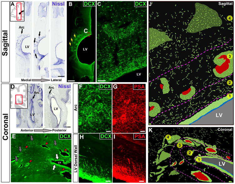
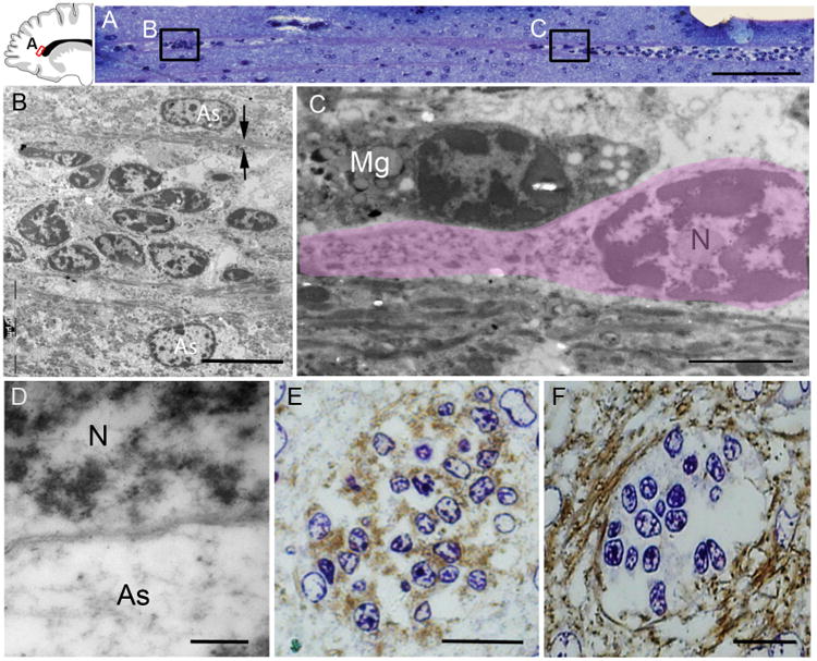
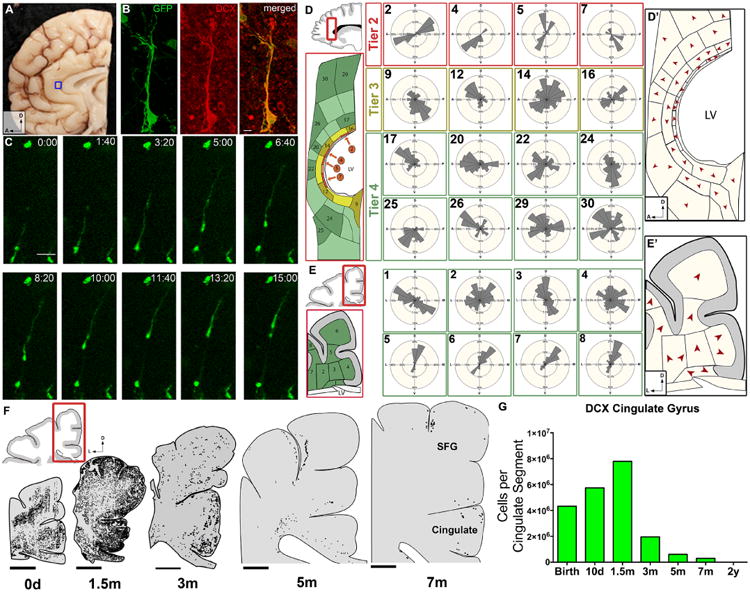
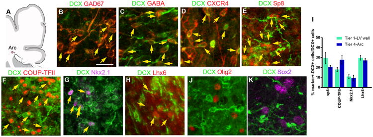
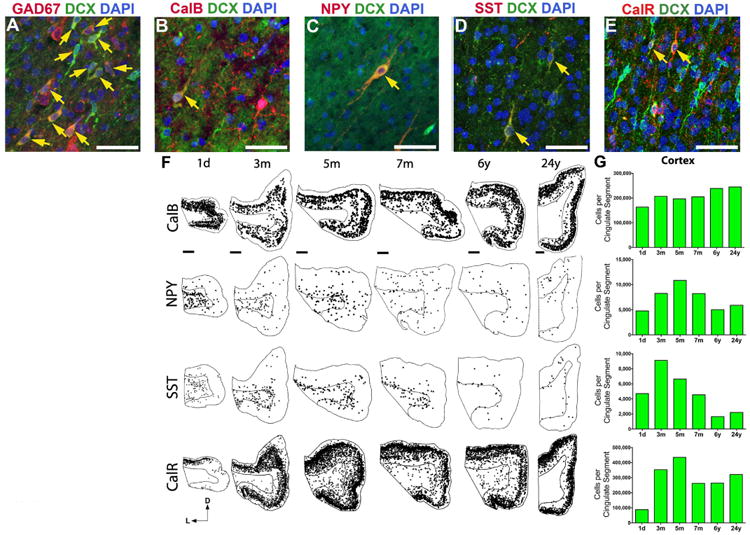
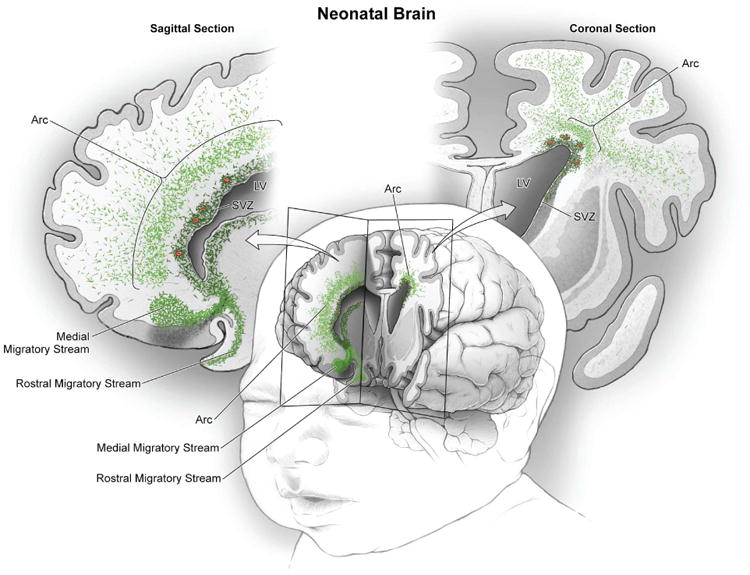
The first few months after birth, when a child begins to interact with the environment, are critical to human brain development. The human frontal lobe is important for social behavior and executive function; it has increased in size and complexity relative to other species, but the processes that have contributed to this expansion are unknown. Our studies of postmortem infant human brains revealed a collection of neurons that migrate and integrate widely into the frontal lobe during infancy. Chains of young neurons move tangentially close to the walls of the lateral ventricles and along blood vessels. These cells then individually disperse long distances to reach cortical tissue, where they differentiate and contribute to inhibitory circuits. Late-arriving interneurons could contribute to developmental plasticity, and the disruption of their postnatal migration or differentiation may underlie neurodevelopmental disorders.


Figures

Fig. 1
Fig. 1
Migrating young neurons in the infant frontal lobe are widely distributed in four tiers. (A) Serial Nissl-stained sections (taken at birth) reveal cell-dense collections around the anterior body of the lateral ventricle (black arrows, defined here as the Arc); LV, lateral ventricle. (B and C) The cells in these densities (yellow arrows) and next to the ventricular wall express DCX. (D) Coronal sections (38 GW) showing cell densities close to the ventricular wall (eyebrow-shaped, black arrows). (E) Dense aggregates of DCX+ cells around the walls of the lateral ventricles (white arrows), around blood vessels (red arrowhead), and in the parenchyma within the Arc (gray arrows). (F to I) DCX+ cells also express PSA-NCAM; (F) and (G) show cells within the Arc; (H) and (I) show cells next to the ventricular walls. (J and K) Schematic drawings of traced DCX+ cells (in green) illustrating how cells within the Arc are organized into four tiers (see text). Blood vessels are shown in red; light green clusters correspond to DCX+ cellular densities seen in (B) and (E). Scale bars, 2 mm [(A) and (B)], 50 μm (C), 1 mm (D), 25 μm [(F) to (I)].
Fig. 2
Fig. 2. Arc cells have ultra-structural features of migrating young neurons
(A) Toluidine blue staining of a semithin sagittal section from a 1-month-old brain showing a chain of cells around a blood vessel in tier 3 (see Fig. 1). Locations of images in (B) and (C) are shown. (B) Electron microscopy shows that this chain is made up of elongated cells with ultrastructural features of young migrating neurons; the chain is flanked by astrocytes (As) whose expansions (arrows) contain intermediate filaments. (C) An elongated migrating neuron (outlined in pink) next to a microglial cell (Mg). Migrating young neurons (N) frequently had an elongated morphology, a leading process, poly-ribosomes, and no intermediate filaments. (D) The cytoplasm of astrocytes is lighter and contains intermediate filaments. (E and F) 3,3′-Diaminobenzidine (DAB) staining of semithin coronal sections (adjacent to those used for electron microscopy) shows DCX expression within the chain and GFAP expression surrounding them; the counterstain is toluidine blue. Scale bars, 50 μm (A), 10 μm (B), 2 μm (C), 200 nm (D), 15 μm [(E) and (F)].
Fig. 3
Fig. 3. Migration and directionality of young neurons in the infant brain
(A) Boxed region shows area of the neonatal brain that was imaged in (B) and (C) in the cingulate gyrus. (B) DCX+ adenoGFP-labeled cell with migratory morphology. (C) Time-lapse sequence (15 hours) of adenoGFP-labeled cell revealing leading process extension, nucleokinesis, and trailing process retraction. This cell traveled ∼100 μm, migrating anteriorly in the sagittal plane. (D and E) Vector mapping of orientation of DCX+ cell leading processes, in sagittal and coronal sections; note how directionality changes in the different tiers. See figs. S6 and S7 for complete analysis. (D′ and E′) Red arrowheads indicate the modal (most frequent) direction of DCX+ cells' leading process. (F) Spatiotemporal mapping of DCX+ cells in coronal cortical sections; between birth and 1.5 months, many DCX+ cells have moved from the periventricular and parenchymal regions into the developing cortex of the cingulate and superior frontal gyrus. DCX+ cells then rapidly decrease at 3 and 5 months, but a few DCX+ cells with clear migratory morphology remain at 7 months. (G) Quantification of DCX+ cells in the cingulate gyrus (white matter and gray matter). Scale bars, 10 μm (B), 50 μm (C), 5 mm (F). Directional axes: D, dorsal; L, lateral; A, anterior.
Fig. 4
Fig. 4. Interneuron and subpallial marker expression in migrating DCX+ cells in the infant brain
(A) Schematic of coronal section indicating the Arc area that was analyzed at the dorsolateral edge of the ventricle; see fig. S2 for marker expression next to the walls of the lateral ventricle. (B to D) DCX+ cells express GAD67, GABA, and the cytokine receptor CXCR4 present in migrating interneurons. (E to H) Subpopulations of DCX+ cells express different transcription factors associated with ventral telencephalic origin, including Sp8, COUP-TFII, Nkx2.1, or Lhx6 associated with the CGE or MGE. (I) Quantification of DCX+ cells expressing Sp8, COUP-TFII, Nkx2.1, and Lhx6. Bars show means ± SEM of counts performed on three or four individual cases. (J and K) DCX+ cells do not express Olig2 or Sox2. Scale bar, 20 μm.
Fig. 5
Fig. 5. Interneuron subtype development in the cingulate gyrus
(A to E) Many DCX+ cells in the neonatal cingulate cortex express GAD67 (A), and sub-populations also coexpress interneuron subtype markers: calbindin (CalB) (B), neuropeptide Y (NPY) (C), somatostatin (SST) (D), and calretinin (CalR) (E). DAPI, 4′,6-diamidino-2-phenylindole. Yellow arrows point to DCX+ cells that coexpress the indicated subtype markers. (F) Spatiotemporal distribution of interneuron subtypes from birth to 24 years. NPY+ and SST+ cells are located primarily in the white matter at birth but shift to the cortex over time. CalR+ and CalB+ are already expressed in cells throughout the cortex at all ages, but their number continues to increase during the first five postnatal months. (G) Stereological quantification of interneuron subtypes in the cingulate cortex from birth to 24 years. The number of NPY+, SST+, CalB+, and CalR+ cells increases between birth and 5 months, coinciding with the arrival of DCX+ cells in the cingulate cortex (see Fig. 3G). Scale bars, 50 μm [(A) to (E)], 2 mm [(F), 1 day to 6 years], 1 mm [(F), 24 years]. Directional axes: D, dorsal; L, lateral.
Fig. 6
Fig. 6. Migratory streams of young neurons in the frontal lobe of the early postnatal human brain
In the frontal lobe of the neonatal human brain, cut in sagittal and coronal planes in this schematic, large numbers of young migrating neurons persist (shown in green) (see Figs. 1 to 3). Multiple concentric tiers of migrating cells are observed around the anterior pole of the lateral ventricle (see Fig. 1). Close to the ventricular wall, migrating young neurons are largely oriented tangentially; dense subpopulations are also clustered around blood vessels (red). Farther out, young neurons are more dispersed, many now oriented radially; they appear to migrate long distances through the developing white matter to reach the cortex. Ventrally, we also illustrate the RMS and the MMS, which target the olfactory bulb and medial prefrontal cortex, respectively (20).

Comment in

References

    1. Gandhi SP, Yanagawa Y, Stryker MP. Delayed plasticity of inhibitory neurons in developing visual cortex. Proc Natl Acad Sci USA. 2008;105:16797–16802. doi: 10.1073/pnas.0806159105. - DOI - PMC - PubMed
    1. Toyoizumi T, et al. A theory of the transition to critical period plasticity: Inhibition selectively suppresses spontaneous activity. Neuron. 2013;80:51–63. doi: 10.1016/j.neuron.2013.07.022. - DOI - PMC - PubMed
    1. Marín O, Rubenstein JL. A long, remarkable journey: Tangential migration in the telencephalon. Nat Rev Neurosci. 2001;2:780–790. doi: 10.1038/35097509. - DOI - PubMed
    1. Nery S, Fishell G, Corbin JG. The caudal ganglionic eminence is a source of distinct cortical and subcortical cell populations. Nat Neurosci. 2002;5:1279–1287. doi: 10.1038/nn971. - DOI - PubMed
    1. Ma T, et al. Subcortical origins of human and monkey neocortical interneurons. Nat Neurosci. 2013;16:1588–1597. doi: 10.1038/nn.3536. - DOI - PubMed

Substances