. 2017 May 15:612:29-35.
doi: 10.1016/j.gene.2016.11.010. Epub 2016 Nov 12.

Ribosome biogenesis is dynamically regulated during osteoblast differentiation

Affiliations

Ribosome biogenesis is dynamically regulated during osteoblast differentiation

Cynthia L Neben et al. Gene. .

Abstract

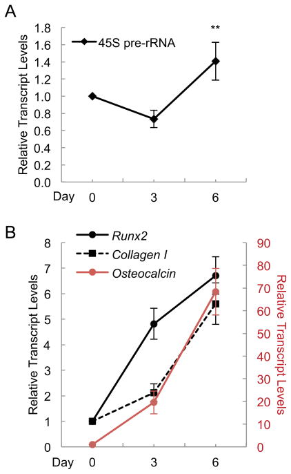
Changes in ribosome biogenesis are tightly linked to cell growth, proliferation, and differentiation. The rate of ribosome biogenesis is established by RNA Pol I-mediated transcription of ribosomal RNA (rRNA). Thus, rRNA gene transcription is a key determinant of cell behavior. Here, we show that ribosome biogenesis is dynamically regulated during osteoblast differentiation. Upon osteoinduction, osteoprogenitor cells transiently silence a subset of rRNA genes through a reversible mechanism that is initiated through biphasic nucleolar depletion of UBF1 and then RNA Pol I. Nucleolar depletion of UBF1 is coincident with an increase in the number of silent but transcriptionally permissible rRNA genes. This increase in the number of silent rRNA genes reduces levels of ribosome biogenesis and subsequently, protein synthesis. Together these findings demonstrate that fluctuations in rRNA gene transcription are determined by nucleolar occupancy of UBF1 and closely coordinated with the early events necessary for acquisition of the osteoblast cell fate.

Keywords: Bone; Osteoblast; Osteoprogenitor; RNA Pol I; Ribosome biogenesis; UBF1; rRNA.


Figures

Fig. 1
Fig. 1. rRNA expression fluctuates during osteoblast differentiation
(A) Levels of 45S precursor rRNA relative to β-actin in MC3T3-E1 cells, detected by qPCR, were reduced 27% at day 3 compared to day 0 and elevated 1.4-fold at day 6 compared to day 0 (n = 4). (B) Transcript levels of Runx2, Collagen I, and Osteocalcin, detected by qPCR, were up-regulated upon osteoinduction in MC3T3-E1 cells (n = 4). Error bars represent SD. **P<0.01.
Fig. 2
Fig. 2. Nucleolar localization of UBF1 and POLR1D is altered following osteoinduction
Immunofluorescent detection of UBF1 and the RNA Pol I subunit POLR1D in MC3T3-E1 cells during osteoinduction. (A) At day 0, prior to osteoinduction, UBF1 (green, A) and POL1D (red, A′) were highly enriched and colocalized (yellow, A″) in mature nucleoli. (B) On day 2 of osteoinduction, the number and intensity of UBF1 nucleolar foci was reduced (B), while nucleolar PORL1D localization remained high (B′). Thus, colocalization of UBF1 and POLR1D was reduced (B″). (C) By day 3, differentiating osteoblasts showed a reduction in the number and intensity of POLR1D nucleolar foci (C′) compared to day 0. Nucleolar localization of UBF1 remained depleted (C). The remaining colocalization of UBF1 and POLR1D was confined to nucleoli that were much reduced in size compared to their undifferentiated counterparts at day 0 (C″). (D) ChIP showed that UBF1 was significantly reduced at the rRNA gene UCE and 5′-ETS at day 3 of osteoinduction compared to day 0 (n = 3). Upstream enhancer element (UCE), 5′-external transcribed spacer (5′-ETS), and 28S transcribed region (28S). Error bars represent SD. *P<0.05.
Fig. 3
Fig. 3. CpG methylation and the positioning of the promoter-bound nucleosome are altered during osteoblast determination
NOMe was used to identify endogenous CpG methylation sites (black circles) as well as accessible GpC dinucleotides (teal circles) in MC3T3-E1 rRNA gene promoter clones. The pink bar represents the promoter-bound nucleosome. (A) At day 0, 11% of the 18 clones analyzed were partially methylated. GpC accessibility indicated that the TSS was accessible in 61% of these 18 clones. (B) On day 2 of osteoinduction, 11% of the 18 clones analyzed were partially methylated and 5.5% were hypermethylated. The percentage of clones with TSS accessibility reduced to 39%. (C) By day 3, 6.3% of clones were partially methylated and 6.3% were hypermethylated. Of these 16 clones, 62% were accessible at the TSS.
Fig. 4
Fig. 4. Translation potential is decreased following osteoinduction
(A) Polysome profiling of MC3T3-E1 cells showed decreased synthesis of the 40S (11%) and 60S subunits (32%), as well as the abundance of the 80S ribosomes (18%) and polysomes (21%) at day 3 compared to day 0 (n = 3). (B) Protein synthesis, as measured by O-propargyl-puromycin incorporation, was reduced 27% by day 3 of osteoinduction compared to day 0 (n = 3). Error bars represent SD. **P<0.01.

References

    1. Ali SA, Dobson JR, Lian JB, Stein JL, van Wijnen AJ, Zaidi SK, Stein GS. A RUNX2-HDAC1 co-repressor complex regulates rRNA gene expression by modulating UBF acetylation. J Cell Sci. 2012;125:2732–9. - PMC - PubMed
    1. Ali SA, Zaidi SK, Dacwag CS, Salma N, Young DW, Shakoori AR, Montecino MA, Lian JB, van Wijnen AJ, Imbalzano AN, Stein GS, Stein JL. Phenotypic transcription factors epigenetically mediate cell growth control. Proc Natl Acad Sci U S A. 2008;105:6632–7. - PMC - PubMed
    1. Ali SA, Zaidi SK, Dobson JR, Shakoori AR, Lian JB, Stein JL, van Wijnen AJ, Stein GS. Transcriptional corepressor TLE1 functions with Runx2 in epigenetic repression of ribosomal RNA genes. Proc Natl Acad Sci U S A. 2010;107:4165–9. - PMC - PubMed
    1. Bacalini MG, Pacilli A, Giuliani C, Penzo M, Trere D, Pirazzini C, Salvioli S, Franceschi C, Montanaro L, Garagnani P. The nucleolar size is associated to the methylation status of ribosomal DNA in breast carcinomas. BMC Cancer. 2014;14:361. - PMC - PubMed
    1. Belin S, Beghin A, Solano-Gonzalez E, Bezin L, Brunet-Manquat S, Textoris J, Prats AC, Mertani HC, Dumontet C, Diaz JJ. Dysregulation of ribosome biogenesis and translational capacity is associated with tumor progression of human breast cancer cells. PLoS One. 2009;4:e7147. - PMC - PubMed

LinkOut - more resources