. 2016 Jul 18:2:18.
doi: 10.1186/s40942-016-0042-y. eCollection 2016.

Expanded spectral domain-OCT findings in the early detection of hydroxychloroquine retinopathy and changes following drug cessation

Affiliations

Expanded spectral domain-OCT findings in the early detection of hydroxychloroquine retinopathy and changes following drug cessation

David R Lally et al. Int J Retina Vitreous. .

Abstract

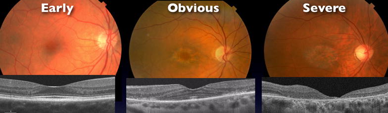
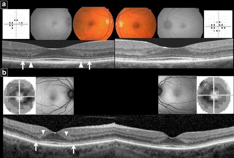
Purpose: To report expanded SD-OCT findings of HCQ retinopathy that may assist the clinician in earlier diagnosis. To characterize structural changes of HCQ retinopathy with SD-OCT after drug cessation.

Methods: Setting: Private practice and academic institution. Patient Population: Patients at New England Eye Center and Ophthalmic Consultants of Boston in Boston, MA diagnosed with HCQ retinopathy and followed after drug cessation. Retrospective clinical data review by the Boston Image Reading Center. Main Outcome Measures: SD-OCT findings suggestive of HCQ retinopathy before parafoveal ellipsoid disruption. Change in SD-OCT morphological appearance and retinal thickness of each of the nine subfields corresponding to the Early Treatment of Diabetic Retinopathy Study areas.

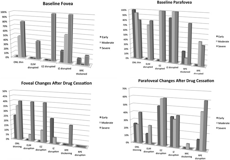
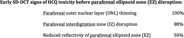
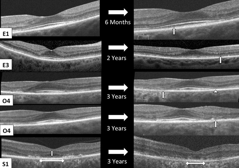
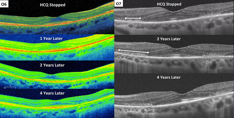
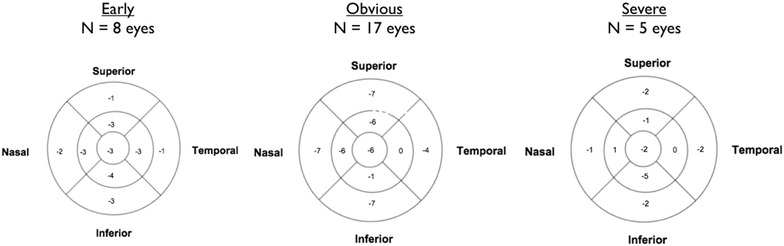
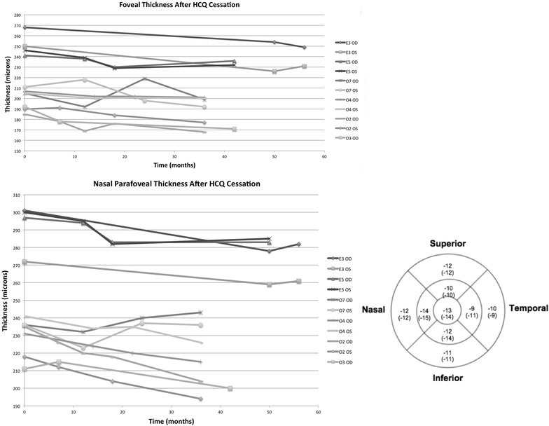
Results: Thirty eyes with HCQ retinopathy were followed with SD-OCT after drug cessation. Findings before disruption of the parafoveal EZ included parafoveal outer nuclear layer (ONL) thinning, disruption of the parafoveal interdigitation zone, and reduced reflectivity of the parafoveal EZ. In early toxicity, 75 % developed progression after drug cessation, including disruption of the parafoveal EZ and retinal pigment epithelium and thinning of the ONL. Eyes with obvious toxicity had greater inferior outer ring thinning 12 months after drug cessation compared to early toxicity (p = 0.002, 95 % CI -2 to -8 μm). In obvious toxicity, the nasal inner subfield showed more thinning than the temporal inner subfield at 12 months after drug cessation (p = 0.018, 95 % CI -1 to -8 μm).

Conclusions: Once HCQ retinopathy is diagnosed and the medication is discontinued, structural retinal changes commonly occur.


Figures

Fig. 1
Fig. 1
Fundus photograph and SD-OCT example of each group. Cohort divided into three groups based on BIRC’s grading of ellipsoid zone at the time of drug cessation. Early—no disruption of parafoveal or foveal EZ; Obvious—disruption of parafoveal EZ with intact foveal EZ; Severe—disruption of both foveal and parafoveal EZ
Fig. 2
Fig. 2
(Top row) reading center’s grading of SD-OCTs at time of HCQ discontinuation in fovea and parafovea. (Bottom row) reading center’s grading of interval changes in SD-OCT appearance of most recent follow-up exam compared to time of HCQ cessation. If thinning, disruption, or thickening was present at the time of drug cessation and an interval change was noted, the change was progressive or worsening thinning, disruption, or thickening
Fig. 3
Fig. 3
SD-OCT findings of early toxicity preceding the development of parafoveal EZ disruption
Fig. 4
Fig. 4
Examples of two Early patients. a E1, fundus photograph shows mild parafoveal RPE changes in each eye with unremarkable FAF. HVF 10-2 shows incomplete parafoveal ring scotoma in right eye and parafoveal ring scotoma in left eye. SD-OCT of each eye shows no clear disruption of parafoveal EZ. However, reduced reflectivity of the parafoveal EZ (arrow) on both sides of the fovea is observed and enhances the appearance of the foveal EZ. Abrupt discontinuation of the parafoveal interdigitation zone (IZ) (arrowhead) is also observed on both sides of the fovea. b E4, Fundus autoflourescence is unremarkable. HVF 10-2 shows incomplete parafoveal ring scotomas in each eye. No clear disruption of the parafoveal EZ is seen on SD-OCT. However, the reflectivity of the parafoveal EZ is diminished (arrow) and enhances the appearance of the foveal EZ. There is also thinning of the parafoveal outer nuclear layer causing broadening of reflectivity of the parafoveal Henle’s fiber layer (arrowhead) on both sides of the fovea
Fig. 5
Fig. 5
Examples of changes in SD-OCT appearance from the time of diagnosis of HCQ retinopathy (left column) to after drug cessation (right column). E1, left eye, shows progressive interdigitation zone disruption (arrow). E4, right eye, shows development of parafoveal EZ disruption (arrow). O4, right eye, shows progressive RPE atrophy of the temporal parafovea (arrow) and progressive EZ disruption of the nasal parafovea (arrowhead). O4, left eye, shows progressive disruption of the ELM, EZ, and RPE in the temporal parafovea (arrow). S1, left eye, shows progressive loss of the foveal ELM (arrow), foveal RPE migration into outer retina, and disruption with shortening of foveal RPE (ruler)
Fig. 6
Fig. 6
SD-OCT changes 4 years after HCQ cessation. O6, right eye, shows development of parafoveal RPE atrophy that becomes evident 2 years after drug cessation and progresses at 4 year follow up. O7, left eye, shows progressive parafoveal RPE atrophy (arrow) with broadening of RPE migration into outer retina in the nasal parafovea (ruler)
Fig. 7
Fig. 7
Median change in thickness in each ETDRS subfield at 12 months after HCQ cessation
Fig. 8
Fig. 8
Subanalysis for eyes with at least 3 years of follow-up after drug cessation (E3, E5, O2, O3 OD, O4, O7). (Top left) Subfoveal thickness change after drug cessation. (Bottom left) Example of nasal inner ring ETDRS subfield thickness change after drug cessation. (Bottom right) Mean change (Median change) in thickness in each ETDRS subfield

References

    1. Rynes RI. Antimalarial drugs in the treatment of rheumatological diseases. Rheumatology. 1997;36(7):799–805. doi: 10.1093/rheumatology/36.7.799. - DOI - PubMed
    1. Shearer RV, Dubois EL. Ocular changes induced by long-term hydroxychloroquine (plaquenil) therapy. Am J Ophthalmol. 1967;64:245–252. doi: 10.1016/0002-9394(67)92518-4. - DOI - PubMed
    1. Wolfe F, Marmor MF. Rates and predictors of hydroxychloroquine retinal toxicity in patients with rheumatoid arthritis and systemic lupus erythematosus. Arthritis Care Res (Hoboken) 2010;62:775–784. doi: 10.1002/acr.20133. - DOI - PubMed
    1. Marmor MF, Kellner U, Lai TTY, for the American Academy of Ophthalmology et al. Revised recommendations on screening for chloroquine and hydroxychloroquine retinopathy. Ophthalmology. 2011;118:415–422. doi: 10.1016/j.ophtha.2010.11.017. - DOI - PubMed
    1. Carr RE, Henkind P, Rothfield N, et al. Ocular toxicity of antimalarial drugs: long-term follow-up. Am J Ophthalmol. 1968;66:738–744. doi: 10.1016/0002-9394(68)91300-7. - DOI - PubMed