

Macular pigment in retinal health and disease
- PMID: 27847637
- PMCID: PMC5088450
- DOI: 10.1186/s40942-016-0044-9
Macular pigment in retinal health and disease
Abstract
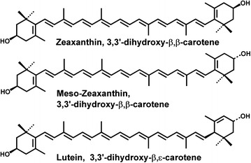
Lutein and zeaxanthin, two carotenoid pigments of the xanthophyll subclass, are present in high concentrations in the retina, especially in the macula. They work as a filter protecting the macula from blue light and also as a resident antioxidant and free radical scavenger to reduce oxidative stress-induced damage. Many observational and interventional studies have suggested that lutein and zeaxanthin may reduce the risk of various eye diseases, especially late forms of AMD. In vitro and in vivo studies indicate that they could protect various ocular cells against oxidative damage. Recent research has shown that in addition to traditional mechanisms, lutein and zeaxanthin can influence the viability and function of cells through various signal pathways or transcription factors: for instance, they can affect immune responses and inflammation, and have anti-angiogenic and anti-tumor properties. This review covers the basic aspects and results of recent studies regarding the effects of lutein, zeaxanthin and other carotenoids, such as meso-zeaxanthin, on the eye in different clinical and experimental models and the management of various ocular diseases using these molecules.
Figures
References
-
- Snodderly DM, Auran JD, Delori FC. The macular pigment. II. Spatial distribution in primate retinas. Invest Ophthalmol Vis Sci. 1984;25:674–685. - PubMed
Publication types
LinkOut - more resources
Full Text Sources
Other Literature Sources
