. 2016 Sep 15:2:22.
doi: 10.1186/s40942-016-0046-7. eCollection 2016.

Choroidal thickness in older patients with central serous chorioretinopathy

Affiliations

Choroidal thickness in older patients with central serous chorioretinopathy

Caio V Regatieri et al. Int J Retina Vitreous. .

Abstract

Background: To investigate the choroidal thickness in older patients with central serous chorioretinopathy (CSCR) compared to age-matched normal subjects.

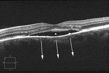
Methods: Fifteen patients (30 eyes) with CSCR, all aged ≥60 years, and 21 age-matched normal subjects (21 eyes) underwent high-definition raster scanning using SD-OCT. Both eyes from CSCR patients were included in the analysis. The eyes in patients with CSCR were divided into two groups: active CSCR (17 eyes) if there was foveal-involving subretinal fluid and inactive contralateral eye group (13 eyes). Choroidal thickness was measured from the posterior edge of the retinal pigment epithelium to the choroidal-scleral junction at 500 µm intervals up to 2500 µm temporal and nasal to the fovea (11 locations).

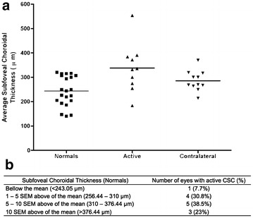
Results: The mean age of the patients with CSCR was 68.87 ± 6.83 years (mean ± standard deviation). Reliable measurements of choroidal thickness were obtainable in 70.6 % of eyes examined. The choroid was statistically significantly thicker in eyes with both active CSCR (P < 0.001) and inactive contralateral eyes (P < 0.01) when compared to normal age-matched eyes. The subfoveal choroid was 95 µm (P < 0.01) thicker in eyes with active CSCR (338.05 ± 31.42 µm) compared with normal eyes (243.05 ± 13.39 µm). The subfoveal choroid thickness in the inactive contralateral eyes was numerically greater than normal, and it was not statistically significantly thicker compared to the normal eyes (difference-55.68 µm, P > 0.05).

Conclusion: Choroid in older patients with active CSCR was thicker than the choroid in age-matched normal eyes. It is important to consider CSCR as a differential diagnosis of serous retinal detachment in elderly patients with thickened choroid and to consider SD-OCT as an imaging modality by which to evaluate the choroidal thickness.

Keywords: Central serous chorioretinopathy; Choroid; Optical coherence tomography.


Figures

Fig. 1
Fig. 1
High definition 1-line optical coherence tomography B-scan image from a 72-year-old man with active central serous chorioretinopathy. Serous detachment of the neurosensory retina (asterisk). In this eye the choroid may be so thick that it is not possible to visualize the choroidal-sclera interface (arrows)
Fig. 2
Fig. 2
Graph of mean choroidal thickness in normal eyes, eyes with active central serous chorioretinopathy (CSCR) and inactive contralateral eye. Mean choroidal thickness at each of the 11 locations measured at 500 µm (0.5 mm) intervals temporal (T) and nasal (N), centered on the fovea. Active: eyes with active CSCR; contralateral: inactive contralateral eyes. P value represents the result of statistical analyses (ANOVA)
Fig. 3
Fig. 3
Representative images of an elderly patient with active chronic central serous chorioretinopathy (CSCR) for 8 months. a Fundus color photograph shows pigmentary changes on the macula area, which corresponds to a leakage point on the dye-based angiographies. b Fluorescein angiography image shows a focal area of leakage (white arrow). c Late phase of an indocyanine green angiography shows multiple areas of choroidal hyperpermeability. d SD OCT foveal B-scan shows subretinal and intraretinal fluid. The perpendicular green line was drawn from posterior edge of retinal pigment epithelium to choroidal–scleral junction to demonstrate the measurement. The green arrow heads point the choroidal–scleral junction
Fig. 4
Fig. 4
a Scatter plot of subfoveal choroidal thickness in normal eyes, eyes with active central serous chorioretinopathy (CSCR) and the inactive contralateral eye. Active: eyes with active CSCR; contralateral: inactive contralateral eyes. b Distribution of subfoveal choroidal thickness of eyes presenting with active CSCR, comparing with normal eyes. SEM standard error of the mean

References

    1. Hussain D, Gass JD. Idiopathic central serous chorioretinopathy. Indian J Ophthalmol. 1998;46(3):131–137. - PubMed
    1. Gemenetzi M, De Salvo G, Lotery AJ. Central serous chorioretinopathy: an update on pathogenesis and treatment. Eye (Lond) 2010;24(12):1743–1756. doi: 10.1038/eye.2010.130. - DOI - PubMed
    1. Guyer DR, Yannuzzi LA, Slakter JS, Sorenson JA, Ho A, Orlock D. Digital indocyanine green videoangiography of central serous chorioretinopathy. Arch Ophthalmol. 1994;112(8):1057–1062. doi: 10.1001/archopht.1994.01090200063023. - DOI - PubMed
    1. Piccolino FC, Borgia L, Zinicola E, Zingirian M. Indocyanine green angiographic findings in central serous chorioretinopathy. Eye (Lond) 1995;9(Pt 3):324–332. doi: 10.1038/eye.1995.63. - DOI - PubMed
    1. Kuroda S, Ikuno Y, Yasuno Y, et al. Choroidal thickness in central serous chorioretinopathy. Retina. 2013;33(2):302–308. doi: 10.1097/IAE.0b013e318263d11f. - DOI - PubMed

LinkOut - more resources