Review
. 2016 Nov 1:2:25.
doi: 10.1186/s40942-016-0050-y. eCollection 2016.

Novel perspectives on swept-source optical coherence tomography

Affiliations
Review

Novel perspectives on swept-source optical coherence tomography

Fabio Lavinsky et al. Int J Retina Vitreous. .

Abstract

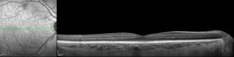
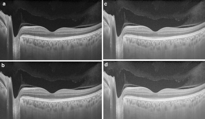
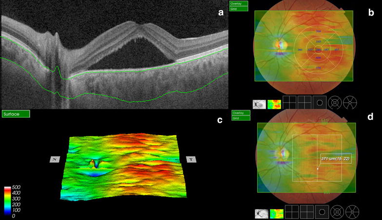
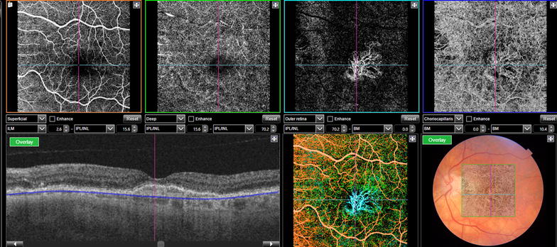
Technologies for multimodal digital imaging of vitreoretinal diseases have improved the accuracy of diagnosis and the depth of the knowledge of the mechanisms of disease and their response to treatments. Optic coherence tomography (OCT) has become a mandatory tool for the management and for the follow-up of retinal pathologies. OCT technology evolved in the last two decades from time-domain to spectral domain and recently to the swept-source OCTs (SS-OCT). SS-OCT improved the depth of imaging and the scan speed, thus adding novel algorithms and features such as for vitreous and vitreoretinal interface evaluation, choroid segmentation and mapping, OCT angiography and En-face OCT. The multimodal approach using SS-OCT is expected to advance the understanding of retinal pathologies such as age related macular degeneration, diabetic maculopathy, central serous chorioretinopathy, the pachychoroid spectrum and macular telangiectasia. Surgical vitreoretinal diseases such as vitreo-macular traction syndrome, epiretinal membrane, retinal detachment, proliferative vitreoretinal retinopathy and diabetic traction retinal detachment also will be better understood and documented with SS-OCT. This technology also provides great utility for a broad spectrum of ophthalmic pathologies including glaucoma, uveitis, tumors and anterior segment evaluation.


Figures

Fig. 1
Fig. 1
Spectral domain OCT with enhanced depth imaging (EDI) mode
Fig. 2
Fig. 2
Swept source-OCT high resolution single scan using Enhanced Vitreous Visualizationtm (EVV) shows different windows for vitreous visualization, from EVV 0 (a), 1 (b), 3 (c) and 5 (d)
Fig. 3
Fig. 3
Swept source-OCT single scan shows automatic segmentation layers (a) with ETDRS thickness maps (b), 3D reconstruction (c) and point-by-point automatic measurement of choroidal thickness (d)
Fig. 4
Fig. 4
OCT angiography of a patient with exudative age related macular degeneration after treatment with anti-VEGF, however it is still possible to identify choroidal neovascularization bellow the RPE with no signs of inner or subretinal fluid
Fig. 5
Fig. 5
Enface OCT image of a patient with chronic central serous chorioretinopathy at the level of the RPE showing several hypereflective dots under the subretinal fluid and a larger hypereflective spot corresponding to the leaking point on fluorescein angiography
Fig. 6
Fig. 6
Swept source-OCT high resolution single scan of a patient with macular telangiectasia type 2 with corresponding fundus imaging (a), and OCT angiography (b) of same patient that shows vascular tortuosity and dilation in superficial and deep layers with no signs of vascular changes on outer retina or choriocapillaris
Fig. 7
Fig. 7
Color fundus imaging of a patient with proliferative diabetic retinopathy (a) and single scan swept source OCT image shows partial vitreous detachment with hyperreflective thickening (b) that corresponds to retinal neovascularization clearly visible with OCT angiography with vitreous segmentation (c, d) and superficial segmentation (e, f)

References

    1. Huang D, Swanson EA, Lin CP, Schuman JS, Stinson WG, Chang W, et al. Optical coherence tomography. Science. 1991;254:1178–1181. doi: 10.1126/science.1957169. - DOI - PMC - PubMed
    1. Schuman JS, Puliafito CA, Fujimoto JG, Duker JS. Optical coherence tomography of ocular diseases. 3. Thorofare, NJ: SLACK incorporated; 2013.
    1. Hee MR, Izatt JA, Swanson EA, et al. Optical coherence tomography of the human retina. Arch Ophthalmol. 1995;113:325–332. doi: 10.1001/archopht.1995.01100030081025. - DOI - PubMed
    1. Hee MR, Puliafito CA, Duker JS, et al. Characterization of epiretinal membranes using optical coherence tomography. Ophthalmology. 1998;105:360–370. doi: 10.1016/S0161-6420(98)93601-6. - DOI - PubMed
    1. Schuman JS, Pedut-Kloizman T, Pakter H, et al. Optical coherence tomography and histologic measurements of nerve fiber layer thickness in normal and glaucomatous monkey eyes. Invest Ophthalmol Vis Sci. 2007;48(8):3645–3654. doi: 10.1167/iovs.06-0876. - DOI - PMC - PubMed

LinkOut - more resources