. 2016 Nov 11;2(11):e1600990.
doi: 10.1126/sciadv.1600990. eCollection 2016 Nov.

In vivo characterization of distinct modality-specific subsets of somatosensory neurons using GCaMP

Affiliations

In vivo characterization of distinct modality-specific subsets of somatosensory neurons using GCaMP

Edward C Emery et al. Sci Adv. .

Abstract

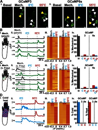
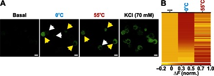
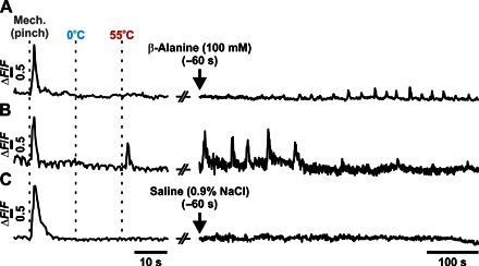
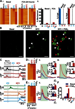
Mechanistic insights into pain pathways are essential for a rational approach to treating this vast and increasing clinical problem. Sensory neurons that respond to tissue damage (nociceptors) may evoke pain sensations and are typically classified on the basis of action potential velocity. Electrophysiological studies have suggested that most of the C-fiber nociceptors are polymodal, responding to a variety of insults. In contrast, gene deletion studies in the sensory neurons of transgenic mice have frequently resulted in modality-specific deficits. We have used an in vivo imaging approach using the genetically encoded fluorescent calcium indicator GCaMP to study the activity of dorsal root ganglion sensory neurons in live animals challenged with painful stimuli. Using this approach, we can visualize spatially distinct neuronal responses and find that >85% of responsive dorsal root ganglion neurons are modality-specific, responding to either noxious mechanical, cold, or heat stimuli. These observations are mirrored in behavioral studies of transgenic mice. For example, deleting sodium channel Nav1.8 silences mechanical- but not heat-sensing sensory neurons, consistent with behavioral deficits. In contrast, primary cultures of axotomized sensory neurons show high levels of polymodality. After intraplantar treatment with prostaglandin E2, neurons in vivo respond more intensely to noxious thermal and mechanical stimuli, and additional neurons (silent nociceptors) are unmasked. Together, these studies define polymodality as an infrequent feature of nociceptive neurons in normal animals.

Keywords: GCaMP imaging; Pain modality; nociception; polymodal nociceptor.


Figures

Fig. 1
Fig. 1. Assessment of GCaMP3 and GCaMP6s activity and sensitivity in DRG neurons in vitro.
(A and B) Confocal images showing changes in fluorescence from GCaMP3-expressing (A) and GCaMP6s-expressing (B) DRG neurons following the application of KCl. Scale bars, 20 μm. (C) Fluorescence response profile of an individual GCaMP6s-expressing neuron in response to single depolarizing pulses. (D) Heatmap and corresponding fluorescence response of a single GCaMP3-expressing neuron in response to depolarizing pulses at variable frequency stimulations. Inset: Averaged responses shown as a function of frequency stimulation [action potential frequency (AP freq.) in hertz; *P < 0.05, **P < 0.01, and ***P < 0.001, repeated-measures analysis of variance (ANOVA); n = 7]. norm., normalized.
Fig. 2
Fig. 2. Assessment of GCaMP3 activity and sensitivity in DRG neurons in vivo.
(Ai) Extracellular electrophysiological recording from an intact L4 DRG in vivo showing individual action potentials being elicited by brief (20 ms) 100-mA pulses (denoted by black arrows). The upper black trace shows activity from a single neuron extracted from the raw signal (green). (Aii) Summary of the average number of action potentials elicited from individual neurons in response to 1-, 10-, and 100-mA pulses (20 ms; n = 48). (Bi) Example fluorescence trace of a single GCaMP3 neuron in response to a train of 100-mA pulses (20 ms, 0.5 Hz). Responses from all neurons analyzed are shown in (Bii) (n = 7). (Biii) Summary of the average change in fluorescence (ΔF/F) for each GCaMP3 neuron for sequential current stimulations (100 mA, 20 ms, 0.5 Hz). The average number of action potentials elicited per current injection is overlaid in red (n = 48). (C) In vivo confocal images showing the levels of GCaMP3 fluorescence from L4 DRG neurons in response to the stimuli applied to the plantar surface. Numbered arrows indicate activated neurons within each application frame. Scale bar, 20 μm. (D) Raw traces of each neuron labeled with a numbered arrowhead in (C). The vertical dotted lines represent the points at which the stimuli were applied to the plantar surface. Gray, von Frey; blue, 0°C water; red, 55°C water; green, light brush. The arrows mark the points at which the frames in (C) were taken for each stimulus event.
Fig. 3
Fig. 3. Basal sensory modality testing in mice expressing GCaMP3 and GCaMP6s.
(A) In vivo confocal images showing levels of GCaMP3 (Ai) and GCaMP6s (Aii) fluorescence from L4 DRG neurons at rest (basal) and in response to mechanical (pinch), cold (0°C), and heat (55°C) stimuli applied to the plantar surface. Unimodal (white arrowheads) and polymodal (yellow arrowheads) neurons are shown. Scale bars, 10 μm. Diagram of application area of the pinch stimulus (Bi) (gray), von Frey stimuli (Ci) (green dots), and cold/heat stimuli (Di) (red/blue stripes). Example raw traces from L4 DRG neurons in response to mechanical [(Bii) pinch; (Cii) von Frey] and 0° and 55°C stimuli (dotted lines indicate application of stimuli; 3 and 6 refer to GCaMP3 and GCaMP6s traces, respectively). Gray- and green-colored traces indicate modality-specific and polymodal response profiles, respectively. (Dii) Example raw traces from L4 DRG neurons in response to thermal stimuli (dotted lines indicate application of stimuli) (3, GCaMP3; 6, GCaMP6s). Red- and blue-colored traces indicate neurons responding to 55° and 0°C stimuli, respectively. Heatmap response profiles for every DRG neuron tested for mechanical [(Biii) pinch; (Ciii) von Frey] and 0° and 55°C stimuli for both GCaMP3-expressing (left; n = 17 to 31) and GCaMP6s-expressing neurons (right; n = 17 to 45). (Diii) Heatmap response profiles for every neuron responsive to 0° or 55°C stimuli for both GCaMP3-expressing (left; n = 146) and GCaMP6s-expressing (right; n = 100) neurons. Summary of DRG neurons [M, mechanical; (Biv) pinch; (Civ) von Frey] also responding to 0°C (C), 55°C (H), or both 0° and 55°C (C/H) stimuli from mice expressing GCaMP3 or GCaMP6s, respectively. (Div) Summary of cold-sensitive neurons also responding to a heat stimulus and of heat-sensitive neurons also responding to a cold stimulus from mice expressing GCaMP3 or GCaMP6s, respectively. Recordings were made from 13 animals expressing GCaMP3 and 8 animals expressing GCaMP6s of both sex.
Fig. 4
Fig. 4. Assessment of cold and heat sensitivity in cultured DRG neurons in vitro.
(A) In vitro images showing changes in GCaMP3 fluorescence from cultured DRG neurons at rest (basal) and in response to perfused 0°C extracellular solution, 55°C extracellular solution, and KCl. Unimodal (white arrowheads) and polymodal (yellow arrowheads) neurons are shown. Scale bars, 10 μm. (B) Heatmap response profiles for every neuron responsive to 0° or 55°C stimuli. Each row displays the maximum normalized fluorescent signal acquired during each application window (n = 171).
Fig. 5
Fig. 5. Assessment of sensory modality responses of β-alanine–sensitive neurons in vivo.
Example GCaMP3 traces of individual DRG neurons following the application of modality-specific stimuli (mechanical pinch, 0°C water, or 55°C water) and their respective sensitivity to the intraplantar administration of β-alanine (100 mM). Most of the β-alanine–sensitive neurons (n = 5 or 7) were only responsive to mechanical stimuli (A), with few responding to both mechanical and heat stimuli (B) (n = 2 or 7). A typical trace following saline (0.9% NaCl) administration is also shown (C).
Fig. 6
Fig. 6. The effect of inflammatory agents on the sensory modalities response of DRG neurons in vivo.
(Ai) Heatmap response profiles for individual GCaMP3-expressing mechanically sensitive (pinch) neurons tested for sensitivity toward mechanical (pinch), 0°, and 55°C stimuli (left), as well as neurons responsive to 0° or 55°C stimuli (right), before (basal; n = 31 and n = 146, respectively) or 24 hours after administration of FCA (n = 90 or n = 102, respectively). (Aii) Summary of mechanically sensitive neurons responding to thermal stimuli as well as thermal-sensitive neurons responding to 55° or 0°C before and 24 hours after the administration of FCA, respectively. Baseline (n = 13 animals) and after FCA (n = 10 animals). (B) In vivo confocal images showing levels of GCaMP3 fluorescence from L4 DRG neurons that respond to 55°C before (pre-PGE2) and 10 min after administration of PGE2 (55°C + PGE2). Basally responsive (white arrowheads) and basally silent (red arrowheads) neurons are shown. Scale bars, 50 μm. (C) Example raw traces of neuronal modality profiles before and after PGE2 administration. (Di) Heatmap response profile for individual neurons responsive to 55°C before or after PGE2 administration. Trace showing the average change in GCaMP3 fluorescence following peripheral noxious heat stimulation (green application bar) from basally heat-responsive neurons (Dii) or basally silent neurons (Diii). AUC, area under the curve. (Ei) Heatmap response profile for individual neurons responsive to 0°C before or after PGE2 administration. (Eii) Trace showing the average change in GCaMP3 fluorescence following peripheral noxious cold stimulation (0°C; green application bar) from basally cold-responsive neurons. Changes in relative maximal fluorescence and AUC (insets). n.s., not significant. (F) Summary of the number of cells displaying discrete modalities before and after PGE2 administration (n = 75) [*P < 0.05, **P < 0.01, and ***P < 0.001 (comparisons made to baseline); #P < 0.05 and ###P < 0.001 (comparisons made between groups)]. Pre- and post-PGE2 recordings were made from five animals. M, mechano; C, cold; H, heat; M/H, mechano-heat; M/C/H, mechano-cold-heat; C/H, cold and hot.

References

    1. Nahin R. L., Estimates of pain prevalence and severity in adults: United States, 2012. J. Pain 16, 769–780 (2015). - PMC - PubMed
    1. Emery E. C., Luiz A. P., Wood J. N., Nav1.7 and other voltage-gated sodium channels as drug targets for pain relief. Expert Opin. Ther. Targets 20, 975–983 (2016). - PMC - PubMed
    1. Davis J. B., Gray J., Gunthorpe M. J., Hatcher J. P., Davey P. T., Overend P., Harries M. H., Latcham J., Clapham C., Atkinson K., Hughes S. A., Rance K., Grau E., Harper A. J., Pugh P. L., Rogers D. C., Bingham S., Randall A., Sheardown S. A., Vanilloid receptor-1 is essential for inflammatory thermal hyperalgesia. Nature 405, 183–187 (2000). - PubMed
    1. Gasser H. S., The classification of nerve fibers. Ohio J. Sci. 41, 145–159 (1941).
    1. Djouhri L., Lawson S. N., Aβ-fiber nociceptive primary afferent neurons: A review of incidence and properties in relation to other afferent A-fiber neurons in mammals. Brain Res. Rev. 46, 131–145 (2004). - PubMed

Substances