. 2017 Jul;10(4):971-982.
doi: 10.1038/mi.2016.98. Epub 2016 Nov 16.

Genetic deletion of the bacterial sensor NOD2 improves murine Crohn's disease-like ileitis independent of functional dysbiosis

Affiliations

Genetic deletion of the bacterial sensor NOD2 improves murine Crohn's disease-like ileitis independent of functional dysbiosis

D Corridoni et al. Mucosal Immunol. 2017 Jul.

Abstract

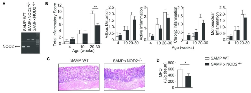
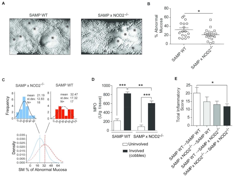
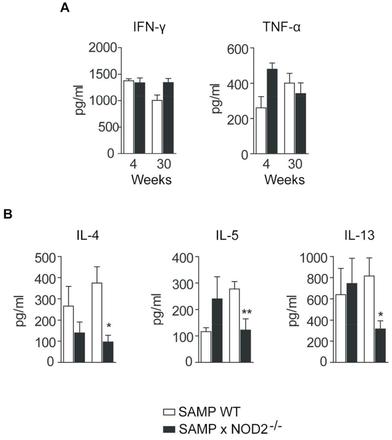
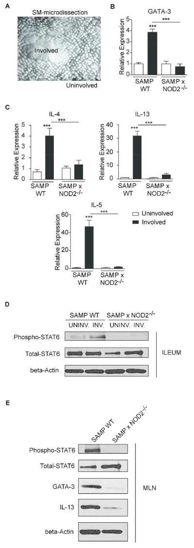
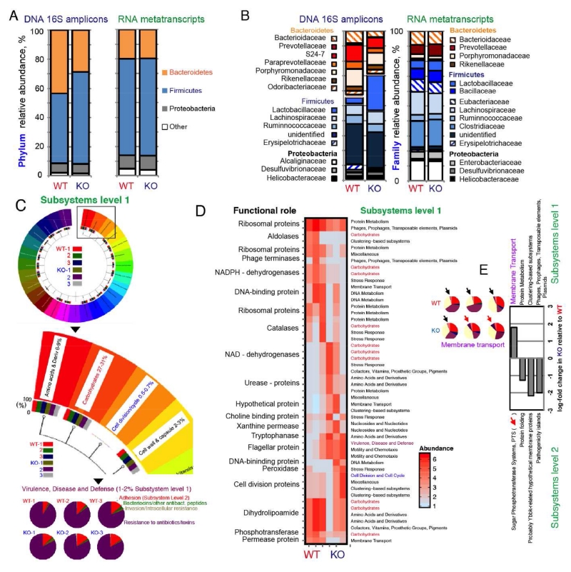
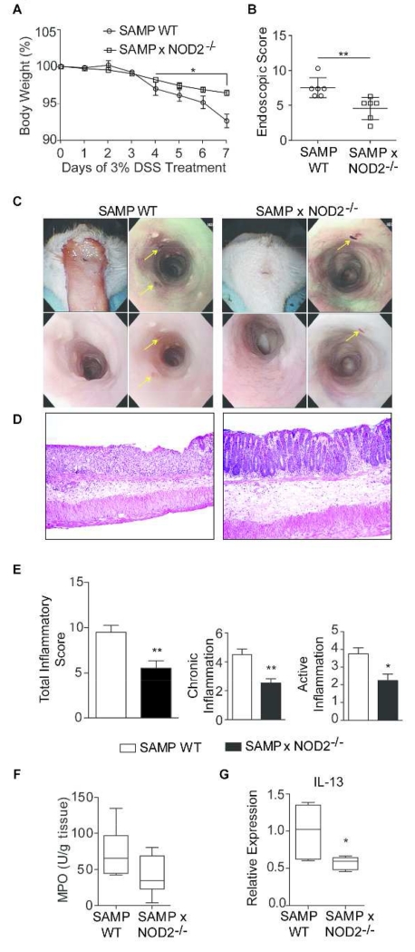
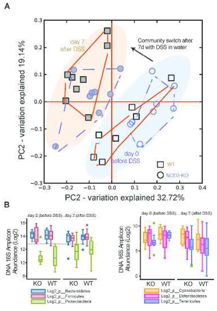
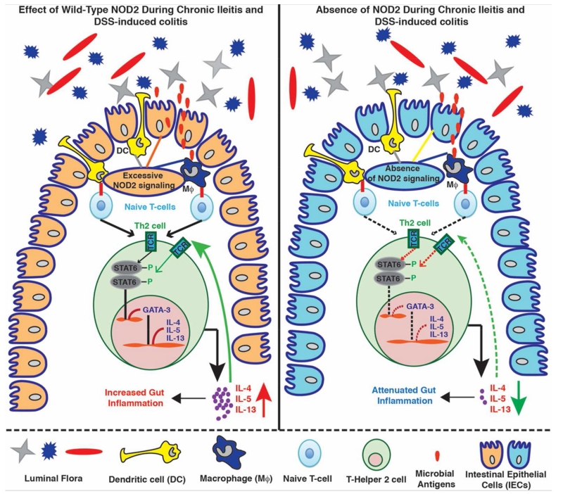
Although genetic polymorphisms in NOD2 (nucleotide-binding oligomerization domain-containing 2) have been associated with the pathogenesis of Crohn's disease (CD), little is known regarding the role of wild-type (WT) NOD2 in the gut. To date, most murine studies addressing the role of WT Nod2 have been conducted using healthy (ileitis/colitis-free) mouse strains. Here, we evaluated the effects of Nod2 deletion in a murine model of spontaneous ileitis, i.e., the SAMP1Yit/Fc (SAMP) strain, which closely resembles CD. Remarkably, Nod2 deletion improved both chronic cobblestone ileitis (by 50% assessed, as the % of abnormal mucosa at 24 wks of age), as well as acute dextran sodium sulfate (DSS) colitis. Mechanistically, Th2 cytokine production and Th2-transcription factor activation (i.e., STAT6 phosphorylation) were reduced. Microbiologically, the effects of Nod2 deletion appeared independent of fecal microbiota composition and function, assessed by 16S rRNA and metatranscriptomics. Our findings indicate that pharmacological blockade of NOD2 signaling in humans could improve health in Th2-driven chronic intestinal inflammation.


Figures

Figure 1
Figure 1. SAMP × NOD2−/− mice show decreased severity of inflammation compared to SAMP WT mice
(A) PCR genotyping of mouse tail DNA showing the generation of SAMP × NOD2−/− mice using speed congenic techniques. (B) Ileal total inflammatory scores at different time points (4, 10 and 20-30-wk old), as determined by the sum of chronic inflammation, active inflammation, villous distortion and mononuclear inflammation. ** P < 0.001 (n=17-24), unpaired Student’s t test; data are represented as mean ± SEM. (C) Representative histopathological sections show significant villous blunting and a significant increase of inflammatory cells in the lamina propria of 20-30-wk old SAMP WT compared to SAMP × NOD2−/− mice. (D) MPO activity in ilea of SAMP WT and SAMP × NOD2−/− mice at 30 wks of age. * P < 0.05 (n=13), pairwise Mann–Whitney test; data are represented as mean ± SEM.
Figure 2
Figure 2. Deletion of Nod2 in SAMP mice decreases the extension of abnormal mucosa
(A) Three-dimensional stereomicroscopic (3D-SM) images of fixed specimens to visualize the architecture of ilea in SAMP WT and SAMP × NOD2−/− mice. (B) Percentage of 3D abnormal mucosa in ilea of SAMP WT and SAMP × NOD2−/− mice. * P < 0.05 (n=17-18), Student’s t test; data are represented as mean ± SEM. (C) Density plot analysis of data with a bimodal distribution for SAMP WT (with ~ 50% of mice affected with severe disease) and normalized (predictable Gaussian) healthy population of SAMP × NOD2−/− mice. (D) MPO activity in uninvolved and involved areas of ilea from SAMP WT and SAMP × NOD2−/− mice. ** P < 0.001, *** P < 0.0001 (n=7), pairwise Mann–Whitney test; data are represented as mean ± SEM. (E) Ileo-colitis total inflammatory scores of the following groups of chimeric mice: SAMP WT BM→SAMP WT, SAMP × NOD2−/−BM→SAMP WT, SAMP WT BM→SAMP × NOD2−/−, and SAMP × NOD2−/− BM→SAMP × NOD2−/−. * P <0.05. (n=7), Student’s t test; data are represented as mean ± SEM.
Figure 3
Figure 3. Decreased production of Th2 cytokines from MLN cells of SAMP × NOD2−/− mice
Secretion levels of Th1-type cytokines (A), TNF-α and IFN-γ, and Th2-type cytokines (B), IL-4, IL-5 and IL-13 from MLN CD4+ T cells isolated from pre-inflamed (4-wk old) and inflamed (30-wk old) SAMP WT and SAMP × NOD2−/−, following polyclonal activation with anti-CD3/anti-CD28. * P < 0.05, ** P < 0.01 (n=6-10), Mann–Whitney test; data are represented as mean ± SEM.
Figure 4
Figure 4. NOD2 regulates Th2-mediated chronic intestinal inflammation
(A) Representative 3D-SM image of involved and uninvolved areas of SAMP WT and SAMP × NOD2−/− mice from which samples were derived. (B) GATA-3, (C) IL-4, IL-5, and IL-13 expression during chronic ileitis (30 wks). *** P < 0.0001 (n=6-12), Student’s t test; data are represented as mean ± SEM. Protein lysates from (D) ilea and (E) MLNs from 30-wk-old SAMP WT and SAMP × NOD2−/− mice immunoblotted with antibodies against phosphorylated-STAT6, total-STAT6, GATA-3, IL-13, and actin (n>3).
Figure 5
Figure 5. Gut metatranscriptome analysis indicates the Nod2 deletion did not have a significant impact on fecal microbiota composition and function
Analysis on samples collected 7 days following IsPreFeH (3 mice/group). (A) Phylum-level composition based on 16S rRNA amplicon libraries and phylogenomic taxonomic inference from functional mRNA metatranscriptome data. (B) Family-level composition based on 16S rRNA amplicon libraries and phylogenomic taxonomic inference from functional mRNA metatranscriptome data (C) MG-RAST visualization of normalized transcript functional abundance (%) from the 6 individual metatranscriptomes. (D) Metatranscriptome cluster analysis of most expressed roles (specific enzymatic reactions). (E) Log-fold changes for the only 4 significantly detected functional roles (4 out of 193 level 2 categories). Pie charts, relative metatranscript abundance for the sugar phosphotransferase system.
Figure 6
Figure 6. Following IsPreFeH, SAMP × NOD2−/− mice show reduced severity of DSS colitis compared to WT mice
After 4 days of exposure to a composite mixture of feces and bedding material (orally gavaged), SAMP WT and NOD2−/− mice were treated with 3% DSS in their drinking water for 7 days (n=6/group). (A) Changes in body weight during DSS treatment (AUC, Student’s t test P < 0.05). (B) Endoscopic score based on a distal-proximal endoscopic colitis scoring system on day 7. (Mann–Whitney, ** P < 0.01); data are represented as mean ± SEM. (C) Endoscopic images of perianal, distal and proximal colon after DSS treatment show severe inflammation in SAMP WT mice (thickened appearance of the mucosa, and numerous ulcers - yellow arrows) compared to mild inflammation in SAMP × NOD2−/− mice. (D) Representative histopathology images at same magnification show thickening and increased cellular infiltration in lamina propria of SAMP WT mice. (E) Histological, total, chronic and active colonic inflammation scores (Mann–Whitney, ** P < 0.01, * P < 0.05). (F) MPO activity in colonic tissue after DSS treatment. (G) IL-13 in colonic tissue after DSS treatment (Mann–Whitney, * P < 0.05); data are represented as mean ± SEM.
Figure 7
Figure 7. 16S rRNA microbial community analysis shows that bacterial community changes before and after DSS-induced colitis do not predict the presence of Nod2 deletion in SAMP mice
Following the fecal homogenization protocol, fecal samples from SAMP WT and SAMP × NOD2−/− mice were collected before and after 7 days of 3% DSS treatment (n=3-6/group, 2-3 experiments). (A) Principal coordinate analysis of 16S rRNA microbial community data before and after DSS. (B) Box plot of the normalized 16S amplicon Log2 abundance for the six most abundant bacterial phyla for all experimental mice.
Figure 8
Figure 8. Working hypothesis for the role of NOD2 signaling in experimental IBD
In the presence of WT NOD2 signaling, SAMP mice with spontaneous chronic ileitis or DSS-induced colitis show increased production of Th2 cytokines, such as IL-4, IL-5, and IL-13, which is mediated by excessive NOD2 signaling (left panel). In the absence of NOD2 signaling following NOD2 genetic deletion, there is inhibition of Th2 cytokine production with attenuated gut inflammation (right panel). Mechanistically, these effects are mediated by the activity of Th2 transcription factors, STAT6 and GATA-3 (left and right panel).

References

    1. Kanneganti TD, Lamkanfi M, Nunez G. Intracellular NOD-like receptors in host defense and disease. Immunity. 2007;27:549–559. - PubMed
    1. Chen G, Shaw MH, Kim YG, Nunez G. NOD-like receptors: role in innate immunity and inflammatory disease. Annu. Rev. Pathol. 2009;4:365–398. - PubMed
    1. Abbott DW, Yang Y, Hutti JE, Madhavarapu S, Kelliher MA, Cantley LC. Coordinated regulation of Toll-like receptor and NOD2 signaling by K63-linked polyubiquitin chains. Mol. Cell Biol. 2007;27:6012–6025. - PMC - PubMed
    1. Philpott DJ, Sorbara MT, Robertson SJ, Croitoru K, Girardin SE. NOD proteins: regulators of inflammation in health and disease. Nat. Rev. Immunol. 2014;14:9–23. - PubMed
    1. Yang Y, Yin C, Pandey A, Abbott D, Sassetti C, Kelliher MA. NOD2 pathway activation by MDP or Mycobacterium tuberculosis infection involves the stable polyubiquitination of Rip2. J. Biol. Chem. 2007;282:36223–36229. - PubMed

MeSH terms