. 2016 Nov 16:6:37305.
doi: 10.1038/srep37305.

Mitigation of radiation-induced hematopoietic injury by the polyphenolic acetate 7, 8-diacetoxy-4-methylthiocoumarin in mice

Affiliations

Mitigation of radiation-induced hematopoietic injury by the polyphenolic acetate 7, 8-diacetoxy-4-methylthiocoumarin in mice

Kavya Venkateswaran et al. Sci Rep. .

Erratum in

Abstract

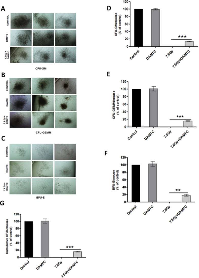
Protection of the hematopoietic system from radiation damage, and/or mitigation of hematopoietic injury are the two major strategies for developing medical countermeasure agents (MCM) to combat radiation-induced lethality. In the present study, we investigated the potential of 7, 8-diacetoxy-4-methylthiocoumarin (DAMTC) to ameliorate radiation-induced hematopoietic damage and the associated mortality following total body irradiation (TBI) in C57BL/6 mice. Administration of DAMTC 24 hours post TBI alleviated TBI-induced myelo-suppression and pancytopenia, by augmenting lymphocytes and WBCs in the peripheral blood of mice, while bone marrow (BM) cellularity was restored through enhanced proliferation of the stem cells. It stimulated multi-lineage expansion and differentiation of myeloid progenitors in the BM and induced proliferation of splenic progenitors thereby, facilitating hematopoietic re-population. DAMTC reduced the radiation-induced apoptotic and mitotic death in the hematopoietic compartment. Recruitment of pro-inflammatory M1 macrophages in spleen contributed to the immune-protection linked to the mitigation of hematopoietic injury. Recovery of the hematopoietic compartment correlated well with mitigation of mortality at a lethal dose of 9 Gy, leading to 80% animal survival. Present study establishes the potential of DAMTC to mitigate radiation-induced injury to the hematopoietic system by stimulating the re-population of stem cells from multiple lineages.


Figures

Figure 1
Figure 1. DAMTC mitigates TBI-induced lethality in mice.
(A,B) Protective effects of DAMTC (i.e. administered prior to irradiation) against lethal TBI (9 Gy). Mice were injected DAMTC intra-peritoneally and irradiated 1 or 4 hours later. Kaplan-Meier survival curve depicts the 30 day survival (n = 20 in all the groups i.e. TBI, Vehicle + TBI, DAMTC + TBI). (C,D) Mitigative effects of DAMTC administered 24 hours post TBI is represented in the Kaplan-Meier survival curves, when mice were exposed to lethal TBI (9 Gy; (C)) and sub-lethal TBI (7.6 Gy; (D)). In both (C) and (D) n = 20 for all the groups (TBI, TBI + Vehicle, TBI + DAMTC). (E) Dose response of TBI on mice and DMF evaluation of DAMTC at LD50/30. Mice were subjected to varying TBI doses (5–11 Gy) and treated with DAMTC or vehicle at 24 hours following irradiation and monitored for survival through 30 days (n = 10). (F) Table summarises the findings of Mantel-Cox (log-rank) and Gehan-Breslow-Wilcoxon statistics for comparing survival responses between TBI and TBI + DAMTC in experiments used for generating the DMF shown in (E). *P < 0.05; **P < 0.01; ***P < 0.001.
Figure 2
Figure 2. DAMTC mitigates TBI-induced pancytopenia in mice.
Alterations in the hematological indices in the peripheral blood by DAMTC. Mice were subjected to TBI (7.6 Gy) followed by the administration of DAMTC at 24 hours and assessed for blood cell parameters. Blood counts showing (A) WBC (B) Lymphocyte at days 3, 7, 14, 21 post TBI. (C,D) WBC, lymphocyte levels showing the effects of DAMTC observed on day 21 (C) and day 28 (D). Blood counts showing (E) RBC, (F) hemoglobin, (G) platelet at days 3, 7, 14, 21 post TBI (n = 10). All error bars indicate SEM. *P < 0.05; **P < 0.01; ***P < 0.001.
Figure 3
Figure 3. DAMTC mediates stimulation of the splenic progenitor cells to mitigate TBI-induced hematopoietic damage in mice.
Mice were subjected to TBI (7.6 Gy) followed by the administration of DAMTC at 24 hours and assessed for endogenous splenic colonies on day 10. (A) Spleen was fixed in Bouin’s solution to enumerate the CFUs. Representation of CFU-S in TBI (n = 12) and DAMTC + TBI (n = 12). (B) Spleen weight and (C) spleen-body weight ratio (n = 12). All error bars indicate SEM. *P < 0.05; **P < 0.01; ***P < 0.001.
Figure 4
Figure 4. DAMTC mitigates TBI-induced BM failure in mice.
Effects of DAMTC on bone marrow (BM) cellularity in TBI mice. (A) Panels show H&E staining of mouse femurs. Representative images are shown for naïve, DAMTC, TBI, and TBI + DAMTC treatments. For the upper panel, scale bar = 50 μm with an original magnification of ×400. For the lower panel, scale bar = 200 μm with an original magnification of ×100. (B) Total nucleated cell numbers of BM seen in naïve, TBI, TBI + DAMTC mice at day 10 and (C) day 45 post TBI (cells from ten animals were examined in each group; n = 10). All error bars indicate SEM. *P < 0.05; **P < 0.01; ***P < 0.001. Imaging of H&E stained femurs was done on day 10 post TBI.
Figure 5
Figure 5. DAMTC facilitates expansion of hematopoietic progenitors in the BM of TBI mice.
Effects of DAMTC on BM hematopoietic progenitor cells (HPCs) in TBI mice. Panels show colonies of hematopoietic progenitors (A) CFU-GM, (B) CFU-GEMM and (C) BFU-E after performing ex-vivo culturing on day 10 post TBI. Representative images of colonies from naïve, DAMTC and TBI + DAMTC mice are shown (cells from ten animals were examined in each group; n = 10). Percentages of (D) CFU-GM, (E) CFU-GEMM, (F) BFU-E and (G) cumulative CFUs are shown. All error bars indicate SEM. *P < 0.05; **P < 0.01; ***P < 0.001. Imaging of BM hematopoietic CFUs was done on day 12 of ex-vivo culture. Original magnification, ×40 (AC).
Figure 6
Figure 6. DAMTC mitigates TBI-induced cytogenetic damage in BM.
Effects of DAMTC on TBI-mediated MN-PCEs in mouse BM. BM smears were evaluated for MN induction by May-Grϋnwald-Geimsa staining. Representation of frequency of MN-PCEs per 103 PCEs at (A) 24 hours (B) 48 hours and (C) 72 hours following DAMTC treatment in naïve, DAMTC, TBI (3 Gy) and TBI + DAMTC mice (n = 10 in all groups). All error bars indicate SEM. *P < 0.05; **P < 0.01; ***P < 0.001.
Figure 7
Figure 7. DAMTC mitigates TBI-induced cytogenetic damage in the peripheral blood of TBI mice.
Effects of DAMTC on TBI-induced MN-PCEs in mouse blood. Blood smears were evaluated for MN induction by May-Grϋnwald-Geimsa staining. Representation of frequency of MN-PCEs per 2 × 103 erythrocytes at (A) 24, 48 and 72 hours following DAMTC treatment in naïve, DAMTC, TBI (3 Gy) and TBI + DAMTC mice (n = 10 in all groups). Blood smears were stained with acridine orange (AO) to assess the effects of DAMTC on TBI-mediated cytotoxicity and genotoxicity. (B) Panels showing representative images of AO stained blood smears from naïve, TBI and TBI + DAMTC mice (Original magnification, ×400). Arrow heads and arrows represent PCE and MN-PCE respectively. (C) Frequency of PCEs and MN-PCEs per 2 × 103 cells at 24, 48 and 72 hours visualised by AO staining. All error bars indicate SEM. *P < 0.05; **P < 0.01; ***P < 0.001.
Figure 8
Figure 8. Reduction in the radiation-induced apoptosis and stimulation of cell proliferation in the BM by DAMTC.
Effects of DAMTC on TBI-mediated apoptotic death and compromised proliferative ability in BM was evaluated by analysis of hypo-diploid cells and S-phase cells respectively in the BM. (A) Frequency of apoptotic and S-phase cells at days 3 and 21 post TBI (7.6 Gy) in mice (n = 10 in all groups), along with representative DNA flow cytograms. (B) Average values of apoptotic and S-phase cells and representative DNA flow cytograms observed at 24, 48 and 72 hours following treatment in naïve, TBI (3 Gy) and TBI + DAMTC mice (n = 10 in all groups). All error bars indicate SEM. *P < 0.05; **P < 0.01; ***P < 0.001.
Figure 9
Figure 9. DAMTC-facilitates the induction of M1 macrophages in the spleen of TBI mice.
Splenocytes were isolated at 24, 48 and 72 hours following treatment and stained for macrophagic markers (F4/80 and CD11b) and antigen-presenting/co-stimulatory markers (MHC-II and CD86). Results were analysed as double-positive cells. Representation of frequency of (A) F4/80+MHC-II+ cells (B) F4/80+CD86+ cells (C) CD11b+MHC-II+ cells and (D) CD11b+CD86+ cells (n = 10 for naïve, DAMTC, TBI (3 Gy) and TBI + DAMTC groups). All error bars indicate SEM. *P < 0.05; **P < 0.01; ***P < 0.001.

Similar articles

Cited by

References

    1. Park E. et al. Dieckol rescues mice from lethal irradiation by accelerating hemopoiesis and curtailing immunosuppression. International Journal of Radiation Biology 86, 848–859 (2010). - PubMed
    1. Hall E. J. & Giaccia A. J. Radiobiology for the Radiobiologist. PA: Lippincott Williams and Wilkins 6th ed. Philadelphia (2006).
    1. Fliedner T. M. et al. Multi-organ involvement as a pathogenetic principle of the radiation syndromes: a study involving 110 case histories documented in SEARCH and classified as the bases of haematopoietic indicators of effect. British Journal of Radiology Supplement 27, 1–8 (2005).
    1. Hill R. P. Radiation effects on the respiratory system. Br. J. Radiol. Suppl. 27, 75–81 (2005).
    1. Moulder J. E. & Cohen E. P. Radiation-induced multi-organ involvement and failure: the contribution of radiation effects on the renal system. British Journal of Radiology Supplement 27, 82–88 (2005).

Publication types

MeSH terms

Substances