

Storage pool diseases illuminate platelet dense granule biogenesis
- PMID: 27849413
- PMCID: PMC5994342
- DOI: 10.1080/09537104.2016.1243789
Storage pool diseases illuminate platelet dense granule biogenesis
Abstract
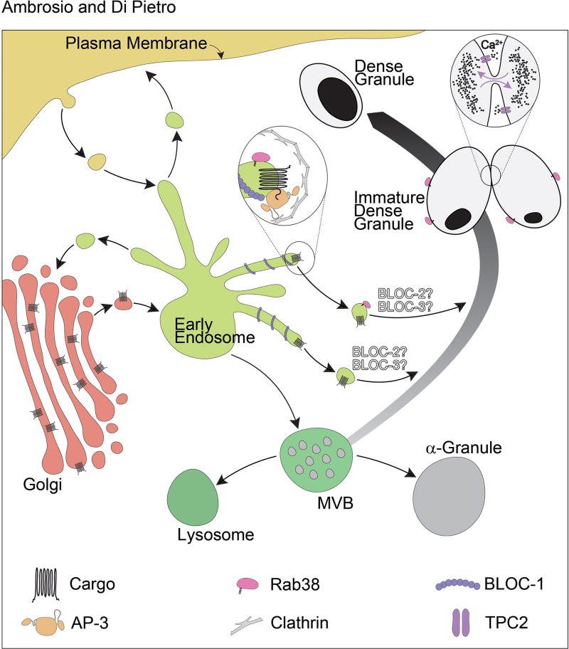
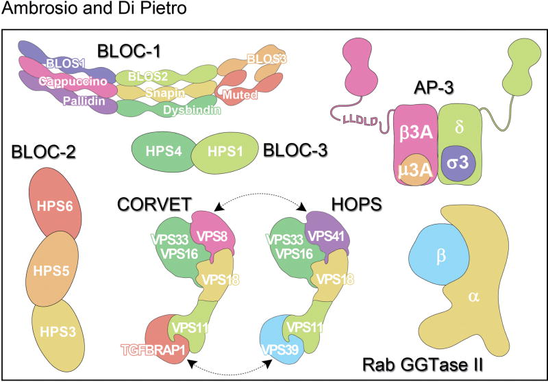
Platelet dense granules (DGs) are membrane bound compartments that store polyphosphate and small molecules such as ADP, ATP, Ca2+, and serotonin. The release of DG contents plays a central role in platelet aggregation to form a hemostatic plug. Accordingly, congenital deficiencies in the biogenesis of platelet DGs underlie human genetic disorders that cause storage pool disease and manifest with prolonged bleeding. DGs belong to a family of lysosome-related organelles, which also includes melanosomes, the compartments where the melanin pigments are synthesized. These organelles share several characteristics including an acidic lumen and, at least in part, the molecular machinery involved in their biogenesis. As a result, many genes affect both DG and melanosome biogenesis and the corresponding patients present not only with bleeding but also with oculocutaneous albinism. The identification and characterization of such genes has been instrumental in dissecting the pathways responsible for organelle biogenesis. Because the study of melanosome biogenesis has advanced more rapidly, this knowledge has been extrapolated to explain how DGs are produced. However, some progress has recently been made in studying platelet DG biogenesis directly in megakaryocytes and megakaryocytoid cells. DGs originate from an endosomal intermediate compartment, the multivesicular body. Maturation and differentiation into a DG begins when newly synthesized DG-specific proteins are delivered from early/recycling endosomal compartments. The machinery that orchestrates this vesicular trafficking is composed of a combination of both ubiquitous and cell type-specific proteins. Here, we review the current knowledge on DG biogenesis. In particular, we focus on the individual human and murine genes encoding the molecular machinery involved in this process and how their deficiencies result in disease.
Keywords: AP-3 complex; BLOC; HOPS; Hermansky–Pudlak syndrome; Rab38; protein traffic.
Conflict of interest statement
The authors declare that they have no conflict of interest.
Figures
References
-
- Thon JN, Italiano JE. Platelets: production, morphology and ultrastructure. Handb Exp Pharmacol. 2012:3–22. - PubMed
-
- Koseoglu S, Flaumenhaft R. Advances in platelet granule biology. Curr Opin Hematol. 2013;20:464–471. - PubMed
-
- King SM, Reed GL. Development of platelet secretory granules. Semin Cell Dev Biol. 2002;13:293–302. - PubMed
Publication types
MeSH terms
Grants and funding
LinkOut - more resources
Full Text Sources
Other Literature Sources
Research Materials
Miscellaneous