. 2017 Jul;66(7):1286-1296.
doi: 10.1136/gutjnl-2016-312268. Epub 2016 Nov 14.

Palbociclib (PD-0332991), a selective CDK4/6 inhibitor, restricts tumour growth in preclinical models of hepatocellular carcinoma

Affiliations

Palbociclib (PD-0332991), a selective CDK4/6 inhibitor, restricts tumour growth in preclinical models of hepatocellular carcinoma

Julien Bollard et al. Gut. 2017 Jul.

Abstract

Objective: Advanced hepatocellular carcinoma (HCC) is a lethal malignancy with limited treatment options. Palbociclib, a well-tolerated and selective CDK4/6 inhibitor, has shown promising results in the treatment of retinoblastoma (RB1)-positive breast cancer. RB1 is rarely mutated in HCC, suggesting that palbociclib could potentially be used for HCC therapy. Here, we provide a comprehensive characterisation of the efficacy of palbociclib in multiple preclinical models of HCC.

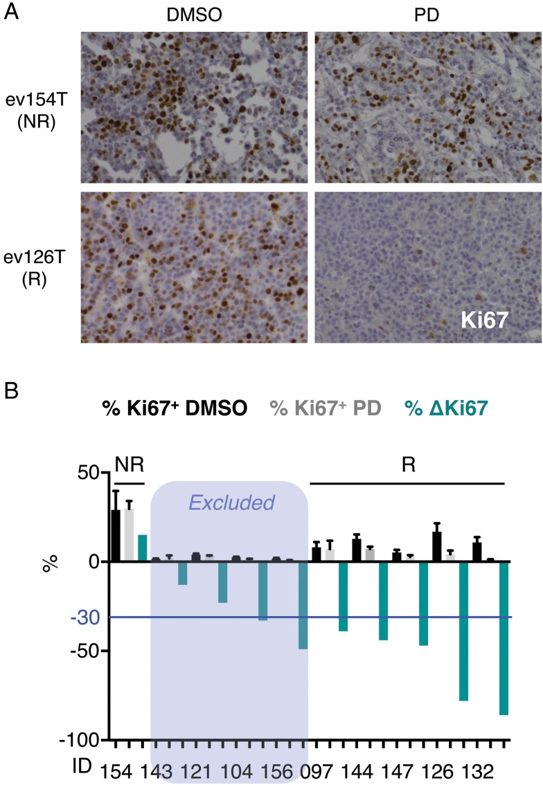
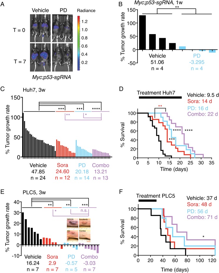
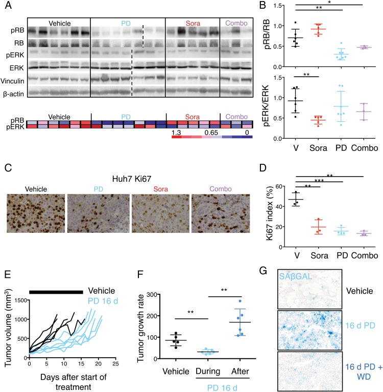
Design: The effects of palbociclib on cell proliferation, cellular senescence and cell death were investigated in a panel of human liver cancer cell lines, in ex vivo human HCC samples, in a genetically engineered mouse model of liver cancer, and in human HCC xenografts in vivo. The mechanisms of intrinsic and acquired resistance to palbociclib were assessed in human liver cancer cell lines and human HCC samples by protein and gene expression analyses.

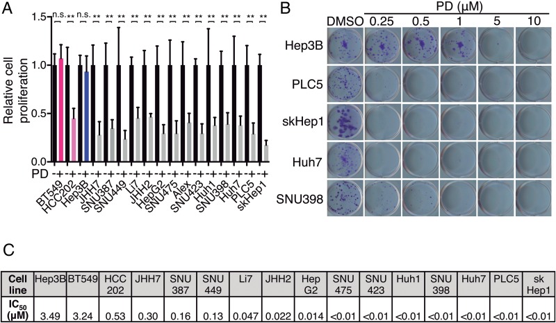
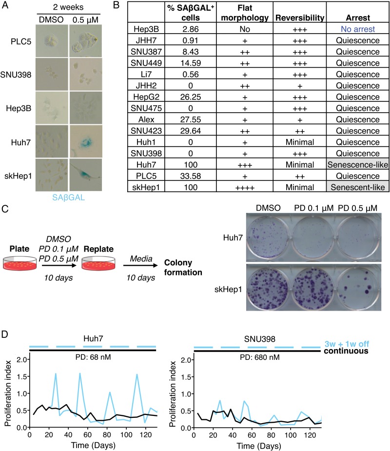
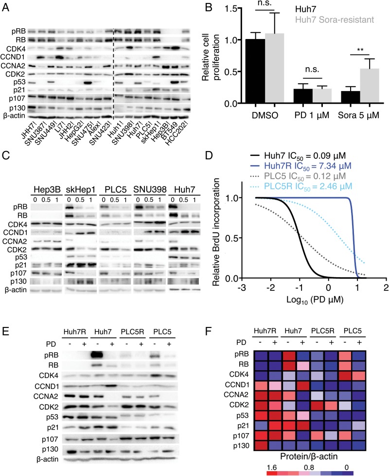
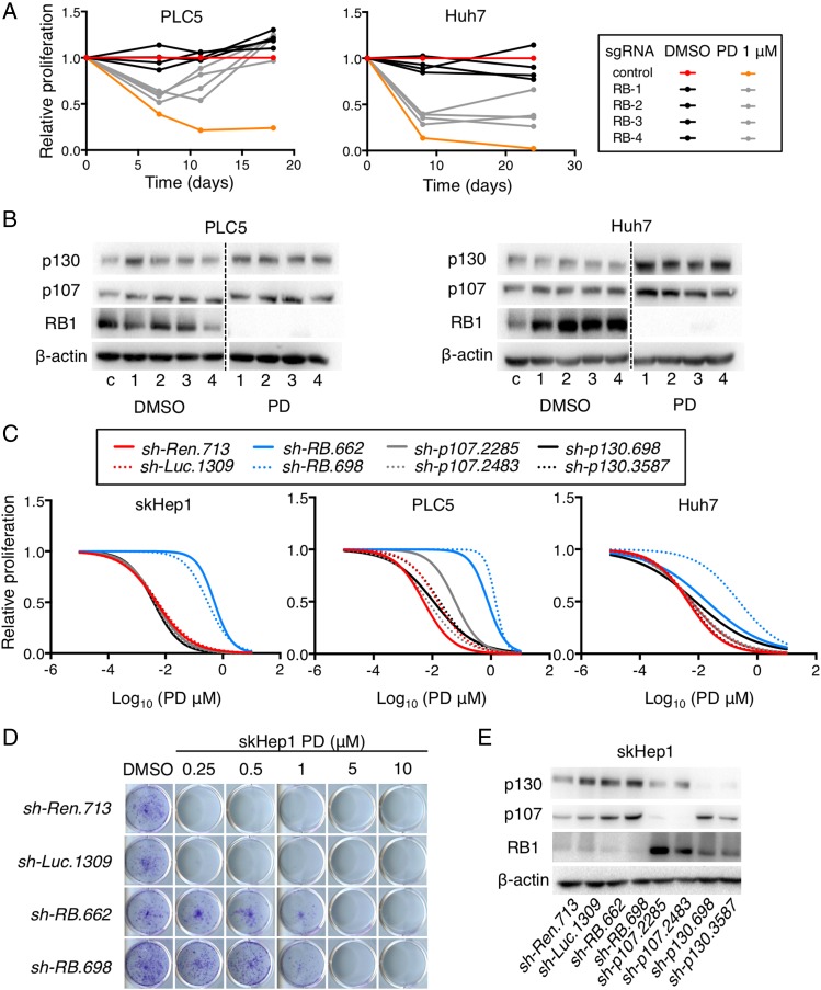
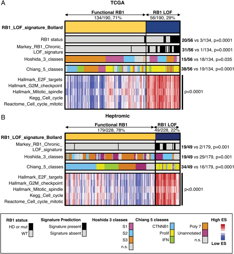
Results: Palbociclib suppressed cell proliferation in human liver cancer cell lines by promoting a reversible cell cycle arrest. Intrinsic and acquired resistance to palbociclib was determined by loss of RB1. A signature of 'RB1 loss of function' was found in <30% of HCC samples. Palbociclib, alone or combined with sorafenib, the standard of care for HCC, impaired tumour growth in vivo and significantly increased survival.

Conclusions: Palbociclib shows encouraging results in preclinical models of HCC and represents a novel therapeutic strategy for HCC treatment, alone or particularly in combination with sorafenib. Palbociclib could potentially benefit patients with RB1-proficient tumours, which account for 70% of all patients with HCC.

Keywords: CELL CYCLE; HEPATOCELLULAR CARCINOMA.


Conflict of interest statement

Competing interests: None declared.

Figures

Figure 1
Figure 1
Palbociclib inhibits the proliferation of human liver cancer cell lines. (A) Number of cells, relative to dimethyl sulfoxide (DMSO)-treated condition, after 3 days of treatment with 1 μM palbociclib (PD). BT549 and HCC202 (in pink) are two breast cancer cell lines used as retinoblastoma (RB1)-negative and RB1-positive controls, respectively. Hep3B, indicated in blue, was the only hepatocellular carcinoma (HCC)-resistant cell line. The mean+SD is shown. (B) Crystal violet staining of colonies from five representative cell lines treated during 2 weeks with the indicated doses of PD. (C) IC50 values calculated by quantifying the extracted crystal violet in (B).
Figure 2
Figure 2
Palbociclib (PD) induces a reversible cell cycle arrest in human liver cancer cell lines. (A) Representative pictures (×200 magnification) of senescence-associated β-galactosidase (SAβGAL) staining in five representative cell lines treated with PD. Blue indicates positive staining. (B) Table summarising the assays to evaluate cellular senescence. (C) Reversibility assay. Left, schematic. Right, crystal violet staining after replating cells that were pretreated as indicated. (D) Proliferation of cells, relative to dimethyl sulfoxide (DMSO)-treated cells, treated continuously (black) or discontinuously (blue) with PD, over time. W, week.
Figure 3
Figure 3
Retinoblastoma (RB1) loss of function correlates with resistance to palbociclib (PD) in human liver cancer cell lines. (A) Immunoblotting analysis of indicated proteins (basal levels) in the panel of liver cancer cell lines. BT549 and HCC202 are two breast cancer cell lines used as RB1-negative and RB1-positive controls, respectively. The dashed line separates independent gels. (B) Number of cells, relative to dimethyl sulfoxide (DMSO)-treated condition, after 3 days of treatment with 1 μM PD or 5 μM sorafenib (Sora). The mean+SD is shown. (C) Immunoblotting of different proteins after treatment with the indicated doses of PD during 3 days for five representative cell lines. (D) Dose–response curves for different doses of PD. The corresponding IC50 value of each cell line is included. (E) Immunoblotting of designated proteins after treatment with 0.5 μM of PD for 3 days in the parental or resistant (R) cell lines. (F) Heatmap showing protein levels relative to β-actin. Red indicates high while blue indicates low, and it is relative in each row.
Figure 4
Figure 4
Loss of retinoblastoma (RB1) confers resistance to palbociclib (PD) in human liver cancer cell lines. (A) Proliferation of cells, relative to control cells treated with dimethyl sulfoxide (DMSO), at different time points. (B) Immunoblotting analysis of indicated proteins at the end of the experiment in (A). The dashed line separates different portions of the same gel. (C) Dose–response curves for different doses of PD and cells infected with different shRNAs. (D) Crystal violet staining of colonies from a representative cell line infected with control or RB1 shRNAs and treated during 2 weeks with the indicated doses of PD. (E) Immunoblotting analysis of indicated proteins (basal levels) of cells in (C). c, control. 1–4 represent the different single-guide RNAs for RB1.
Figure 5
Figure 5
Retinoblastoma (RB1) loss of function (RB1_LOF) signature in human hepatocellular carcinoma (HCC) patients. (A) Heatmap showing the distribution of different gene sets in 190 human HCC patients from The Cancer Genome Atlas (TCGA). The proportions of patients for ‘RB1_LOF’ presence or absence are also included. The values for Hoshida class 2 and Chiang proliferation are included. (B) Same as in (A) but for a different patient cohort including 278 HCC patients. HD, homozygous deletion; IFN, interferon; mut, mutation; WT, wild-type; ES, enrichment score; n.s., not significant.
Figure 6
Figure 6
Palbociclib (PD) is effective in organotypic ex vivo human hepatocellular carcinoma (HCC) samples. (A) Immunostaining for Ki67 (×200 magnification), a marker of cell proliferation, in representative responder (R) and non-responder (NR) ex vivo human HCC samples treated with PD for 2 days. (B) Percentage of Ki67-positive cells in ex vivo human HCC samples treated with dimethyl sulfoxide (DMSO) (black) or PD (grey) for 2 days. The mean+SD is shown. The percentage of change in Ki67 staining is shown in green. The blue line indicates the threshold (–30%) used to assign responsiveness to PD.
Figure 7
Figure 7
Palbociclib (PD), alone or in combination with sorafenib (Sora), has potent antitumour effects in vivo. (A) Bioluminescence imaging of representative mice (Myc;p53-sgRNA) before and after 1 week of treatment. The colour scale is shown on the right. (B) The percentage of tumour growth rate (per day) per each individual mouse measured by bioluminescence imaging. The average tumour growth rate per group is shown as well as the number of mice. (C) As in (B) but for Huh7 xenografts. (D) Survival curves of the mice in (C). The duration of treatment and median survival per group are indicated at the top. (E) As in (B) but for PLC5 xenografts. (F) Survival curves of the mice in (E). The treatment window and median survival per group are indicated as in (D). combo, combination of sorafenib and palbociclib; d, days; V, vehicle; W, weeks.
Figure 8
Figure 8
Effects of mono or combinatorial therapies in vivo. (A) Top, immunoblotting of designated proteins in tumours isolated from Huh7 xenografted mice after 5 days of treatment. Bottom, heatmap showing phospho-RB (pRB) and phospho-ERK (pERK) levels relative to RB and ERK, respectively, from (A). Red indicates high while blue indicates low, and it is relative in each row. (B) Dot plots with quantification of the relative pRB and pERK levels. The mean±SD is shown. (C) Immunostaining for Ki67 (×200 magnification), a marker of cell proliferation, in representative tumours treated with the indicated treatments for 5 days. (D) Quantification of Ki67 index in the different treatment groups. The mean±SD is shown. (E) Spider plots depicting tumour growth in each group of treatment over time. The black bars represent the treatment period (16 days). (F) Quantification of tumour growth rate in vehicle-treated and palbociclib-treated mice (during the 16 days of treatment and after treatment). The mean±SD is shown. (G) Representative images of senescence-associated β-galactosidase (SAβGAL) staining in vehicle-treated and palbociclib-treated tumours at day 16. A representative image of a tumour treated for 16 days and left untreated for additional 3 days it shown ×100 magnification. Combo, combination of sorafenib and palbociclib; d, days; PD, palbociclib; Sora, sorafenib; V, vehicle; WD, withdrawal.

Comment in

References

    1. Llovet JM, Villanueva A, Lachenmayer A, et al. Advances in targeted therapies for hepatocellular carcinoma in the genomic era. Nat Rev Clin Oncol 2015;12:436 10.1038/nrclinonc.2015.121 - DOI - PubMed
    1. Torre LA, Bray F, Siegel RL, et al. Global cancer statistics, 2012. CA Cancer J Clin 2015;65:87–108. 10.3322/caac.21262 - DOI - PubMed
    1. Llovet JM, Hernandez-Gea V. Hepatocellular carcinoma: reasons for phase III failure and novel perspectives on trial design. Clin Cancer Res 2014;20:2072–9. 10.1158/1078-0432.CCR-13-0547 - DOI - PubMed
    1. Llovet JM, Ricci S, Mazzaferro V, et al. Sorafenib in advanced hepatocellular carcinoma. N Engl J Med 2008;359:378–90. 10.1056/NEJMoa0708857 - DOI - PubMed
    1. Malumbres M, Barbacid M. Cell cycle kinases in cancer. Curr Opin Genet Dev 2007;17:60–5. 10.1016/j.gde.2006.12.008 - DOI - PubMed

MeSH terms