. 2016 Nov 29;113(48):13833-13838.
doi: 10.1073/pnas.1611400113. Epub 2016 Nov 14.

Gram-positive bacteria are held at a distance in the colon mucus by the lectin-like protein ZG16

Affiliations

Gram-positive bacteria are held at a distance in the colon mucus by the lectin-like protein ZG16

Joakim H Bergström et al. Proc Natl Acad Sci U S A. .

Abstract

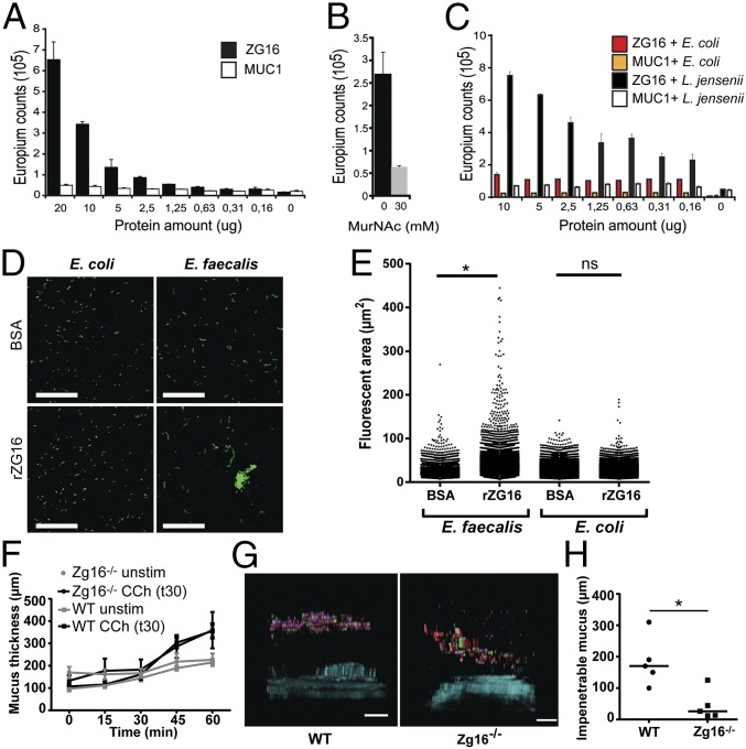
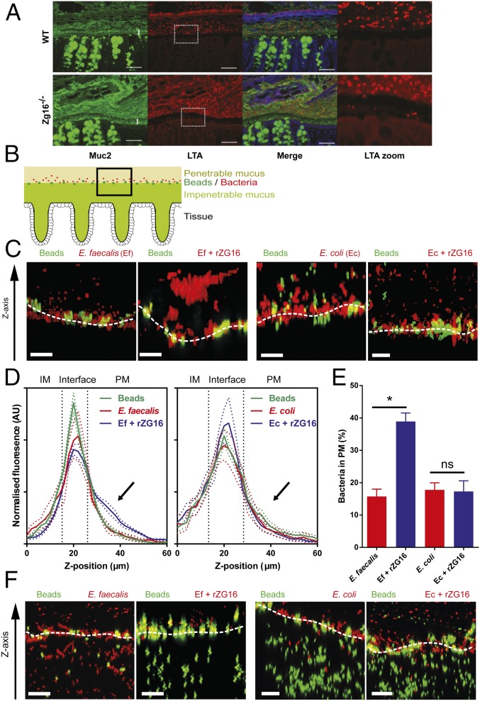
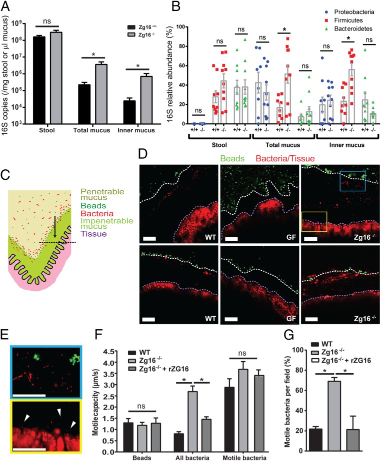
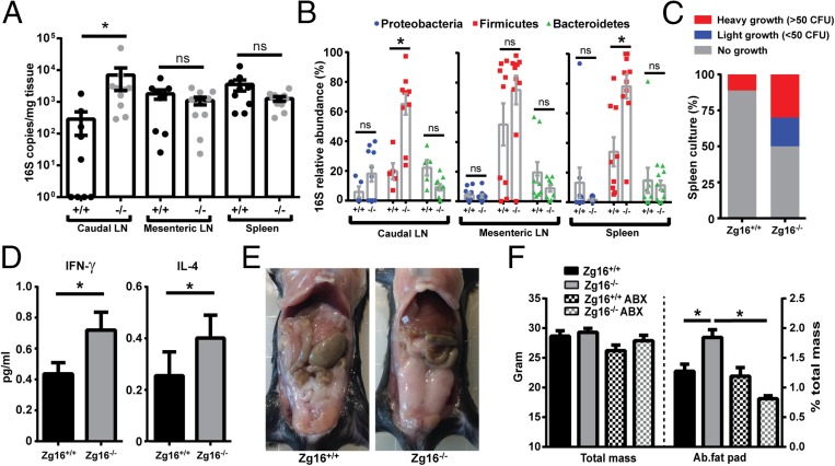
The distal colon functions as a bioreactor and harbors an enormous amount of bacteria in a mutualistic relationship with the host. The microbiota have to be kept at a safe distance to prevent inflammation, something that is achieved by a dense inner mucus layer that lines the epithelial cells. The large polymeric nets made up by the heavily O-glycosylated MUC2 mucin forms this physical barrier. Proteomic analyses of mucus have identified the lectin-like protein ZG16 (zymogen granulae protein 16) as an abundant mucus component. To elucidate the function of ZG16, we generated recombinant ZG16 and studied Zg16-/- mice. ZG16 bound to and aggregated Gram-positive bacteria via binding to the bacterial cell wall peptidoglycan. Zg16-/- mice have a distal colon mucus layer with normal thickness, but with bacteria closer to the epithelium. Using distal colon explants mounted in a horizontal perfusion chamber we demonstrated that treatment of bacteria with recombinant ZG16 hindered bacterial penetration into the mucus. The inner colon mucus of Zg16-/- animals had a higher load of Gram-positive bacteria and showed bacteria with higher motility in the mucus close to the host epithelium compared with cohoused littermate Zg16+/+ The more penetrable Zg16-/- mucus allowed Gram-positive bacteria to translocate to systemic tissues. Viable bacteria were found in spleen and were associated with increased abdominal fat pad mass in Zg16-/- animals. The function of ZG16 reveals a mechanism for keeping bacteria further away from the host colon epithelium.

Keywords: colon; inflammation; mucin; obesity; peptidoglycan.


Conflict of interest statement

The authors declare no conflict of interest.

Figures

Fig. 1.
Fig. 1.
ZG16 binds peptidoglycan and Gram-positive bacteria. Binding experiments were performed using a DELFIA-based assay. (A) Different concentrations of recombinant ZG16-Fc were applied to wells containing insoluble peptidoglycan. ZG16-Fc (filled bars) bound to the peptidoglycan after extensive washing compared with the control MUC1-Fc (open bars). Error bars represent SEM (n = 3). (B) Preincubation with 30 mM MurNAc partially inhibited binding of ZG16-Fc to peptidoglycan. (C) ZG16-Fc binding to the Gram-positive bacteria L. jensenii and Gram-negative bacteria E. coli. The assay was performed in a similar way as for the peptidoglycan binding. (D) Syto9-stained E. faecalis and E. coli cultures were incubated with BSA or rZG16, spread on microscopy slides, and imaged; images show representative field views of BSA (Top) or rZG16 (Bottom) treated bacteria. (Scale bars, 50 µm.) (E) Fluorescent areas from the microscopy images calculated using Imaris software; statistical significance calculated using Tukey’s multiple comparison test (ns, not significant; *P < 0.001). Data representative of n = 3 independent experiments. (F) Mucus measurement of WT (n = 9) and Zg16−/− mice (n = 6). Distal colon tissue was mounted in a horizontal chamber, charcoal was added to visualize the mucus, and the distance to the epithelial cells was measured. The secretagogue carbachol was added after 30 min. (G) Penetrability measurements of WT (n = 5) and Zg16−/− (n = 5) mice. Distal colon tissue was mounted in a horizontal chamber, and fluorescent bacterial-size beads were added on the explant 20 min after mounting. Beads were allowed to sediment for 40 min before the distribution was visualized using confocal microscopy. Images are representative confocal z-stack projections obtained from the experiments. (Scale bars, 100 µm.) (H) Scatter plot of the mean distance of the 20 fluorescent beads closest to the epithelium; lines represent median. *P = 0.016 using Mann–Whitney u test.
Fig. 2.
Fig. 2.
ZG16 alters distal colonic mucus penetration by Gram-positive bacteria in vivo and in vitro. (A) Carnoy-fixed distal colon immunohistochemistry of the mucus layer (anti-MUC2, green) and Gram-positive bacteria (anti-LTA antibody, red) (SI Appendix, Fig. S5). (Scale bars, 50 µm.) (B–F) C57BL/6 WT or Zg16−/− distal colon tissues were mounted in an imaging chamber; Fluorescent beads were applied apically to visualize the interface between the impenetrable (IM) and penetrable (PM) mucus layers; 108 cfu E. faecalis (Ef) or E. coli (Ec) were stained with BacLight Red then treated with 10 µg rZG16 and applied apically to mucus; 3D z-stacks of the mucus surface were acquired by confocal microscopy. (B) Schematic representation of data acquisition region. (C) Confocal z-stacks of WT mucus surface exposed to untreated Ef/Ec (Left) or Ef/Ec treated with rZG16 (Right); White dashed line indicates IM/PM interface. (D) Distribution of beads and untreated or rZG16-treated Ef (Left) or Ec (Right) along the z-stack z axis at the WT IM/PM interface. Colored dashed lines represent SEM from n = 6 mice; black dashed lines separate IM, interface, and PM regions of the z-stack. (E) Quantification of control and rZG16-treated Ef and Ec from the PM z-stack region indicated by the arrows in D. Error bars represent SEM from n = 6 mice. (F) Confocal z-stacks of Zg16−/− mucus surface exposed to untreated Ef/Ec (Left) or Ef/Ec treated with rZG16 (Right). White dashed line indicates IM/PM interface. All images are representative of n = 6 mice. Statistical significance calculated with Tukey’s multiple comparison test (ns, not significant; *P < 0.05). (Scale bars, 20 µm.)
Fig. 3.
Fig. 3.
ZG16 alters the mucus-associated intestinal microbiota as well as the distribution and motile capacity of bacteria within the mucus. (A) Zg16+/+ and Zg16−/− littermate bacterial 16S copy number detected in stool and unflushed (total mucus) or flushed (inner mucus) colonic tissue samples by qPCR. Data were normalized to stool mass or mucus volume (SI Appendix, Fig. S6). (B) Group-specific qPCR showing an increased abundance of Gram-positive Firmicutes in the total and inner mucus of the Zg16−/− in relation to littermate Zg16+/+ mice. (C–E) Conventionally raised C57BL/6 (WT), germ-free (GF), and Zg16−/− distal colon tissues were mounted in an imaging chamber; fluorescent beads were applied apically to visualize the interface between the impenetrable (IM) and penetrable (PM) mucus layers; and bacteria and tissues were visualized in situ using the nucleic acid-binding dye, Syto9. Mucus, bacteria, and tissues were imaged. (C) Schematic representation of chamber-mounted colonic tissues with IM and PM layers, beads, and bacteria indicated. Black dashed line represents the focal plane where confocal optical sections were acquired. (D) Confocal micrographs through WT, GF, and Zg16−/− colonic mucus. White dashed lines indicate the IM/PM interface; purple dashed lines indicate the edge of the colonic tissue. (E) Magnified confocal micrographs from the Inset blue and yellow boxes in D. (Top, blue border) Morphologically distinctive bacteria in the mucus. (Bottom, yellow border) Bacteria (white arrows) near the tissue surface. (F and G) Bacterial motility in WT, Zg16−/−, and Zg16−/− + rZG16 mucus was assessed by recording 1-min time series of optical sections through the colonic mucus as pictured in E (Movies S1–S7). (F) Quantification of motile capacity (maximum velocity) of beads at the IM/PM interface, all bacteria within the mucus, and only bacteria classed as motile within the mucus. (G) Proportion of bacteria determined as motile per imaging field. All error bars are SEM from n = 9 Zg16+/+ and n = 10 Zg16−/−animals. Statistical significances calculated with Mann–Whitney u test (A, B) or Sidak’s (F) or Dunn’s (G) multiple comparison tests (ns, not significant; *P < 0.05). (All scale bars, 50 µm.)
Fig. 4.
Fig. 4.
Zg16−/− mice have an increased systemic bacterial load of viable Gram-positive bacteria and increased abdominal fat pad mass. (A–C) Caudal and mesenteric lymph nodes (LN) and spleen were acquired from littermate Zg16+/+ and Zg16−/− mice. Tissues were weighed and homogenized, and DNA was extracted. (A) Quantification of bacterial 16S copy number in LN and spleen tissue by qPCR. Error bars are SEM from n = 9 Zg16+/+ and n = 10 Zg16−/− mice. (B) Estimation of the relative abundance of different bacterial taxonomic groups (Bacteroidetes, Firmicutes and Proteobacteria) using qPCR in the tissues examined in A. (C) Growth of tissue homogenates from spleen were cultivated anaerobically on brain heart infusion-supplemented agar plates. Isolated bacterial colonies were identified by 16S rDNA sequencing and confirmed to be present in the stool, mucus, and lymphatic samples from the same individual animals (SI Appendix, Fig. S6). (D) Significant differences of IFN-γ and IL-4 in the serum of littermate n = 9 Zg16+/+ and n = 10 Zg16−/− mice. (E) Opened abdomen of 12-wk ZG16+/+ and ZG16−/− mice. (F) Total mass and abdominal fat pads of 12-wk-old male littermate Zg16+/+ (n = 9) and Zg16−/− (n = 10) as well as Zg16+/+ (n = 4) and Zg16−/− (n = 5) after 3 wk of antibiotic treatment. Error bars are SEM. Statistical significance calculated with Mann–Whitney u test (A–D) or unpaired t test with similar variance (F) (ns, not significant; *P < 0.05).

Similar articles

Cited by

References

    1. Bäckhed F, Ley RE, Sonnenburg JL, Peterson DA, Gordon JI. Host-bacterial mutualism in the human intestine. Science. 2005;307(5717):1915–1920. - PubMed
    1. Johansson MEV, et al. The inner of the two Muc2 mucin-dependent mucus layers in colon is devoid of bacteria. Proc Natl Acad Sci USA. 2008;105(39):15064–15069. - PMC - PubMed
    1. Ermund A, Schütte A, Johansson MEV, Gustafsson JK, Hansson GC. Studies of mucus in mouse stomach, small intestine, and colon. I. Gastrointestinal mucus layers have different properties depending on location as well as over the Peyer’s patches. Am J Physiol Gastrointest Liver Physiol. 2013;305(5):G341–G347. - PMC - PubMed
    1. Ambort D, et al. Calcium and pH-dependent packing and release of the gel-forming MUC2 mucin. Proc Natl Acad Sci USA. 2012;109(15):5645–5650. - PMC - PubMed
    1. Johansson MEV, Larsson JM, Hansson GC. The two mucus layers of colon are organized by the MUC2 mucin, whereas the outer layer is a legislator of host-microbial interactions. Proc Natl Acad Sci USA. 2011;108(Suppl 1):4659–4665. - PMC - PubMed

Publication types