. 2016 Nov 16;12(11):e1005917.
doi: 10.1371/journal.ppat.1005917. eCollection 2016 Nov.

Variant Exported Blood-Stage Proteins Encoded by Plasmodium Multigene Families Are Expressed in Liver Stages Where They Are Exported into the Parasitophorous Vacuole

Affiliations

Variant Exported Blood-Stage Proteins Encoded by Plasmodium Multigene Families Are Expressed in Liver Stages Where They Are Exported into the Parasitophorous Vacuole

Aurélie Fougère et al. PLoS Pathog. .

Erratum in

Abstract

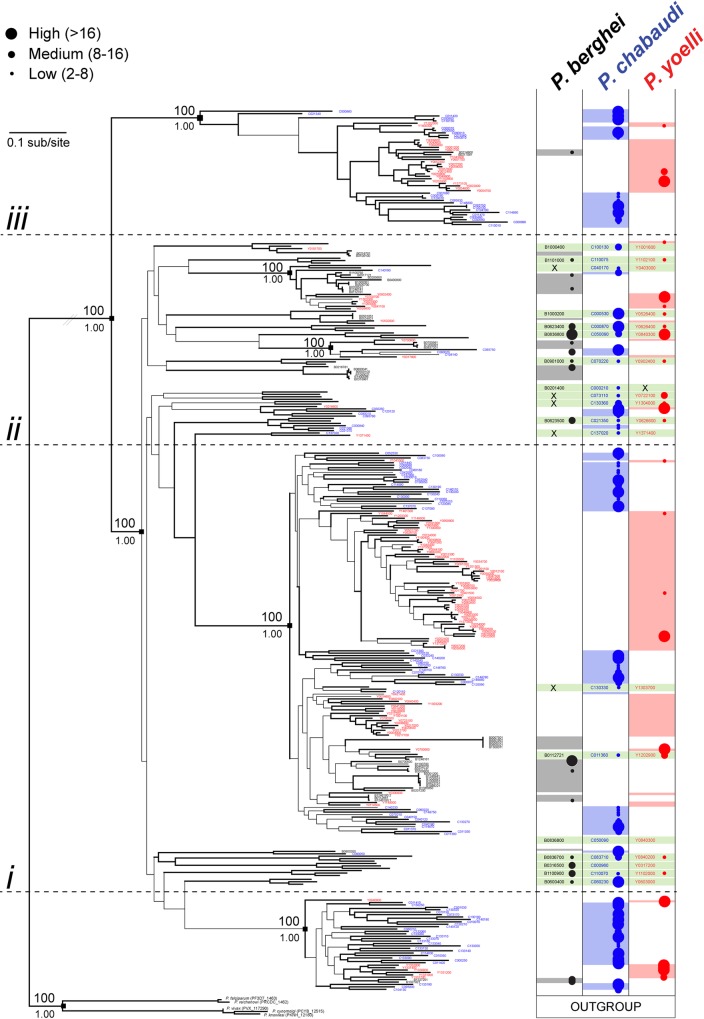
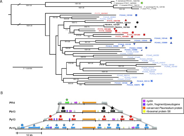
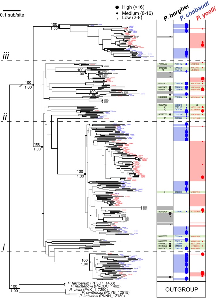
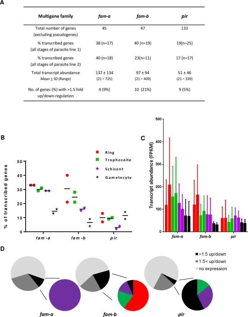
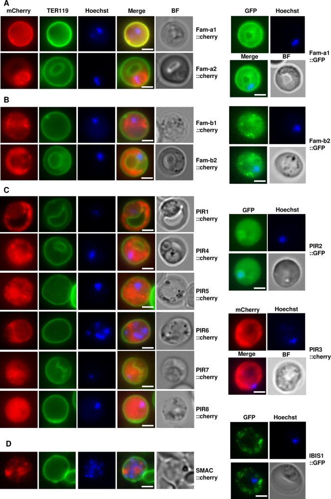
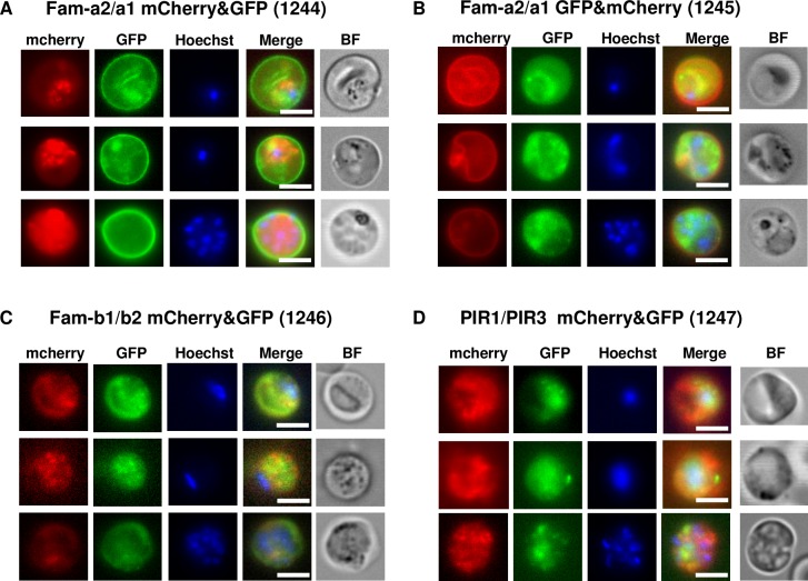
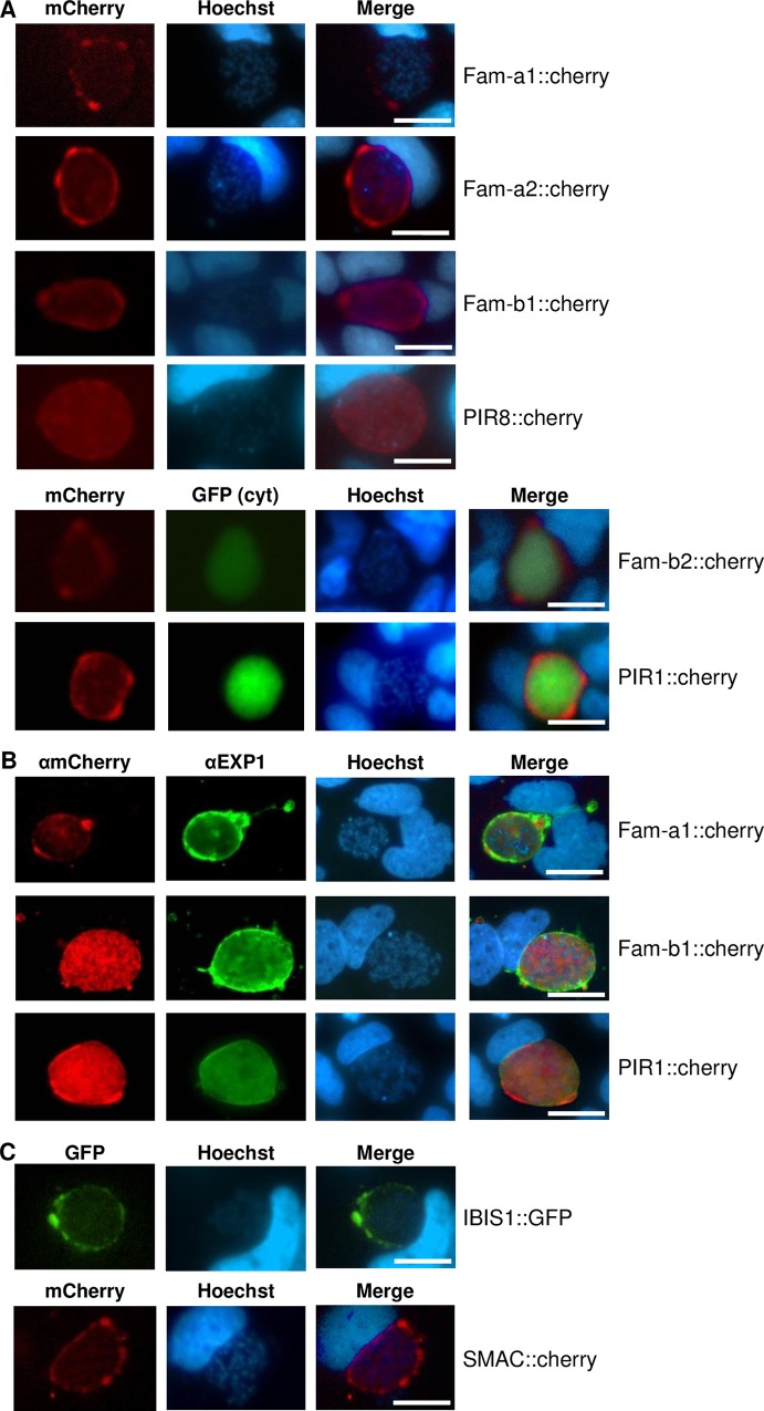
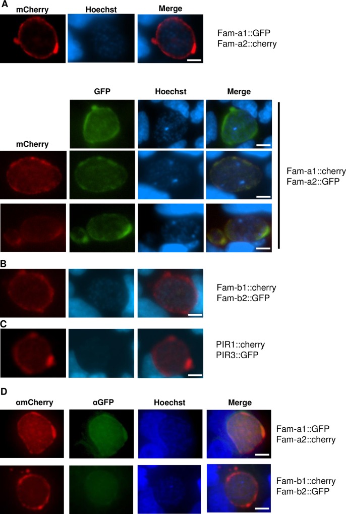
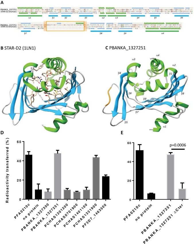
Many variant proteins encoded by Plasmodium-specific multigene families are exported into red blood cells (RBC). P. falciparum-specific variant proteins encoded by the var, stevor and rifin multigene families are exported onto the surface of infected red blood cells (iRBC) and mediate interactions between iRBC and host cells resulting in tissue sequestration and rosetting. However, the precise function of most other Plasmodium multigene families encoding exported proteins is unknown. To understand the role of RBC-exported proteins of rodent malaria parasites (RMP) we analysed the expression and cellular location by fluorescent-tagging of members of the pir, fam-a and fam-b multigene families. Furthermore, we performed phylogenetic analyses of the fam-a and fam-b multigene families, which indicate that both families have a history of functional differentiation unique to RMP. We demonstrate for all three families that expression of family members in iRBC is not mutually exclusive. Most tagged proteins were transported into the iRBC cytoplasm but not onto the iRBC plasma membrane, indicating that they are unlikely to play a direct role in iRBC-host cell interactions. Unexpectedly, most family members are also expressed during the liver stage, where they are transported into the parasitophorous vacuole. This suggests that these protein families promote parasite development in both the liver and blood, either by supporting parasite development within hepatocytes and erythrocytes and/or by manipulating the host immune response. Indeed, in the case of Fam-A, which have a steroidogenic acute regulatory-related lipid transfer (START) domain, we found that several family members can transfer phosphatidylcholine in vitro. These observations indicate that these proteins may transport (host) phosphatidylcholine for membrane synthesis. This is the first demonstration of a biological function of any exported variant protein family of rodent malaria parasites.


Conflict of interest statement

The authors have declared that no competing interests exist.

Figures

Fig 1
Fig 1. Maximum likelihood phylogeny of fam-a gene sequences from Plasmodium spp.
The tree was estimated using RAxML and a GTR+Γ model. Branches subtended by nodes with >75 bootstrap support are shown in bold. Robust basal nodes are indicated by black squares with bootstrap proportions (above node) and Bayesian posterior probabilities (beneath node). At right, coloured blocks indicate the species to which a terminal node belongs. Clades of orthologs that display positional conservation are indicated with green blocks; where a sequence has been lost secondarily in a species is shown by an ‘X’. The tree is rooted using an out-group comprising single copy fam-a orthologs from primate Plasmodium species. The phylogeny is subdivided into four sections: genes located at the conserved, ‘ancestral’ locus on chromosome 13 (below line i); genes found at loci conserved across RMP species (between lines i and ii); and a robust clade of species-specific paralogs derived from a conserved locus on chromosome 6 or 13 (between lines ii and iii); a robust clade of species-specific paralogs derived from a conserved locus on chromosome 8 (above line iii).Transcription levels (shown as different coloured and sized circles) in blood stages are shown for individual genes based on RNAseq data (FPKM values) (from [33] and S1 Table). Expression levels as shown by four different sized circles: Class 1 (smallest circle): 2-8x the threshold level; class 2: 8-16x the threshold; class 3 (largest circle): >16x the threshold.
Fig 2
Fig 2. Maximum likelihood phylogeny and chromosomal location of fam-a genes that have an internal, syntenic chromosomal location
A. The tree was estimated using RAxML and a GTR+Γ model. Nodes support is indicated by bootstrap proportions and Bayesian posterior probabilities. Genes that occupy the conserved, chromosomal-internal locus are labelled in bold and accompanied by a symbol, colour-coded by species (repeated in B). In P. yoelli and P. chabaudi, these genes are paraphyletic with other genes that have been transposed to subtelomeric locations of other chromosomes (these paralogs are labelled in italics). The tree is rooted with the single copy fam-a orthologs from primate Plasmodium spp.B. Chromosomal organisation of the internal copies of fam-a genes on chromosome 14 of P. falciparum 3D7 (Pf14; green), chromosomes 13 of P. berghei ANKA (Pb13; black), P. yoelii yoelii 17X (Py13; red) and P. chabaudi AS (Pc13; blue). Fam-a genes are interspersed with cyclin (lilac), cyclin fragments (violet), ribosomal protein S6 (yellow) and conserved Plasmodium protein (orange). The internal fam-a region is bordered by RNA polymerase III subunit RPC4 (PF3D7_1463400, PBANKA_1327000, PY17X_1330800, PCHAS_133030) and YL1 protein (PF3D7_1464000, PBANKA_1327400, PY17X_1332100, PCHAS_133200) (shown in grey). Synteny between RPC4 and YL1 is shown with grey lines. The arrows indicate the location on forward and reverse strands.
Fig 3
Fig 3. Maximum likelihood phylogeny of fam-b gene sequences from Plasmodium spp.
The tree was estimated using RAxML and a GTR+Γ model. Branches subtended by nodes with >75 bootstrap support are shown in bold. Robust basal nodes are indicated by black squares with bootstrap proportions (above node) and Bayesian posterior probabilities (beneath node). At right, coloured blocks indicate the species to which a terminal node belongs. Clades of orthologs that display positional conservation are indicated with green blocks; where a sequence has been lost secondarily in one species, this is shown by an ‘X’. The phylogeny is subdivided into four sections: divergent genes included conserved loci, placed at the root of the tree (below line i); predominantly P. yoelli species-specific genes P. berghei- and P. yoelli-specific paralogs (between lines i, ii and iii); and predominantly P. chabaudi species-specific genes (above line iii). Transcription levels (shown as different coloured and sized circles) in blood stages are shown for individual genes based on RNAseq data (FPKM values) (from [33] and S1 Table). Expression levels as shown by four different sized circles: Class 1 (smallest circle): 2-8x the threshold level; class 2: 8-16x the threshold; class 3 (largest circle): >16x the threshold.
Fig 4
Fig 4. Transcription of pir, fam-a and fam-b genes based on RNAseq data of different blood stages of two P. berghei ANKA reference lines.
A. Features of transcription of pir, fam-a and fam-b genes in the two P. berghei reference lines (line 1 and line 2) based on RNAseq data (from[33] and shown in S1 Table). Transcribed genes are genes with an FPKM value above the cut-off level of 21. Total transcript abundance is the sum of all FPKM values observed in the different blood stages (see B). The fold up-down regulation is based on the difference in FPKM values of individual genes between blood stages of the two different parasite lines (see S1 Fig). B. Percentage of genes transcribed in the different blood stages (see A). Ring, red; trophozoite, green; schizont, purple; gametocyte, black. C. Total transcript abundance in the different blood stages: mean and standard deviation of total transcript abundance of all FPKM values observed in the different blood stages (see A). D. Percentage of non-transcribed genes (light grey) and genes with less (grey) or more (black) than 1.5x difference in transcript abundance between blood stages of two different parasite lines (see A). The coloured circles show the genes with >1.5 fold down-or upregulation in the four different blood stages (see B).
Fig 5
Fig 5
Expression and export of fluorescently-tagged fam-a (A) fam-b (B) and pir (C) members in blood stages of single-gene tagging (SGT) mutants. In D expression and export is shown of the fluorescently-tagged proteins SMAC and IBIS that are encoded by single copy genes. The plasma membrane of the red blood cell is stained with TER119 antibodies (green) and parasite nuclei are stained with Hoechst. BF: bright field. Scale bar: 2μm.
Fig 6
Fig 6. Simultaneous expression (and export) of two proteins of the same family in a single blood stage parasite (trophozoites or schizonts) of double-gene tagging (DGT) mutants.
These mutants contain the following pairs of genes tagged with either mCherry or GFP: fam-a1/fam-a2 (2 independent mutants; panel A, B), fam-b1/fam-b2 (panel C) and pir1/pir3 (panel D); RMgm ID as indicated in Table 2. Localisation of mCherry-tagged members in the trophozoite stage (upper two rows) and in the schizont (low row) stage. Parasite nuclei are stained with Hoechst, BF bright field. scale bar: 5μm.
Fig 7
Fig 7. Expression of fluorescently-tagged proteins of multigene families in liver stages of single-gene tagging (SGT) mutants at 48hpi in cultured hepatocytes (Huh7).
A. Fluorescence-microscopy analysis of members of the fam-a, fam-b and pir multigene family in live liver-stages. The parasites expressing mCherry-tagged Fam-b2 and PIR1 also express cytoplasmic GFP (cyt GFP; green). B. IFA-analysis of fixed liver-stages using anti-mCherry (red) anti-PbEXP1 (green) antibodies. PbEXP1 is a parasitophorous vacuole membrane resident protein. C Fluorescence-microscopy analysis of expression of SMAC and IBIS, exported proteins encoded by single-copy genes in live liver-stages. Nuclei are stained with Hoechst-33342 (blue); scale bar: 10μm.
Fig 8
Fig 8. Expression of fluorescently-tagged proteins in liver stages of double-gene tagging (DGT) mutants at 48hpi in cultured hepatocytes (Huh7).
Fluorescence-microscopy analysis of members of the fam-a (A), fam-b (B) and pir (C) multigene family in live liver-stages of DGT mutants; we were only able to detect both fluorescently-tagged proteins in only one DGT mutant, fam-a1cherry/fam-a2GFP (in 40–45% of the parasites). D. IFA analysis of fixed liver-stages of DGT mutants using anti-GFP (green) and anti-mCherry (red) antibodies. Nuclei are stained with Hoechst-33342 (blue). Scale bar: 10μm.
Fig 9
Fig 9. Predicted structure of the START domain of fam-a2 and Phosphatidylcholine (PC) transfer activity of selected recombinant Fam-a proteins.
A. Secondary structure of Fam-a protein PBANKA_1327251 threaded against the resolved structure of the STAR-D2 domain in the human phosphatidylcholine transfer protein (1LN1). The orange box identifies an expanded protein loop between β-sheets 7 and 8 in Fam-a. B. Predicted tertiary structure of 1LN1 showing the binding pocket of the STAR-D2 domain in complex with the ligand, dilinoleoyl-phosphatidylcholine (shown as stick model). C. Predicted tertiary structure of PBANKA_1327251 after computational threading against 1LN1. The orange part shows the position of the expanded protein loop in Fam- a as presented in A. Alpha helix (green) and beta pleated sheets (blue) are labelled as described in model 1LN1. D. Phosphatidylcholine (PC) transfer activity of selected recombinant proteins of the P. chabaudi (four proteins) and P. berghei (2 proteins) Fam-A families and of the single P. falciparum Fam-a protein (PF3D7_1463500). PC transfer activity of the recombinant Fam-A proteins carrying a hexahistidine tag at their N termini was tested using a standard PC transfer assay (see the ‘Materials and Methods‘ section). E. Phosphatidylcholine (PC) transfer of full length Fam-a protein PBANKA_1327251 and a mutated form in which the final C-terminal alpha helix of the START domain has been deleted. In D. and E. the P. falciparum phospholipid transfer protein PFA0210c (PF3D7_0104200) was used as a positive control and a sample without Fam-A protein was used as negative control. White bars: control reactions; light grey bars: reactions with P. berghei proteins; dark grey bars: reactions with P. chabaudi proteins; black bars: reaction with P. falciparum protein. All reactions were set up in triplicate. The error bars indicate the standard deviation.

Similar articles

Cited by

References

    1. Goldberg DE, Cowman AF: Moving in and renovating: exporting proteins from Plasmodium into host erythrocytes. Nat Rev Microbiol 2010, 8:617–621. 10.1038/nrmicro2420 - DOI - PubMed
    1. Maier AG, Cooke BM, Cowman AF, Tilley L: Malaria parasite proteins that remodel the host erythrocyte. Nat Rev Microbiol 2009, 7:341–354. 10.1038/nrmicro2110 - DOI - PubMed
    1. Elsworth B, Matthews K, Nie CQ, Kalanon M, Charnaud SC, Sanders PR, Chisholm SA, Counihan NA, Shaw PJ, Pino P, et al.: PTEX is an essential nexus for protein export in malaria parasites. Nature 2014, 511:587–591. 10.1038/nature13555 - DOI - PubMed
    1. Boddey JA, Carvalho TG, Hodder AN, Sargeant TJ, Sleebs BE, Marapana D, Lopaticki S, Nebl T, Cowman AF: Role of plasmepsin V in export of diverse protein families from the Plasmodium falciparum exportome. Traffic 2013, 14:532–550. 10.1111/tra.12053 - DOI - PubMed
    1. Spillman NJ, Beck JR, Goldberg DE: Protein export into malaria parasite-infected erythrocytes: mechanisms and functional consequences. Annu Rev Biochem 2015, 84:813–841. 10.1146/annurev-biochem-060614-034157 - DOI - PubMed

Publication types

Substances