

doi: 10.1117/1.JBO.21.11.114002.
Development of a handheld smart dental instrument for root canal imaging
Affiliations
- PMID: 27851855
- PMCID: PMC8357325
- DOI: 10.1117/1.JBO.21.11.114002
Item in Clipboard
Development of a handheld smart dental instrument for root canal imaging
J Biomed Opt.
.
Abstract
Ergonomics and ease of visualization play a major role in the effectiveness of endodontic therapy. Using only commercial off-the-shelf components, we present the pulpascope—a prototype of a compact, handheld, wireless dental instrument for pulp cavity imaging. This instrument addresses the current limitations of occupational injuries, size, and cost that exist with current endodontic microscopes used for root canal procedures. Utilizing a 15,000 coherent, imaging fiber bundle along with an integrated illumination source and wireless CMOS sensor, we demonstrate images of various teeth with resolution of ?48???m and angular field-of-view of 70 deg.
Figures
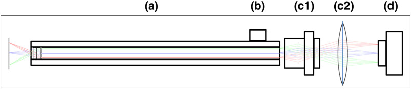
Block diagram showing optical train. Components highlighted are (a) integrated fiber bundle, (b) light source, (c) Relay module consisting of: [c1] objective lens and [c2] tube lens, and (d) camera module.
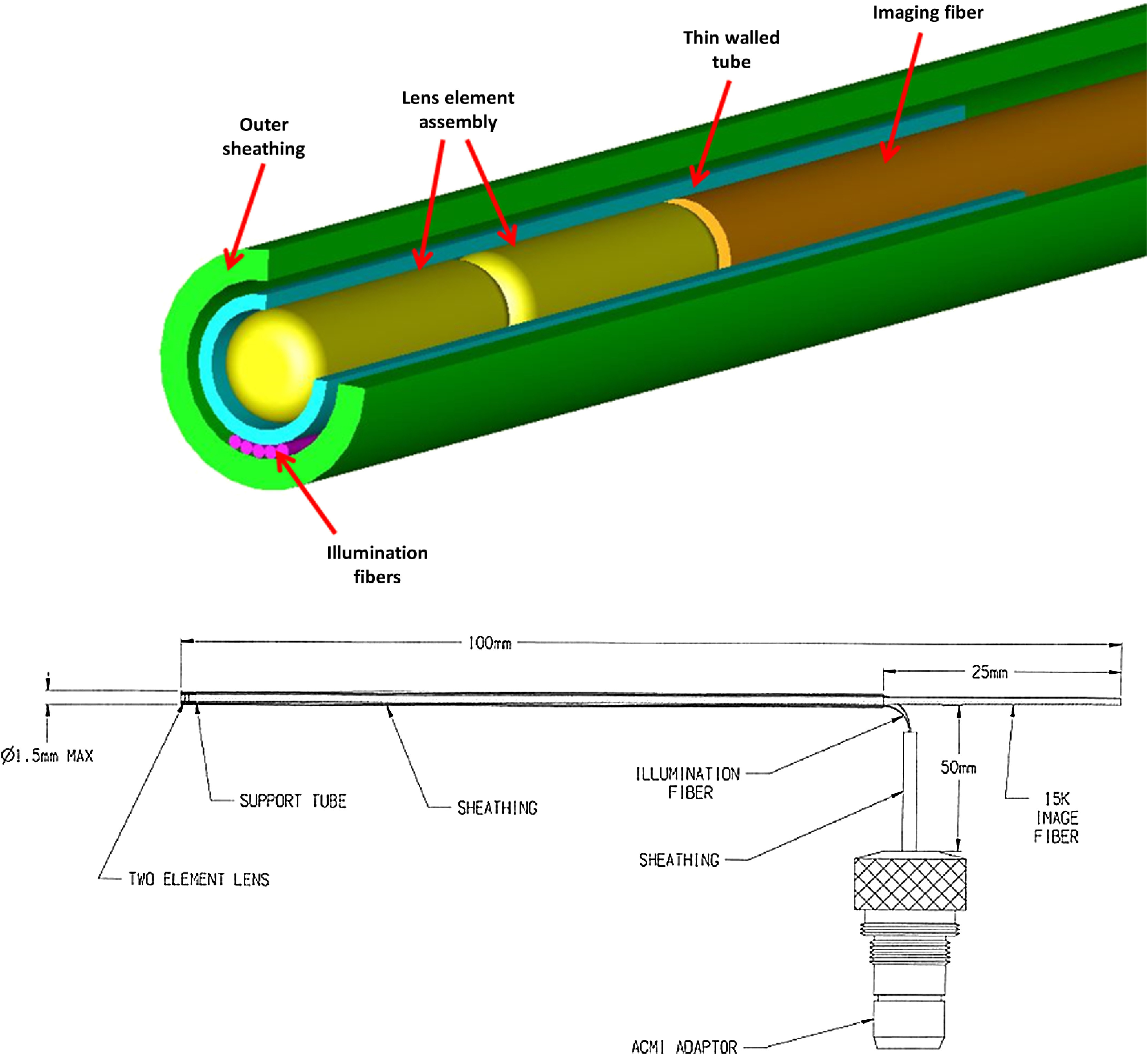
Integrated illumination and imaging fiber bundle (courtesy Myriad Fibers).
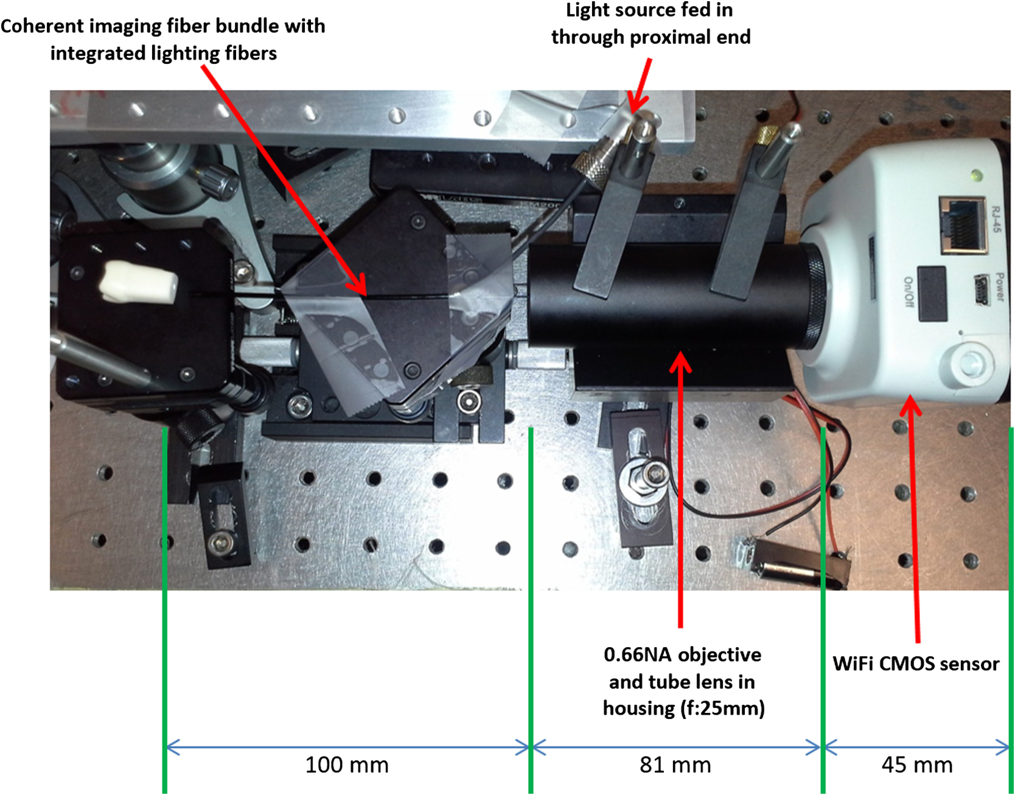
Experimental setup.
3-D design of the pulpascope fiber holder.
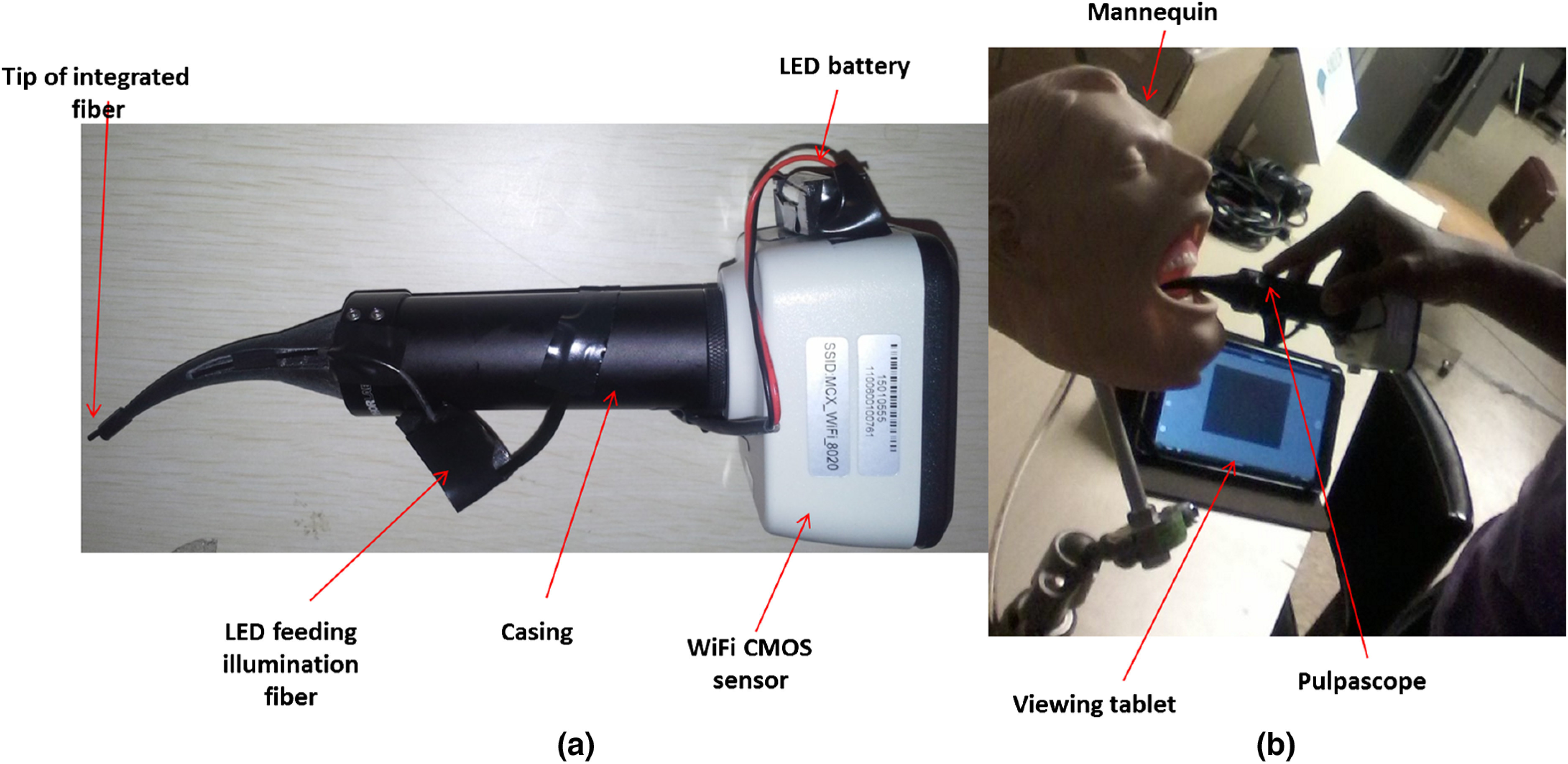
(a) Assembled pulpascope with integrated fiber bundle and the Moticam camera. The LED is attached to the outlet for the illumination fibers. (b) Pulpascope in use. The viewing tablet is shown to the side.
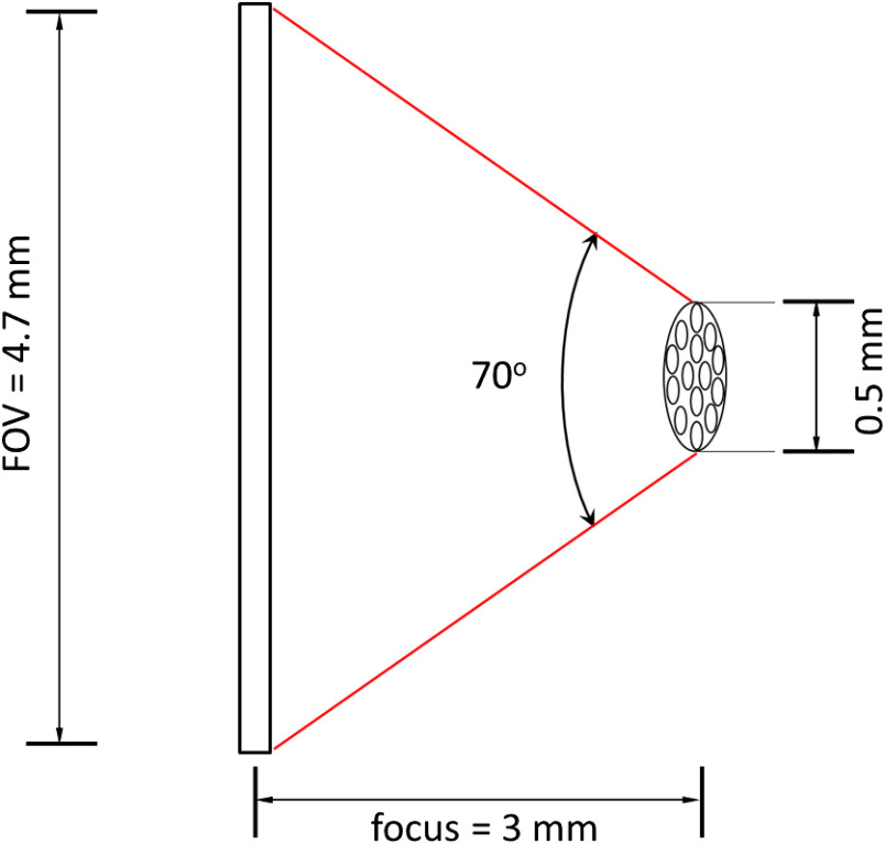
Demagnification from the focal plane to the fiber tip surface by the GRIN lenses.
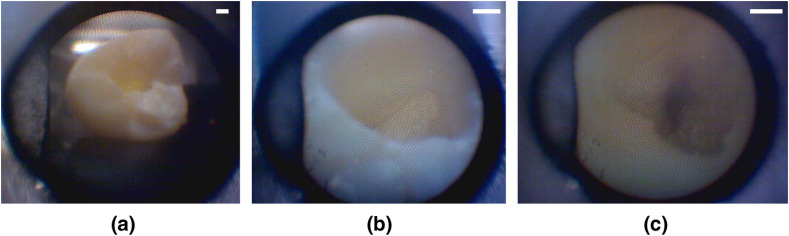
Images taken at distances (a) 7 mm, (b) 3 mm, and (c) inside the tooth. Each scale bar is 1 mm.
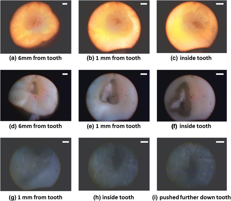
Images of tooth with light from (a)–(c) 30 W halogen bulb, (d)–(f) laboratory flashlight, and (g)–(h) 15 lumens LED. Note that there is no ambient light. Each scale bar is 1 mm.
Similar articles
-
Computer-aided design-based guided endodontic: A novel approach for root canal access cavity preparation.Proc Inst Mech Eng H. 2018 Aug;232(8):787-795. doi: 10.1177/0954411918788104. Epub 2018 Jul 17. Proc Inst Mech Eng H. 2018. PMID: 30014778
-
Factors Affecting the Removal Time of Separated Instruments.J Endod. 2021 Aug;47(8):1245-1252. doi: 10.1016/j.joen.2021.05.003. Epub 2021 May 14. J Endod. 2021. PMID: 34000326
-
In Vitro Evaluation of a Novel Root Canal Endoscope for Visualizing the Apex of Curved Root Canal Models and an Extracted Tooth.J Endod. 2018 Dec;44(12):1856-1861. doi: 10.1016/j.joen.2018.08.014. Epub 2018 Nov 1. J Endod. 2018. PMID: 30390969
-
Effect of combined digital imaging parameters on endodontic file measurements.J Endod. 2012 Oct;38(10):1404-7. doi: 10.1016/j.joen.2012.06.006. Epub 2012 Jul 10. J Endod. 2012. PMID: 22980188
-
Unprepared root canal surface areas: causes, clinical implications, and therapeutic strategies.Braz Oral Res. 2018 Oct 18;32(suppl 1):e65. doi: 10.1590/1807-3107bor-2018.vol32.0065. Braz Oral Res. 2018. PMID: 30365606 Review.
Cited by
-
Image Quality, Radiation Dose, and Patient Comfort Associated with Wireless Sensors in Digital Radiography: A Systematic Review.Dent J (Basel). 2024 Aug 20;12(8):267. doi: 10.3390/dj12080267. Dent J (Basel). 2024. PMID: 39195111 Free PMC article. Review.
References
-
- Michaelides P., “Use of the operating microscope in dentistry,” J. Calif. Dent. Assoc. 24(6), 45–50 (1996). - PubMed
Publication types
MeSH terms
Grants and funding
LinkOut - more resources
Full Text Sources
Other Literature Sources
