. 2016 Nov 15;45(5):999-1012.
doi: 10.1016/j.immuni.2016.10.023.

Aire Inhibits the Generation of a Perinatal Population of Interleukin-17A-Producing γδ T Cells to Promote Immunologic Tolerance

Affiliations

Aire Inhibits the Generation of a Perinatal Population of Interleukin-17A-Producing γδ T Cells to Promote Immunologic Tolerance

Noriyuki Fujikado et al. Immunity. .

Abstract

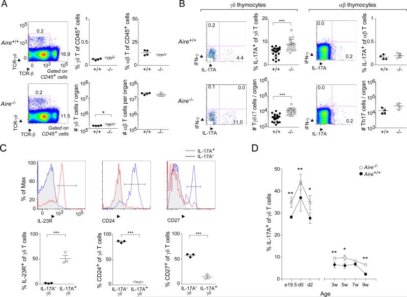
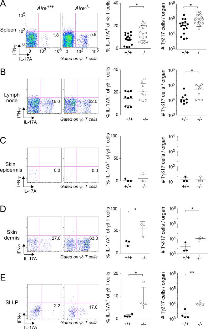
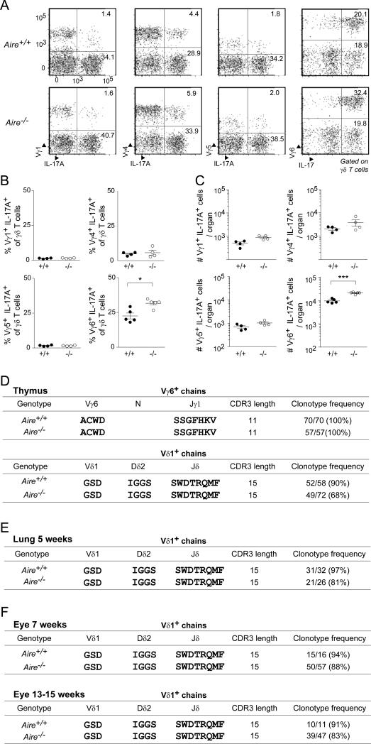
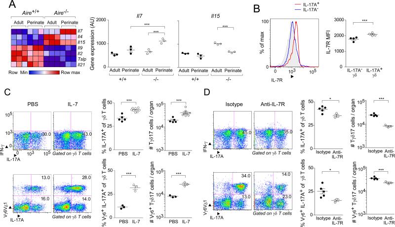
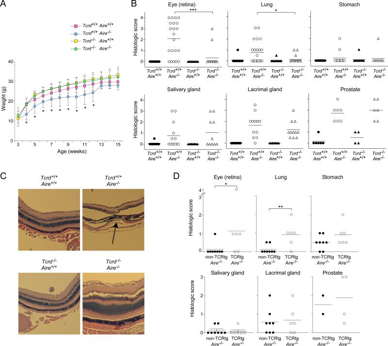
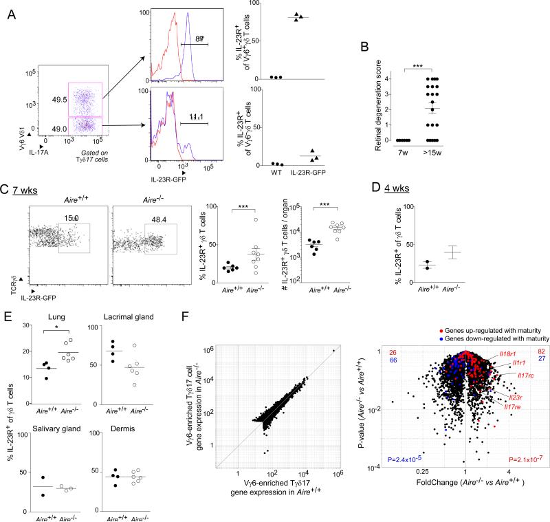
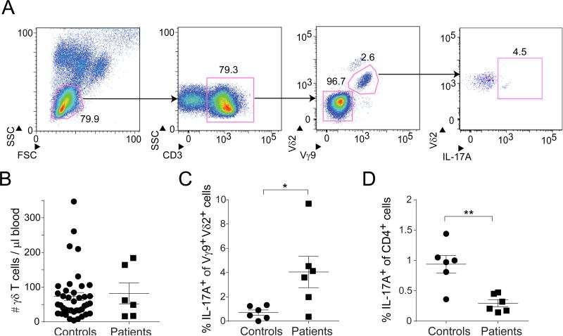
Aire's primary mechanism of action is to regulate transcription of a battery of genes in medullary thymic epithelial cells (mTECs) and, consequently, negative selection of effector T cells and positive selection of regulatory T cells. We found that Aire-deficient mice had expanded thymic and peripheral populations of perinatally generated IL-17A+Vγ6+Vδ1+ T cells, considered to be "early responders" to tissue stress and drivers of inflammatory reactions. Aire-dependent control of Il7 expression in mTECs regulated the size of thymic IL-17A+Vγ6+Vδ1+ compartments. In mice lacking Aire and γδ T cells, certain tissues typically targeted in the "Aire-less" disease, notably the retina, were only minimally infiltrated. IL-17A+Vγ6+Vδ1+ cells were present in the retina of wild-type mice and expanded very early in Aire-deficient mice. A putatively parallel population of IL-17A+Vγ9+Vδ2+ T cells was increased in humans lacking Aire. Thus, Aire exerts multi-faceted autoimmune control that extends to a population of innate-like T cells.


Figures

Fig. 1
Fig. 1. Mice lacking Aire had an increased frequency and number of IL-17A-producing γδ thymocytes
Flow cytometric analysis of γδ thymocyte compartments from Aire+/+ and Aire−/− littermates. Except for panel D, mice were 3-9 weeks of age. A. Comparisons of total γδ and αβ thymocytes. Left-most panel: typical cytofluorometric profiles; top, wild-type (WT); bottom, knock-out (KO); numbers refer to the fraction of CD45+ cells in that gate. Center and right panels: summary data plots for γδ and αβ T cells, respectively (n=4); top, fractional representation; bottom, numbers per thymus. B. Comparisons of cytokine expression by γδ and αβ thymocytes. IFN-γ and IL-17A expression on gated γδ and αβ T cells. In each case, left panels: typical cytofluorometric profiles; numbers refer to the fraction of γδ or αβ cells in the indicated gate. In each case, right panels: summary data plots (n=20 or 4); top, fractional representation; bottom, numbers per thymus. C. Additional markers of IL-17A+ γδ thymocytes. Cells gated as IL-17A+ or IL-17A as per panel B were additionally examined for expression of the indicated markers. Top, typical cytoflurometric histograms; bottom, summary data (n=3). Grey shading indicates isotype-control staining. D. IL-17A+ γδ thymocytes through ontogeny. Summary data for fractional representation (n=3-13). P=*, <0.05; **, <0.01; ***, <0.001 according to Student's t-test. Only significant differences are indicated. Dot plot scales for this figure are all the same.
Fig. 2
Fig. 2. Increased frequency and number of Tγδ17 cells in peripheral tissues of Aire-deficient mice
Designated organs of Aire+/+ and Aire−/− littermates were excised at 3-9 weeks of age and examined by flow cytometry, focusing on Tγδ17 cells. Left panels: typical cytofluorometric profiles. Numbers refer to fraction of total γδ T cells in that gate. SI-LP = small-intestinal lamina propria. Center panels: summary data for fractional representation (n=3-16). Right panels: corresponding summary data for number per organ. Statistics as per Fig. 1. Dot plot scales for this figure are all the same.
Fig. 3
Fig. 3. The expanded Tγδ17 population in Aire−/− mice displayed primarily Vγ6+Vδ1+ TCRs A-C
Flow-cytometric analysis of Vγ usage. Perinatal thymocytes from Aire+/+ and Aire−/− mice were stained for IL-17A and for the indicated Vγ chains. A) Typical cytofluorometric profiles. Numbers refer to fraction of total γδ cells within the designated gate. B) Summary data on fractional representation (n=4-5). C) Corresponding summary data on numbers per thymus. D. Summary of the single-cell sequencing data detailed in Table S1. Single perinatal Vγ6+ thymocytes from Aire+/+ and Aire−/− littermates were sorted, and the CDR3 (in bold) sequences of their Vγ6+ and Vδ1+ chains determined. E. Summary of the lung single-cell sequencing data detailed in Table S2. As per panel D, except Vγ6+ cells were sorted from the lung of 5-week-old littermates. F. Summary of the eye single-cell sequencing data for 7- week and 13-15-week old mice detailed in Table S2. As per panel D, except Il-17+Vγ6+-enriched cells were identified as Vγ1,2,4,5 CD27 for sorting from the eyes of age-matched sets of Aire+/+ and Aire−/− mice (n=5-15). Statistics as per Fig. 1 (see also Tables S1 and S2). Dot plot scales for this figure are all the same.
Fig. 4
Fig. 4. IL-7 droveTγδ17 population expansion in Aire-KO mice
A. Quantification of cytokine gene transcripts from mTECs of perinatal vs adult Aire-WT vs -KO mice. Focused on the family of cytokines that bind to receptors employing the γc chain. Microarray data on independent triplicates of mature mTECs expressing a high level of MHC-II (Yang et al., 2015). Left, heat map; right, expression values for genes deemed of particular interest. B. Flow-cytometric comparison of IL-7R expression by IL-17A+ and IL-17A γδ thymocytes from Aire−/− perinates. Left, histogram of expression; right, mean fluorescence intensity (MFI). C. Boosting IL-7 levels in Aire+/+ mice. Recombinant IL-7 (250ng/g body weight) was ip-injected into day 0 perinates, and thymocytes were analyzed 3 days later. Left panels, typical cytofluormetric profiles; numbers represent the fraction of total γδ T cells within the indicated gate. Center and right panels, summary data on fractional representation and cell numbers, respectively (n=3-8); top, quantification of IL-17A+ γδ thymocytes; bottom, focus on IL-17A+Vγ6+ cells. D. Blocking the effect of IL-7 in Aire-KO mice. On the day of birth, 25μg of the anti-IL-7R mAb, A7R34, was injected; 3 days later, γδ thymocytes were analyzed. Data are presented as in panel C (n=3-5). Statistics as per Fig. 1. (See also Fig. S1). Dot plot scales for this figure are all the same.
Fig 5
Fig 5. γδT cells played a role in the autoimmune disease characteristic of Aire-deficient mice
Littermates carrying a homozygous null mutation of Aire, Tcrd, both or neither on the B6 genetic background were followed until 15 weeks of age, when the indicated organs were taken for histology (n=10 per group). A. Weight curves. B. Histologic scores, employing the scoring system described in Experimental Procedures. Statistics as per Fig. 1. C. Eye histology. Hematoxylin and eosin staining of retinal tissue. Arrow indicates the disrupted retinal layer. D. As per panel B, except mice harboring a homozygous null-mutation of Aire that either did or did not carry a Vγ6+Vδ1+ TCR transgene were followed until 12 weeks of age (n=2-4 for prostate, 6-8 for all other tissues). Statistics as per Fig. 1. (See also Fig. S2)
Fig. 6
Fig. 6. Tγδ17 cells accumulated in the retina prior to autoimmune attack in Aire-deficient mice
A. IL-23 reporter validation. Flow cytometric analysis of thymic Tγδ17 cells from day-of-birth IL-23R-GFP mice. Left and center panels, typical cytofluorometric plots; right panel, summary data (n=3). B. Histologic scores, as per Experimental Procedures, of eye tissue from Aire−/− mice taken at 7 or 15 weeks of age (n=6-20). C. Cytoflurometric comparison of GFP+ Tγδ17 cells from uveoretinal tissue of 7-week-old Aire+/+ and Aire−/− mice carrying the IL-23R-GFP reporter. Left, typical profiles; right, summary data on fractional representation and number (n=6-8). D. Summary data for 4-week-old mice (n=2-3). E. As per panel C, except cells were isolated from lung, lacrimal gland, or dermis (n=2-3 for salivary gland, 4-6 for all other tissues). F. RNA-seq analysis of gene expression in Vγ1,2,4,5 CD27 (i.e. IL-17+ Vγ6+-enriched) γδ T cells isolated from the lungs of 10-week-old Aire+/+ or Aire−/− mice (n=2). Red- or blue-highlighted genes represent loci two-fold up- or down-regulated during Vγ6+ cell thymic maturation (Narayan et al., 2012), respectively. p-values reflect the significance of signature enrichment as calculated by the χ2 -test. Other statistics as per Fig. 1. (See also Fig. S3). Dot plot scales for this figure are all the same.
Fig. 7
Fig. 7. IL-17A+Vγ9+Vδ2+ T cells were enriched in APECED patients
A. Gating strategy. After initial FSC/SSC gating of peripheral blood mononuclear cells, lymphocytes were gated (left panel), CD3+ T cells selected (center-left), Vγ9+Vδ2+ cells delineated (center-right), and IL-17A-producing cells quantified (right panel). B. Summary data for the absolute number of TCRγδ+CD3+ cells per μl of blood. APECED patients, n=6; healthy donors, n =40. C. Summary data on IL-17A production by PMA+ionomycin-stimulated Vγ9+Vδ2+ T cells (n=6). p, <0.05 via the unpaired t-test with Welch's correction. D. Summary data on IL-17A production by PMA+ionomycin-stimulated CD4+ T cells (n=6). p, <0.01 via the unpaired t-test.

References

    1. Anderson MS, Venanzi ES, Chen Z, Berzins SP, Benoist C, Mathis D. The cellular mechanism of Aire control of T cell tolerance. Immunity. 2005;23:227–239. - PubMed
    1. Anderson MS, Venanzi ES, Klein L, Chen Z, Berzins SP, Turley SJ, von Boehmer H, Bronson R, Dierich A, Benoist C, et al. Projection of an immunological self shadow within the thymus by the aire protein. Science. 2002;298:1395–1401. - PubMed
    1. Barbee SD, Woodward MJ, Turchinovich G, Mention JJ, Lewis JM, Boyden LM, Lifton RP, Tigelaar R, Hayday AC. Skint-1 is a highly specific, unique selecting component for epidermal T cells. Proc Natl Acad Sci U S A. 2011;108:3330–3335. - PMC - PubMed
    1. Burzyn D, Kuswanto W, Kolodin D, Shadrach JL, Cerletti M, Jang Y, Sefik E, Tan TG, Wagers AJ, Benoist C, et al. A special population of regulatory T cells potentiates muscle repair. Cell. 2013;155:1282–1295. - PMC - PubMed
    1. Caccamo N, La MC, Orlando V, Meraviglia S, Todaro M, Stassi G, Sireci G, Fournie JJ, Dieli F. Differentiation, phenotype, and function of interleukin-17-producing human Vγ9Vδ2 T cells. Blood. 2011;118:129–138. - PubMed

Publication types

MeSH terms

Substances