. 2016 Nov 15;111(10):2176-2189.
doi: 10.1016/j.bpj.2016.10.003.

Antimicrobial Peptides Share a Common Interaction Driven by Membrane Line Tension Reduction

Affiliations

Antimicrobial Peptides Share a Common Interaction Driven by Membrane Line Tension Reduction

J Michael Henderson et al. Biophys J. .

Abstract

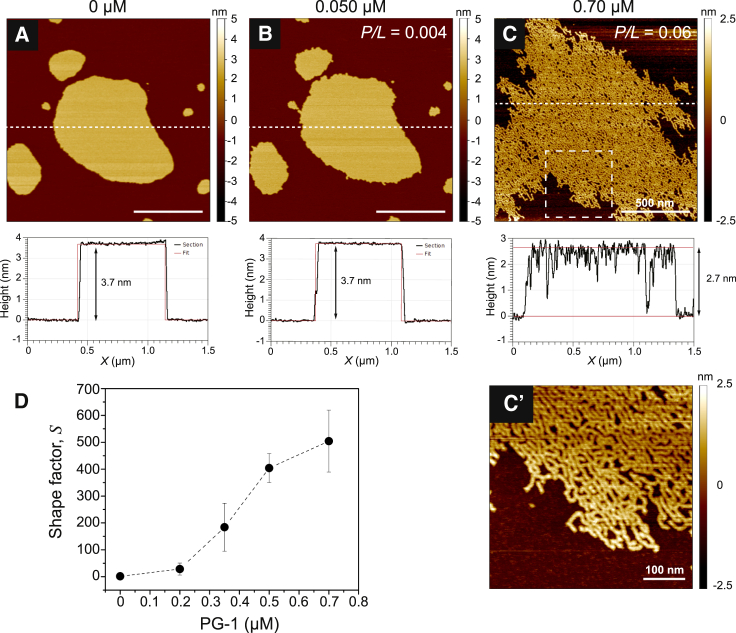
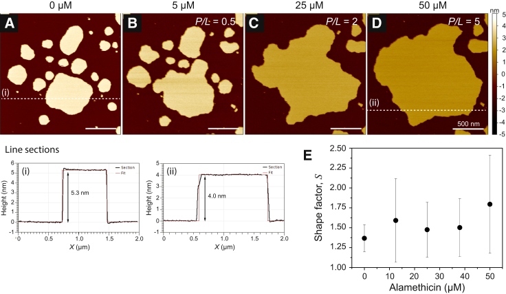
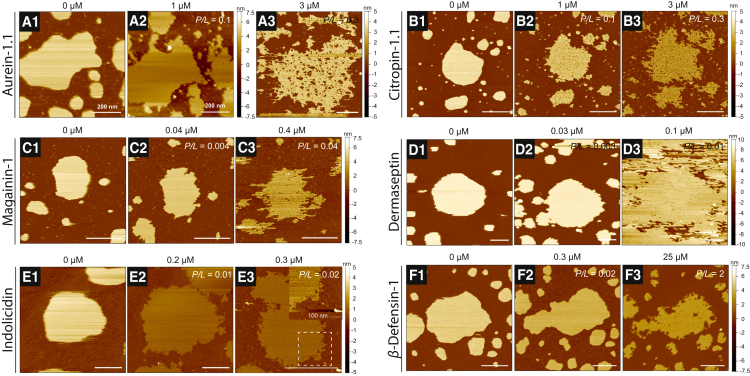
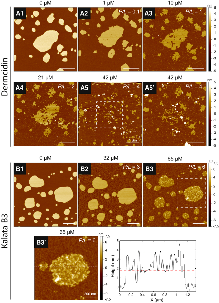
Antimicrobial peptides (AMPs) are a class of host-defense molecules that neutralize a broad range of pathogens. Their membrane-permeabilizing behavior has been commonly attributed to the formation of pores; however, with the continuing discovery of AMPs, many are uncharacterized and their exact mechanism remains unknown. Using atomic force microscopy, we previously characterized the disruption of model membranes by protegrin-1 (PG-1), a cationic AMP from pig leukocytes. When incubated with zwitterionic membranes of dimyristoylphosphocholine, PG-1 first induced edge instability at low concentrations, then porous defects at intermediate concentrations, and finally worm-like micelle structures at high concentrations. These rich structural changes suggested that pore formation constitutes only an intermediate state along the route of PG-1's membrane disruption process. The formation of these structures could be best understood by using a mesophase framework of a binary mixture of lipids and peptides, where PG-1 acts as a line-active agent in lowering interfacial bilayer tensions. We have proposed that rather than being static pore formers, AMPs share a common ability to lower interfacial tensions that promote membrane transformations. In a study of 13 different AMPs, we found that peptide line-active behavior was not driven by the overall charge, and instead was correlated with their adoption of imperfect secondary structures. These peptide structures commonly positioned charged residues near the membrane interface to promote deformation favorable for their incorporation into the membrane. Uniquely, the data showed that barrel-stave-forming peptides such as alamethicin are not line-active, and that the seemingly disparate models of toroidal pores and carpet activity are actually related. We speculate that this interplay between peptide structure and the distribution of polar residues in relation to the membrane governs AMP line activity in general and represents a novel, to our knowledge, avenue for the rational design of new drugs.


Figures

Figure 1
Figure 1
DMPC membrane structural transformations induced by PG-1’s line-active behavior. (A) In the absence of PG-1, the bilayer was compact and nearly circular with a smooth and minimized edge. (B) Introduction of 0.050 μM PG-1 caused the bilayer to extend and become roughened, and this remained stable over time. Line sections (dashed white lines) of the bilayer before and after the introduction of 0.050 μM PG-1 showed that the lamellar core remained unaffected, and that PG-1 adsorption at the curved edge resulted in a reduction in the bilayer line tension. (C) With increasing bulk PG-1 concentrations, the lamellar organization of the bilayer was compromised by 0.70 μM PG-1, with the formation of worm-like micelles. Peptide insertion thinned the membrane considerably (by ∼1 nm). To better reveal the bilayer’s transformation in (C), the height data were rescaled to a 5 nm range. The dashed box in (C) indicates a zoomed-in region (500 × 500 nm2) that is shown in (C′). (D) A shape factor analysis of the bilayer morphology (S) as a function of the bulk PG-1 concentration showed that PG-1 had a considerable effect in promoting extended morphologies with increased perimeter lengths beyond initial values near S = 1. Error bars in (D) are SDs from at least three replicate bilayer monitoring experiments. All images were obtained at 30°C. White scale bars are 500 nm unless otherwise indicated. To see this figure in color, go online.
Figure 2
Figure 2
Concentration-dependent interaction of ALM with DMPC bilayer patches. (A) The unperturbed bilayer patches in the absence of ALM displayed smooth edges and compact shapes, as shown in Fig. 1A. (B–D) Through the course of successive injections, the concentration of ALM was raised from 5 μM (B) to 25 μM (C) and finally to 50 μM (D). In contrast to the structural transformations induced by PG-1, ALM had a negligible effect on the bilayer morphology and instead only caused gross lateral expansion of the bilayers. Line sections (dashed white lines) revealed progressive membrane thinning, presumably from peptide insertion, although pores were not observed. (E) Shape factor analysis as a function of increasing ALM concentration. Error bars in (E) are SDs from five replicate bilayer monitoring experiments to confirm repeatability. All images were obtained at 30°C. White scale bars are 500 nm. To see this figure in color, go online.
Figure 3
Figure 3
(A–F) Membrane structural transformations of DMPC bilayer patches induced by the positively charged AMPs aurein-1.1 (A), citropin-1.1 (B), magainin-1 (C), dermaseptin-1 (D), indolicidin (E), and HBD-1 (F). Bilayer patches were first imaged in the absence of peptide (A1, B1, C1, etc.) and then monitored through the course of successive peptide concentrations (increasing from left to right). (A) Increasing concentrations of aurein-1.1 revealed edge instability and a porated membrane (A2). Micrograph (A3) is from a separate experiment showcasing worm-like micelle formation by 3 μM aurein-1.1. (B) Citropin-1.1 exhibited a similar response to aurein-1.1, displaying edge instability and a porated membrane (B2), with worm-like micelle formation (B3) occurring by 3 μM. The concentration responses of magainin-1, dermaseptin-1, and indolicidin were similar, as the onset of micellization (C3, D3, and E3) occurred in the same range below 0.5 μM. The bilayer patch monitored through images (E1) and (E2) was inadvertently smeared, and a nearby patch (E3) on the substrate was chosen to obtain a high-resolution image of worm-like micelles induced by indolicidin. To better reveal the self-assembled structures, the dashed box in (E3) indicates a zoomed-in region shown in the inset (data scaled to a 5 nm range). (F) Although bilayer edge instability was observed at 0.3 μM HBD-1 (F2), a significantly higher amount of peptide was needed to observe detergent behavior (F3). All images were obtained at 30°C. White scale bars are 500 nm unless otherwise indicated. To see this figure in color, go online.
Figure 4
Figure 4
(A and B) Membrane structural transformations of DMPC bilayer patches induced by the negatively charged AMPs DCD (A) and KB3 (B). Bilayer patches in the absence of peptide (A1 and B1) were monitored through the course of increasing peptide concentrations. (A) DCD exhibited a noticeable line-active behavior from an observed expanded bilayer edge (A2). The number and size of porous defects within the bilayer core grew (A3 and A4), and eventually the integrity of the membrane was fully compromised as micellization resulted (A5). (B) KB3 exhibited less activity than DCD. Bilayer edge instability was not observed until 32 μM KB3 (B2). Detergent behavior was less evident and the membrane was decorated with peripherally bound peptide (B3). A line section in (B3′) shows that the features are ∼2 nm high from the surrounding surface. The dashed boxes in (A5) and (B3) indicate zoomed-in regions that are shown in (A5′) and (B3′). All images were obtained at 30°C. White scale bars are 500 nm unless otherwise indicated. To see this figure in color, go online.
Figure 5
Figure 5
(A–C) Membrane structural transformations of DMPC bilayer patches induced by the neutral AMPs pardaxin-1 (A), caerin-1.3 (B), and histatin-2 (C). Bilayer patches were first imaged in the absence of peptide (A1, B1, and C1) and then monitored through the course of successive peptide concentrations (increasing from left to right). (A) With increasing pardaxin-1, the bilayer underwent structural transformations exhibiting edge instability (A2), porous defects (A3), and finally worm-like micelles (A4) within a low concentration regime similar to that of PG-1, magainin-1, dermaseptin-1, and indolicidin. (B) The line activity of caerin-1.3 was evident from the bilayer edge instability observed at 4 μM (B2) that caused porous defects to form within the core of the bilayer (B3) and led to micellization (B4). The bilayer patch monitored through images (B1)(B3) was damaged from its softened state, and a nearby patch (B4) on the substrate was chosen to obtain a high-resolution image of the micellized state at 19 μM caerin-1.3. To better reveal caerin-induced membrane transformations, the dashed boxes in (B3) and (B4) indicate zoomed-in regions shown in the inset (data scaled to a 5 nm range). (C) Edge instability and membrane thinning were observed upon introduction of 1 μM histatin-2 (C2). (C3) Zoomed-out image showing bilayer solubilization in the surrounding area. A dashed box is included to indicate the scanning area corresponding to images (C1) and (C2). The sample was translated laterally to a new area (C4) to capture the presence of worm-like micelle structures. All images were obtained at 30°C. White scale bars are 500 nm unless otherwise indicated. To see this figure in color, go online.

Similar articles

Cited by

References

    1. Hughes J.M. Preserving the lifesaving power of antimicrobial agents. JAMA. 2011;305:1027–1028. - PubMed
    1. Zasloff M. Antimicrobial peptides of multicellular organisms. Nature. 2002;415:389–395. - PubMed
    1. Henderson J.M., Lee K.Y.C. Promising antimicrobial agents designed from natural peptide templates. Curr. Opin. Solid State Mater. Sci. 2013;17:175–192.
    1. Baumann G., Mueller P. A molecular model of membrane excitability. J. Supramol. Struct. 1974;2:538–557. - PubMed
    1. Christensen B., Fink J., Mauzerall D. Channel-forming properties of cecropins and related model compounds incorporated into planar lipid membranes. Proc. Natl. Acad. Sci. USA. 1988;85:5072–5076. - PMC - PubMed

Substances