. 2016 Nov 15;111(10):2309-2316.
doi: 10.1016/j.bpj.2016.10.004.

Shape Transformation of the Nuclear Envelope during Closed Mitosis

Affiliations

Shape Transformation of the Nuclear Envelope during Closed Mitosis

Qian Zhu et al. Biophys J. .

Abstract

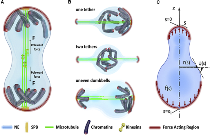
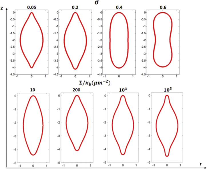
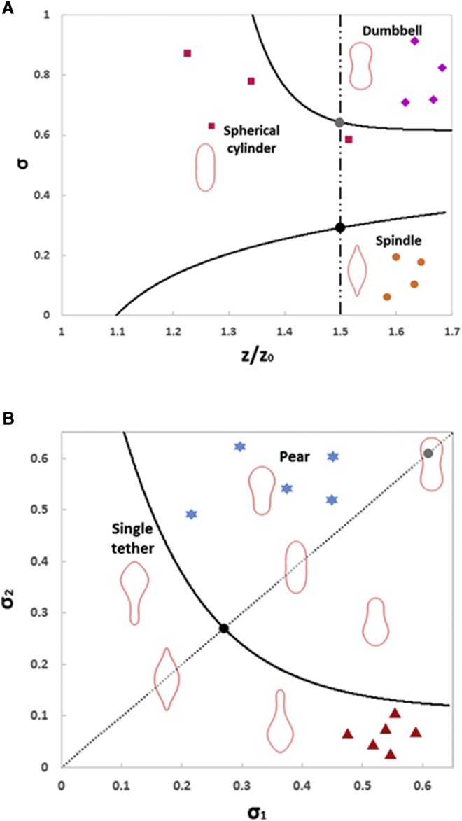
The nuclear envelope (NE) in lower eukaryotes such as Schizosaccharomyces pombe undergoes large morphology changes during closed mitosis. However, which physical parameters are important in governing the shape evolution of the NE, and how defects in the dividing chromosomes/microtubules are reflected in those parameters, are fundamental questions that remain unresolved. In this study, we show that improper separation of chromosomes in genetically deficient cells leads to membrane tethering or asymmetric division in contrast to the formation of two equal-sized daughter nuclei in wild-type cells. We hypothesize that the poleward force is transmitted to the nuclear membrane through its physical contact with the separated sister chromatids at the two spindle poles. A theoretical model is developed to predict the morphology evolution of the NE where key factors such as the work done by the poleward force and bending and surface energies stored in the membrane have been taken into account. Interestingly, the predicted phase diagram, summarizing the dependence of nuclear shape on the size of the load transmission regions, and the pole-to-pole distance versus surface area relationship all quantitatively agree well with our experimental observations, suggesting that this model captures the essential physics involved in closed mitosis.


Figures

Figure 1
Figure 1
Shape evolution of the NE in a wild-type fission yeast cell and klp5-deleted mutants during anaphase. The top row shows time-lapse sequences of a wild-type cell expressing cut11-RFP (in red, representing the nuclear envelope) and hht1-GFP (in green, representing chromatin). Numbers refer to the time in min while red dots correspond to the SPBs (marked by sid4p). The three bottom rows display time-lapse sequences of klp5Δ cells undergoing aberrant DNA segregation during anaphase. Scale bar, 2 μm. To see this figure in color, go online.
Figure 2
Figure 2
Schematic diagram illustrating the shape transformation of the dividing nucleus driven by the internally generated poleward force. (A) Equal separation of chromosomes in wide-type cells transmits the poleward forces to the NE over a broad region surrounding each pole and results in the formation of a dumbbell-shaped nucleus. (B) Improper separation of chromatids causes the transmission of the poleward force over a small area on the NE, ultimately leading to membrane tethering or the formation of a pear-shaped nucleus (or uneven dumbbell). (C) The axis-symmetric shape of the nucleus can be described by two geometric parameters r and ψ, both as a function of the arc coordinate s. The physical contact between the separated sister chromatids and the NE is assumed to uniformly distribute the poleward force to the membrane (in the vertical direction) within the contact region. To see this figure in color, go online.
Figure 3
Figure 3
Predicted axis-symmetric NE shapes under the fixed poleward force of FR/κb=7.5. The top panel shows the predicted nuclear shapes as σ varies from 0.05 to 0.6 for a given value of Σ/κb=200. The bottom panel shows the transition of the minimum energy shape when the value of Σ/κb increases from 10 to 105μm−2 while σ is fixed at 0.1. To see this figure in color, go online.
Figure 4
Figure 4
The pole-to-pole distance versus the surface area relationship of the dividing nucleus. Square symbols represent data from confocal micrographs of 30 wild-type fission yeasts nuclei where the error bar shows the standard error of the mean. Theoretical prediction is given by the solid curve with Σ/κb=200μm2 and σ = 0.8. Comparison between the predicted and observed nuclear shapes at different dividing stages is included in the insets where, similar to that in Fig. 1, cut11p, hht1p, and sid4p were used as markers for NE (red), chromatin (green), and SPB (red dots), respectively. The prediction corresponding to Σ/κb=105μm2 is also shown by the dashed line. Scale bar, 1 μm. To see this figure in color, go online.
Figure 5
Figure 5
(A) Phase diagram summarizing how the size of the load-transmitting region, along with the pole-to-pole distance, affects the axis-symmetric shape of the NE. (B) The σ1σ2 phase diagram illustrating how the morphology of nucleus undergoing asymmetric division is governed by the sizes of two load-distributing regions, where the pole-to-pole distance is fixed at Z=1.5Z0=3R. Note that the dotted line here corresponds to symmetric division (that is σ1=σ2) and hence carries the same information as the dashed line in (A). Different nuclear shapes observed in our experiment are also indicated in the phase diagram by markers, with square, diamond, circular, triangle, and star symbols corresponding to spherical cylinder, dumbbell, spindle, single tether, and pear, respectively. To see this figure in color, go online.

Similar articles

Cited by

References

    1. Méndez-López I., Worman H.J. Inner nuclear membrane proteins: impact on human disease. Chromosoma. 2012;121:153–167. - PubMed
    1. Scaffidi P., Misteli T. Lamin A-dependent nuclear defects in human aging. Science. 2006;312:1059–1063. - PMC - PubMed
    1. Arnone J.T., Walters A.D., Cohen-Fix O. The dynamic nature of the nuclear envelope: lessons from closed mitosis. Nucleus. 2013;4:261–266. - PMC - PubMed
    1. Yam C., He Y., Oliferenko S. Divergent strategies for controlling the nuclear membrane satisfy geometric constraints during nuclear division. Curr. Biol. 2011;21:1314–1319. - PubMed
    1. Jaspersen S.L., Winey M. The budding yeast spindle pole body: structure, duplication, and function. Annu. Rev. Cell Dev. Biol. 2004;20:1–28. - PubMed

MeSH terms

LinkOut - more resources