

Novel mutations c.28G>T (p.Ala10Ser) and c.189G>T (p.Glu63Asp) in WDR62 associated with early onset acanthosis and hyperkeratosis in a patient with autosomal recessive microcephaly type 2
- PMID: 27852057
- PMCID: PMC5346645
- DOI: 10.18632/oncotarget.13279
Novel mutations c.28G>T (p.Ala10Ser) and c.189G>T (p.Glu63Asp) in WDR62 associated with early onset acanthosis and hyperkeratosis in a patient with autosomal recessive microcephaly type 2
Abstract
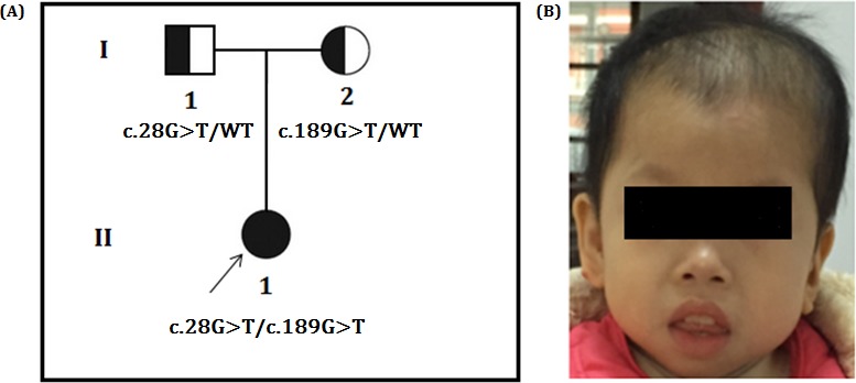
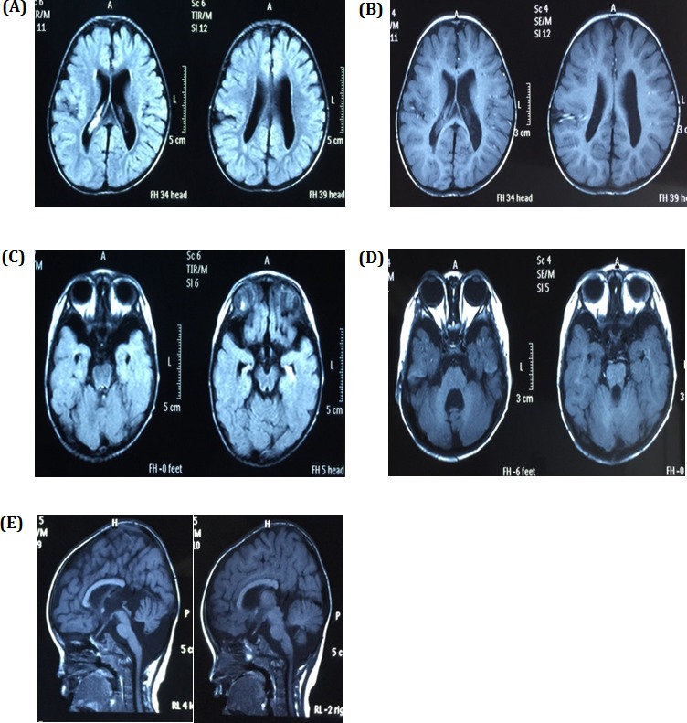
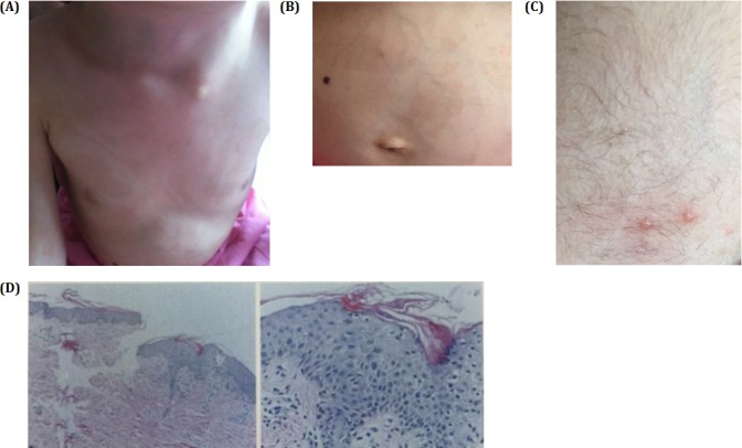
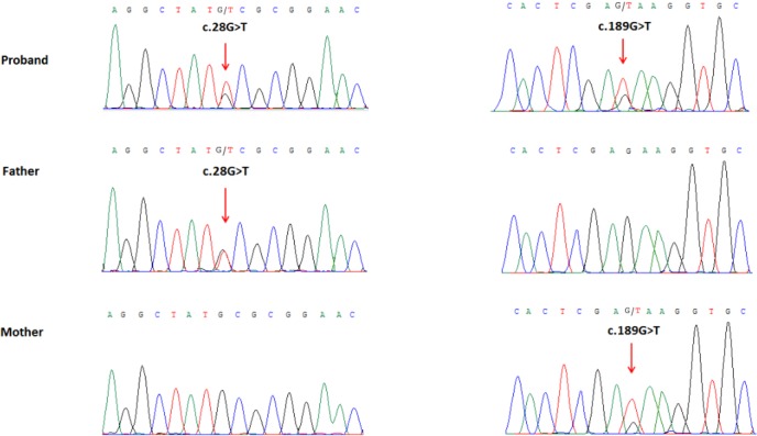
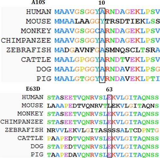
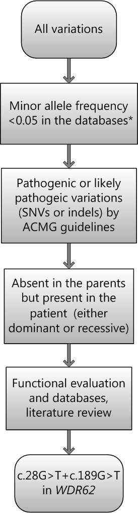
Microcephaly (MCPH) is a developmental disorder characterized by reduced brain size and intellectual disability. A proportion of microcephaly is caused by defects in a single gene. Microcephaly 2 (MCPH2) is one of the most frequent subtypes of MCPH.WD repeat-containing protein 62 gene (WDR62) is the most frequently mutated gene in MCPH2 patients. Phenotypes involving dermatological changes in MCPH2 have not been reported. We have identified and investigated a 5-year-old Chinese girl with markedly reduced brain size (86% of normal size), intellectual disability and psychomotor developmental delay. The patient also exhibited spattered blisters and reduced hair density on her head, anisochromasia with reticular hyperpigmentation and hypopigmentation on the trunk, which she has had since the age of 4 and had been found by her parents. Histological examination of a skin biopsy revealed acanthosis, hyperkeratosis and necrotic keratinocytes. Whole exome and Sanger sequencing identified two novel missense mutations, c.28G>T and c.189G>T, in the WDR62 gene. Both the mutations non-synonymously affect evolutionarily conserved amino acids and are predicted to be disease causing. We report the first case of MCPH2 that also presented with marked dermatological changes. Our findings expand the mutational and phenotypical spectra of MCPH2 and are valuable in the mutation-based pre- and post-natal screening and genetic diagnosis for MCPH2.
Keywords: Pathology Section; WDR62 mutation; compound heterozygosity; exome sequencing; microcephaly 2; novel mutations.
Conflict of interest statement
The authors declare that they have no financial or other conflicts of interest in relation to this research and its publication.
Figures
References
-
- Belligni EF, Dokal I, Hennekam RC. Prenatal and postnatal growth retardation, microcephaly, developmental delay, and pigmentation abnormalities: Naegeli syndrome, dyskeratosis congenita, poikiloderma Clericuzio type, or separate entity? Eur J Med Genet. 2011;54:231–5. - PubMed
-
- Nicholas AK, Khurshid M, Desir J, Carvalho OP, Cox JJ, Thornton G, Kausar R, Ansar M, Ahmad W, Verloes A, Passemard S, Misson JP, Lindsay S, Gergely F, Dobyns WB, Roberts E, Abramowicz M, Woods CG. WDR62 is associated with the spindle pole is mutated in human microcephaly. Nat Gen. 2010;42:1010–1014. doi: 10.1038/ng.682. - DOI - PMC - PubMed
-
- Hofman MA. A biometric analysis of brain size in micrencephalics. J. Neurol. 1984;231:87–93. - PubMed
-
- Farag HG, Froehler S, Oexle K, Ravindran E, Schindler D, Staab T, Huebner A, Kraemer N, Chen W1, Kaindl AM. Abnormal centrosome and spindle morphology in a patient with autosomal recessive primary microcephaly type 2 due to compound heterozygous WDR62 gene mutation. Orphanet J Rare Dis. 2013;8:178. - PMC - PubMed
Publication types
MeSH terms
Substances
Supplementary concepts
LinkOut - more resources
Full Text Sources
Other Literature Sources
