. 2017 Feb;11(1):41-51.
doi: 10.1080/17435390.2016.1262919. Epub 2016 Dec 9.

TIMP1 promotes multi-walled carbon nanotube-induced lung fibrosis by stimulating fibroblast activation and proliferation

Affiliations

TIMP1 promotes multi-walled carbon nanotube-induced lung fibrosis by stimulating fibroblast activation and proliferation

Jie Dong et al. Nanotoxicology. 2017 Feb.

Abstract

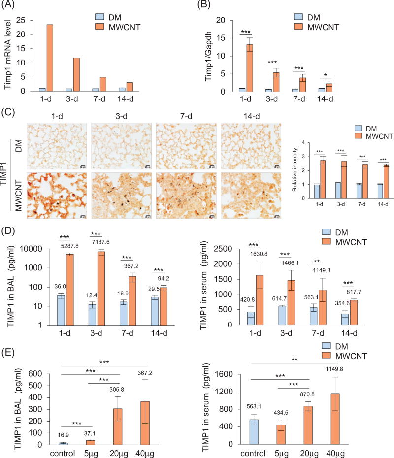
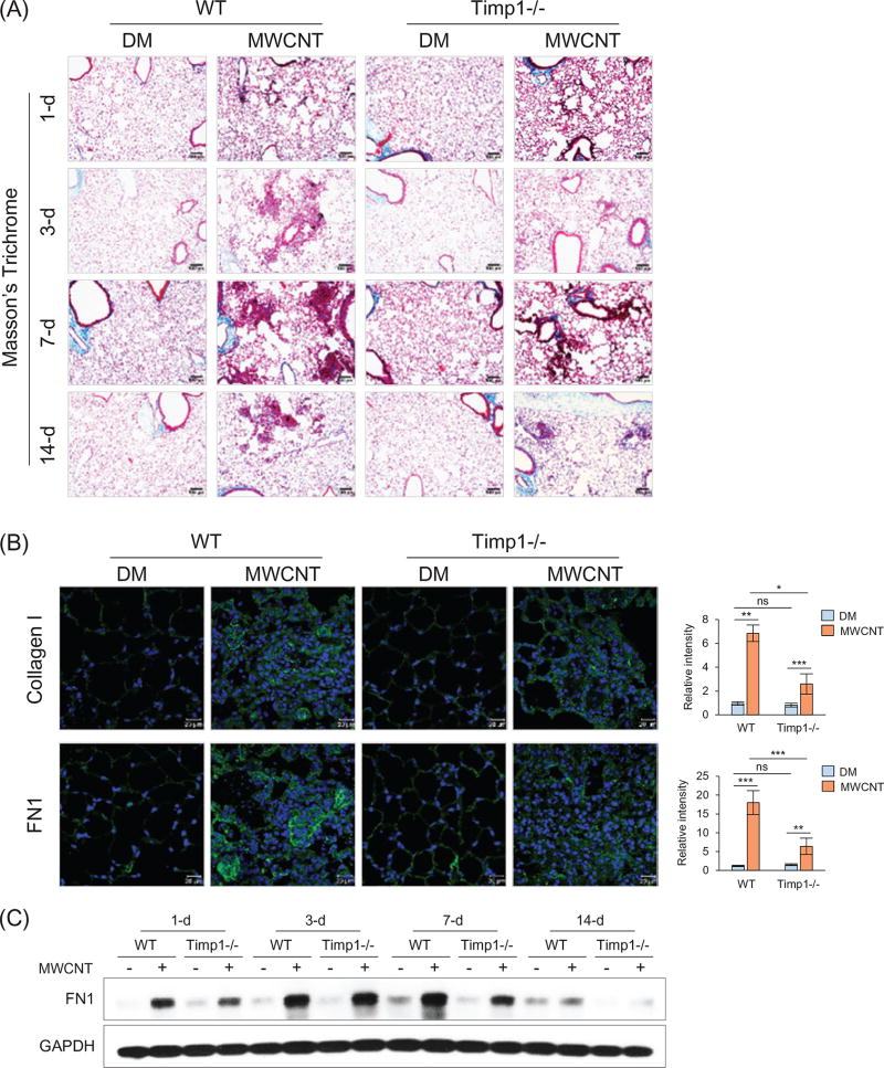
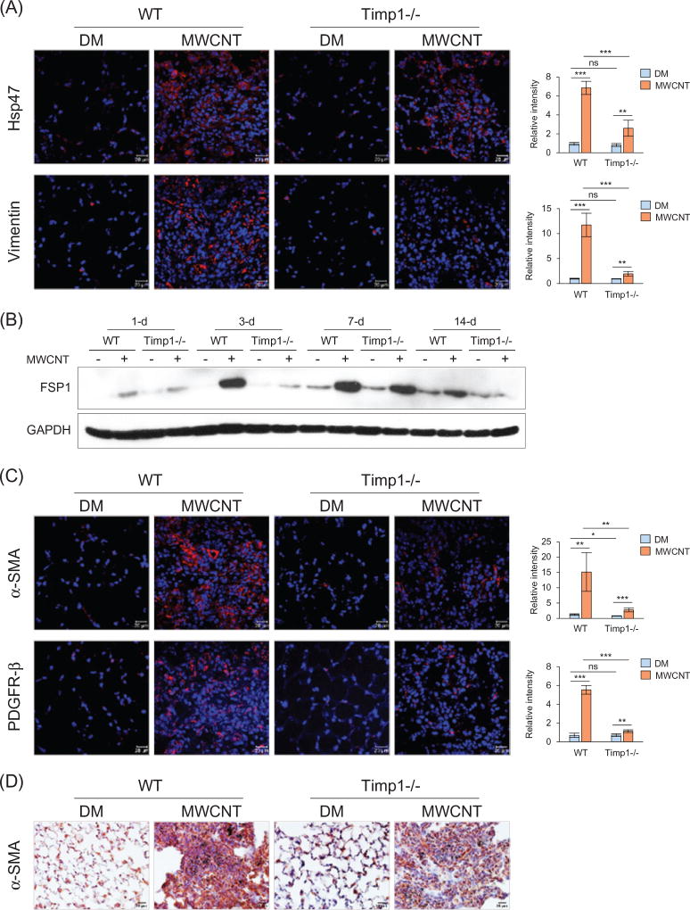
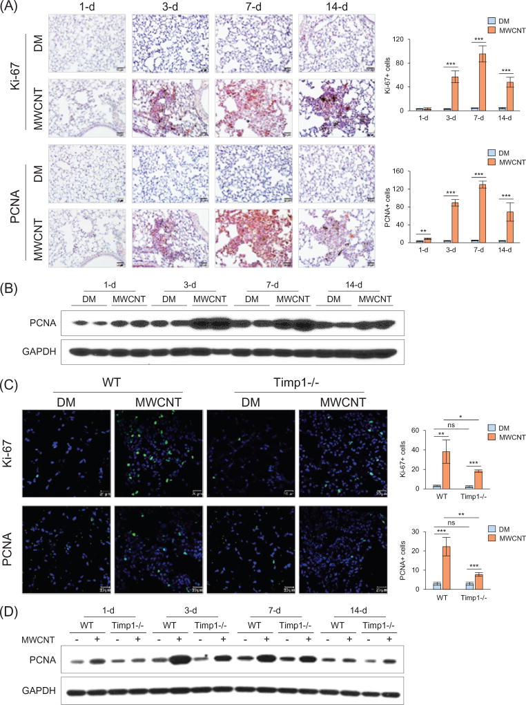
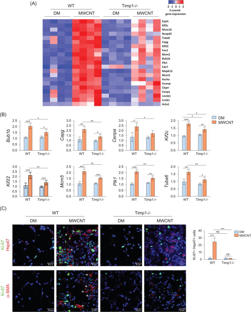
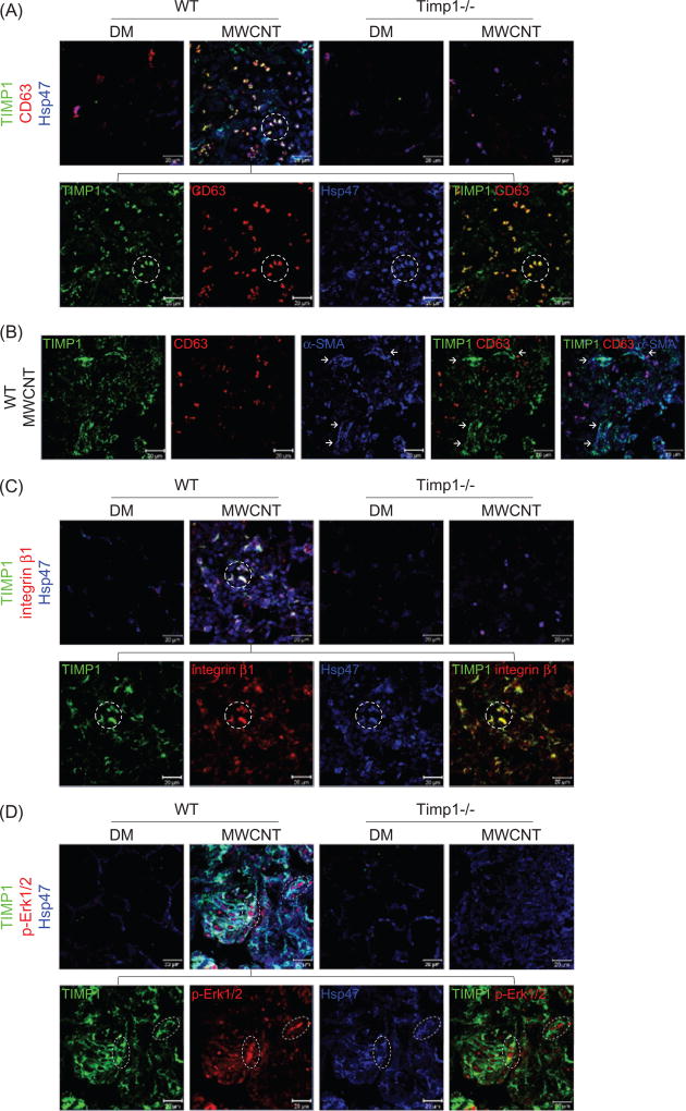
Pulmonary exposure to multi-walled carbon nanotubes (MWCNTs) may cause fibrosing lesions in animal lungs, raising health concerns about such exposure in humans. The mechanisms underlying fibrosis development remain unclear, but they are believed to involve the dysfunction of fibroblasts and myofibroblasts. Using a mouse model of MWCNT exposure, we found that the tissue inhibitor of metalloproteinase 1 (Timp1) gene was rapidly and highly induced in the lungs by MWCNTs in a time- and dose-dependent manner. Concomitantly, a pronounced elevation of secreted TIMP1 was observed in the bronchoalveolar lavage (BAL) fluid and serum. Knockout (KO) of Timp1 in mice caused a significant reduction in fibrotic focus formation, collagen fiber deposition, recruitment of fibroblasts and differentiation of fibroblasts into myofibroblasts in the lungs, indicating that TIMP1 plays a critical role in the pulmonary fibrotic response to MWCNTs. At the molecular level, MWCNT exposure significantly increased the expression of the cell proliferation markers Ki-67 and PCNA and a panel of cell cycle-controlling genes in the lungs in a TIMP1-dependent manner. MWCNT-stimulated cell proliferation was most prominent in fibroblasts but not myofibroblasts. Furthermore, MWCNTs elicited a significant induction of CD63 and integrin β1 in lung fibroblasts, leading to the formation of a TIMP1/CD63/integrin β1 complex on the surface of fibroblasts in vivo and in vitro, which triggered the phosphorylation and activation of Erk1/2. Our study uncovers a new pathway through which induced TIMP1 critically modulates the pulmonary fibrotic response to MWCNTs by promoting fibroblast activation and proliferation via the TIMP1/CD63/integrin β1 axis and ERK signaling.

Keywords: TIMP1; fibroblast; lung fibrosis; multi-walled carbon nanotube; myofibroblast.


Figures

Figure 1
Figure 1
Induction of Timp1 by MWCNTs. WT mice received DM or MWCNTs (40 µg) and were sacrificed on days 1, 3, 7, and 14 post-exposure (A–D). (A) Mouse fibrosis PCR array. Pooled lung tissue total RNA from six mice each group was analyzed. Fold changes are presented. (B) qRT-PCR analysis (n = 4). (C) Immunohistochemistry of TIMP1 (red, scale bar: 20 µm). The relative intensity of positive staining is presented as the mean ± SD (n = 4) on the right. (D) TIMP1 protein levels in the BAL fluid and serum detected by ELISA (mean ± SD, n = 5–6). (E) Dose dependence. WT mice were treated with DM or MWCNTs (5, 20 or 40 µg) for 7 d. TIMP1 protein levels in the BAL fluid and serum were determined by ELISA (mean ± SD, n = 5–6).
Figure 2
Figure 2
Reduced fibrosis in Timp1 KO lungs. WT and Timp1 KO mice received DM or MWCNTs (40 µg). (A) Masson’s Trichrome staining. Time course is shown. Scale bar: 100 µm. (B) Collagen I (upper panel) and FN1 (lower panel) immunofluorescence staining (green indicates positive staining, blue represents nuclear staining) on lung sections from mice sacrificed on day 7 post-exposure. Scale bar: 20 µm. Relative intensity is shown as the mean ± SD (n = 4). (C) Immunoblotting of FN1. Lung proteins from randomly selected samples of each group were analyzed and a representative blotting image is presented.
Figure 3
Figure 3
Recruitment of fibroblasts and myofibroblasts by MWCNTs. Lung sections from mice exposed to DM or 40 µg MWCNTs for 7 d were examined for the expression of fibroblast and myofibroblast markers (A, C and D). (A) Immunofluorescence detection of fibroblast markers Hsp47 and Vimentin. (B) Immunoblotting of FSP1. Lung proteins from randomly selected samples of each group exposed to DM or 40 µg MWCNTs were analyzed and a representative blotting image is presented. (C) Immunofluorescence detection of myofibroblast markers α-SMA and PDGFR-β. (D) Immunohistochemistry staining of α-SMA. Images have scale bars of 20 µm. For (A) and (C), the red color indicates positive staining and blue indicates nuclear staining. Relative intensity is shown as the mean ± SD (n = 4).
Figure 4
Figure 4
Reduced cell proliferation in Timp1 KO lungs. (A and B) Time-dependent stimulation of cell proliferation by MWCNTs (40 µg) in WT lungs on days 1, 3, 7 and 14 post-exposure. (A) Immunohistochemistry of Ki-67 (upper panel) and PCNA (lower panel) on lung sections of WT mice. Red indicates positive staining and blue nuclear counterstaining. Scale bar: 20 µm. The number of cells with positive staining is shown as the mean ± SD (n = 4). (B) Immunoblotting of PCNA in lung tissues of WT mice. Lung proteins from two randomly selected samples of each group were used. (C and D) Comparison of cell proliferation between WT and Timp1 KO lungs exposed to MWCNTs (40 µg). (C) Immunofluorescence of Ki-67 (upper panel) and PCNA (lower panel) on lung sections (7 d post-exposure). Green indicates positive staining and blue nuclear staining. Scale bar: 20 µm. The number of cells with positive staining is shown as the mean ± SD (n = 4). (D) Immunoblotting of PCNA. Lung proteins from randomly selected samples of each group were studied, and a representative blotting image is presented.
Figure 5
Figure 5
TIMP1-mediated stimulation of gene expression and fibroblast proliferation by MWCNTs. Mice were exposed to DM or 40 µg MWCNTs for 7 d. (A) Heat map of cell cycle control genes identified by microarray. Total RNA isolated from individual mice was used (n = 4, for each genotype and treatment group). Red, white and blue indicate high, medium and low expression levels, respectively. (B) Examples of up-regulated genes shown in the heat map are presented with fold changes and p values (n = 4). *p <0.05; ** p <0.01; and ***p< 0.001. (C) Cell proliferation. Proliferation of fibroblasts was examined by double immunofluorescence staining of Ki-67 (green) and Hsp47 (red), with double-positive cells showing green staining in the nucleus and red staining outside of the nucleus (upper panel, scale bar: 20 µm, example cells are indicated by arrows). The number of Ki-67 and Hsp47 double-positive cells is shown as the mean ± SD (n = 4). Proliferation of myofibroblasts was examined by double immunofluorescence staining of Ki-67 (green) and α-SMA (red) (lower panel, scale bar: 20 µm). Blue indicates nuclear staining. Representative myofibroblasts are indicated by arrows, showing strong staining for α-SMA but weak or no co-staining for Ki-67.
Figure 6
Figure 6
Induction and co-localization of TIMP1, CD63, and integrin β1 and activation of ERK. Mice were exposed to DM or 40 µg MWCNTs for 7 d. (A) Induction and co-localization of TIMP1 and CD63 in fibroblasts detected by triple immunofluorescence of TIMP1 (green), CD63 (red) and Hsp47 (blue) on lung sections. Scale bar: 20 lm. Circled cells illustrate representative triple-positive cells. (B) Induction and co-localization of TIMP1 and CD63 in myofibroblasts examined by triple immunofluorescence of TIMP1 (green), CD63 (red) and α-SMA (blue) on lung sections from MWCNT-exposed WT mice. Images of single staining and merged images of double and triple staining are presented, showing the strong induction and co-localization of TIMP1 and α-SMA, but markedly reduced induction of CD63 and co-localization of CD63 with TIMP1 in myofibroblasts. A subset of TIMP1+ cells are co-localized with α-SMA+ CD63− cells (indicated by arrows). Scale bar: 20 µm. (C) Induction and co-localization of TIMP1 and integrin β1 in fibroblasts detected by triple immunofluorescence of TIMP1 (green), integrin β1 (red) and Hsp47 (blue) on lung sections. Example triple-positive cells are circled. Scale bar: 20 µm. (D) The level of p-Erk1/2 in fibroblasts examined by triple immunofluorescence of TIMP1 (green), p-Erk1/2 (red) and Hsp47 (blue) on lung sections (scale bar: 20 µm). Example triple-positive cells are indicated in the oval.

References

    1. Aiso S, Yamazaki K, Umeda Y, Asakura M, Kasai T, Takaya M, et al. Pulmonary toxicity of intratracheally instilled multiwall carbon nanotubes in male Fischer 344 rats. Ind Health. 2010;48:783–95. - PubMed
    1. Bertaux B, Hornebeck W, Eisen AZ, Dubertret L. Growth stimulation of human keratinocytes by tissue inhibitor of metalloproteinases. J Invest Dermatol. 1991;97:679–85. - PubMed
    1. Chesler L, Golde DW, Bersch N, Johnson MD. Metalloproteinase inhibition and erythroid potentiation are independent activities of tissue inhibitor of metalloproteinases-1. Blood. 1995;86:4506–15. - PubMed
    1. De Volder MF, Tawfick SH, Baughman RH, Hart AJ. Carbon nanotubes: present and future commercial applications. Science. 2013;339:535–9. - PubMed
    1. Ding L, Stilwell J, Zhang T, Elboudwarej O, Jiang H, Selegue JP, et al. Molecular characterization of the cytotoxic mechanism of multiwall carbon nanotubes and nano-onions on human skin fibroblast. Nano Lett. 2005;5:2448–64. - PMC - PubMed

MeSH terms

Substances