

Two unusual cases of successful treatment of hypermucoviscous Klebsiella pneumoniae invasive syndrome
- PMID: 27852233
- PMCID: PMC5112683
- DOI: 10.1186/s12879-016-2011-3
Two unusual cases of successful treatment of hypermucoviscous Klebsiella pneumoniae invasive syndrome
Abstract
Background: A few Japanese cases of hypermucoviscous Klebsiella pneumoniae (K. pneumoniae) invasive syndrome have recently been reported. Although extrahepatic complications from bacteremic dissemination have been observed, infected aneurysms are rare. Furthermore, the primary source of infection is generally a liver abscess, and is rarely the prostate. Therefore, we report two atypical cases of hypermucoviscous K. pneumoniae invasive syndrome.
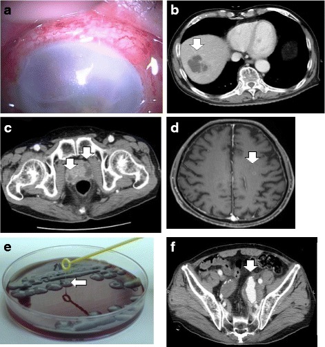
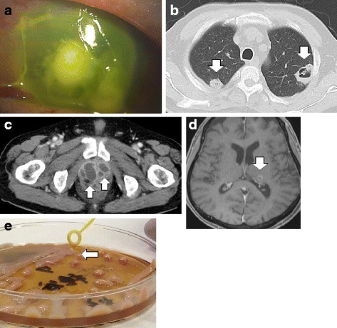
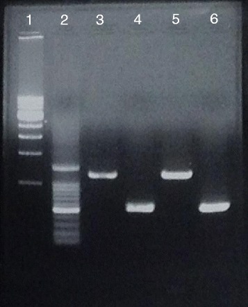
Case presentation: The first case was an 81-year-old Japanese man with no significant medical history, who was referred to our hospital for vision loss in his right eye. Contrast-enhanced whole-body computed tomography revealed abscesses in the liver and the prostate, and an infected left internal iliac artery aneurysm. Contrast-enhanced head magnetic resonance imaging revealed brain abscesses. Cultures of the liver abscess specimen and aqueous humor revealed K. pneumoniae with the hypermucoviscosity phenotype, which carried the magA gene (mucoviscosity-associated gene A) and the rmpA gene (regulator of mucoid phenotype A). We performed enucleation of the right eyeball, percutaneous transhepatic drainage, coil embolization of the aneurysm, and administered a 6-week course of antibiotic treatment. The second case was a 69-year-old Japanese man with diabetes mellitus, who was referred to our hospital with fever, pollakiuria, and pain on urination. Contrast-enhanced whole-body computed tomography revealed lung and prostate abscesses, but no liver abscesses. Contrast-enhanced head magnetic resonance imaging revealed brain abscesses. The sputum, urine, prostate abscess specimen, and aqueous humor cultures revealed K. pneumoniae with the hypermucoviscosity phenotype, which carried magA and rmpA. We performed enucleation of the left eyeball, percutaneous drainage of the prostate abscess, and administered a 5-week course of antibiotic treatment.
Conclusions: Hypermucoviscous K. pneumoniae can cause infected aneurysms, and the prostate can be the primary site of infection. We suggest that a diagnosis of hvKP invasive syndrome should be considered in all patients who present with K. pneumoniae infection and multiple organ abscesses.
Keywords: Abscess; Endophthalmitis; Hypermucoviscous Klebsiella pneumoniae; Infected aneurysm; Prostate; String test; magA; rmpA.
Figures
References
-
- Chang FY, Chou MY, Fan RL, Shaio MF. A clinical study of Klebsiella liver abscess. Taiwan Yi Xue Hui Za Zhi. 1988;87(3):282–7. - PubMed
-
- Maruno T, Ooiwa Y, Takahashi K, Kodama Y, Takakura S, Ichiyama S, et al. A liver abscess deprived a healthy adult of eyesight: endogenous endophthalmitis associated with a pyogenic liver abscess caused by serotype K1 Klebsiella pneumonia. Intern Med. 2013;52(8):919–22. doi: 10.2169/internalmedicine.52.9076. - DOI - PubMed
Publication types
MeSH terms
Substances
LinkOut - more resources
Full Text Sources
Other Literature Sources
