. 2016 Nov 16;18(1):263.
doi: 10.1186/s13075-016-1158-z.

Elevated expression of miR-142-3p is related to the pro-inflammatory function of monocyte-derived dendritic cells in SLE

Affiliations

Elevated expression of miR-142-3p is related to the pro-inflammatory function of monocyte-derived dendritic cells in SLE

Yilun Wang et al. Arthritis Res Ther. .

Abstract

Background: Recent studies have shown that alterations in the function of dendritic cells (DCs) are involved in the pathogenesis of systemic lupus erythematosus (SLE). However, the mechanism of the alteration remains unclear.

Methods: We cultured monocyte-derived DCs (moDCs) in vitro and examined the cytokines and chemokines in the supernatants of moDCs in negative controls (NC) and SLE patients in active phase. We then profiled microRNAs (miRNAs) of LPS-stimulated moDCs in SLE patients and used real-time PCR to verify the differentially expressed miRNAs. A lentiviral construct was used to overexpress the level of miR-142-3p in moDCs of NC. We examined the cytokines and chemokines in the supernatants of moDCs overexpressing miR-142-3p and used Transwell test, flow cytometric analysis and cell proliferation to observe the impact on CD4+ T cells in moDC-CD4+T cell co-culture.

Results: moDCs in patients with SLE secreted increased level of IL-6, CCL2 and CCL5, with attraction of more CD4+ T cells compared with NC. We found 18 differentially expressed microRNAs in moDCs of SLE patients by microarray, and target gene prediction showed some target genes of differentially expressed miRNAs were involved in cytokine regulation. miR-142-3p was verified among the highly expressed miRNAs in the SLE group and overexpressing miR-142-3p in moDCs of the NC group caused an increase of SLE-related cytokines, such as CCL2, CCL5, CXCL8, IL-6 and TNF-α. Moreover, moDCs overexpressed with miR-142-3p resulted in attraction of an increased number of CD4+ T cells and in suppression of the proportion of Tregs in DC-CD4+T cell co-culture whereas the proliferation of CD4+T cells was not altered.

Conclusions: The results demonstrated a role for miR-142-3p in regulating the pro-inflammatory function of moDCs in the pathogenesis of SLE. These findings suggested that miR-142-3p could serve as a novel therapeutic target for the treatment of SLE.

Keywords: MicroRNA; Monocyte-derived DCs; SLE.


Figures
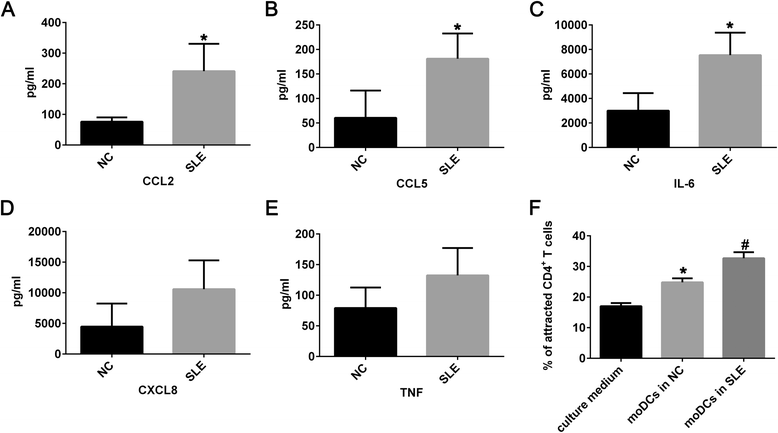
Fig. 1
Fig. 1
Pro-inflammatory function of moDCs in SLE. The supernatants derived from culture medium of moDCs from negative controls (n = 5) and patients with SLE (n = 5) in the presence of LPS (1 μg/ml) for 24 hours was assessed using cytometric bead array including CCL2 (a, *P < 0.001), CCL5 (b, *P = 0.002), IL-6 (c, *P = 0.002), CXCL8 (d, no significant difference) and TNF-α (e, no significant difference). Percentage of CD4+ T cells attracted by supernatants of culture medium, supernatants of moDCs in NC, and supernatants of moDCs in SLE group (f, # P < 0.05 versus supernatants of culture medium and supernatants of moDCs in NC, *P < 0.05 versus supernatants of culture medium). Data were shown as mean ± SD. CCL C-C motif ligand, CXCL C-X-C motif ligand, IL interleukin, moDCs monocyte-derived DCs, NC negative controls, SLE systemic lupus erythematosus, TNF-α tumor necrosis factor alpha
Fig. 2
Fig. 2
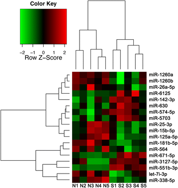
miRNA profiling in LPS-activated moDCs of patients with SLE. The expression levels of mature miRNAs purified from 24-h LPS-activated moDCs were analyzed using miRNA microarrays and hierarchical clustering of statistically significant differential miRNAs with analysis of variance (P < 0.05). N = 10. N1-N5 represented negative controls. S1-S5 represented the patients with SLE
Fig. 3
Fig. 3
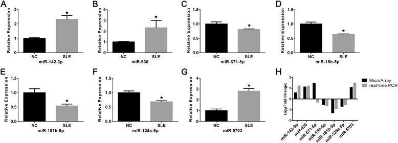
Validation of selected miRNAs by qRT-PCR. The levels of miR-142-3p (*P < 0.001), miR-630 (*P = 0.072), miR-671-5p (*P = 0.021), miR-15b-5p (*P = 0.005), miR-181b-5p (*P < 0.001), miR-125a-5p (*P < 0.001), and miR-5703 (*P < 0.001) were determined in LPS-activated moDCs of 15 negative controls (NCs) and 15 patients with SLE (ag). Data were shown as mean ± SD. The change patterns between microarray analysis and qRT-PCR were shown in (h). NC negative controls, PCR polymerase chain reaction, SLE systemic lupus erythematosus
Fig. 4
Fig. 4
Transfection efficiency of overexpressing miR-142-3p in moDCs. a Morphology and GFP fluorescence of moDCs transfected with empty lentivirus vector (VEC) and miR-142-3p overexpressing lentivirus (LV). Scale bar = 10 μm. b The level of miR-142-3p expressed in moDCs of negative controls (NC), VEC or LV group was assessed by quantitative real-time PCR. Data were expressed as mean ± SD. Each experiment was conducted at least three times. # P < 0.005 versus NC and VEC
Fig. 5
Fig. 5
Overexpression of miR-142-3p promoted pro-inflammatory function of moDCs. The level of exocrine secretion of TNF-α (a, *P = 0.013), IL-6 (b, *P = 0.03), CCL2 (c, *P = 0.014), CCL5 (d, *P < 0.001) and CXCL8 (e, *P = 0.004) in the supernatant derived from moDCs in miR-142-3p lentivirus (LV) group compared with empty lentivirus vector (VEC). N = 3 in each group. Data were shown as means ± SD. CCL C-C motif ligand, CXCL C-X-C motif ligand, IL interleukin, TNF-α tumor necrosis factor alpha
Fig. 6
Fig. 6
Elevated expression level of miR-142-3p affected moDCs-CD4+ T cells interaction. a Percentage of CD4+ T cells attracted by supernatants of moDCs in NC or empty lentiviral vector (VEC) or lentivirus (LV) group measured by a Transwell chamber. # P < 0.05 versus NC and VEC. b The percentage of CD4+CD25+Foxp3+ Tregs among CD4+ T cells co-cultured with moDCs was assessed using flow cytometry and data of one representative experiment (c) out of three independent experiments was shown. Foxp3+ cells were gated from CD4+CD25+ T cells. Data were shown as means ± SD of three independent experiments. # P < 0.05 versus NC and VEC. d and e The level of exocrine secretion of IL-17 (# P < 0.001 versus NC and VEC) and IL-10 (# P < 0.05 versus NC and VEC) in the supernatants derived from co-culture medium of CD4+T cells- moDCs. N = 3 in each group. Data were shown as means ± SD. IL interleukin

Similar articles

Cited by

References

    1. Zhu Z, Liang Z, Liany H, Yang C, Wen L, Lin Z, Sheng Y, Lin Y, Ye L, Cheng Y, et al. Discovery of a novel genetic susceptibility locus on X chromosome for systemic lupus erythematosus. Arthritis Res Ther. 2015;17:349. doi: 10.1186/s13075-015-0857-1. - DOI - PMC - PubMed
    1. Feng JB, Ni JD, Yao X, Pan HF, Li XP, Xu JH, Pan FM, Xu SQ, Ye DQ. Gender and age influence on clinical and laboratory features in Chinese patients with systemic lupus erythematosus: 1,790 cases. Rheumatol Int. 2010;30(8):1017–23. doi: 10.1007/s00296-009-1087-0. - DOI - PubMed
    1. Patel DR, Richardson BC. Epigenetic mechanisms in lupus. Curr Opin Rheumatol. 2010;22(5):478–82. doi: 10.1097/BOR.0b013e32833ae915. - DOI - PMC - PubMed
    1. Gaipl US, Munoz LE, Grossmayer G, Lauber K, Franz S, Sarter K, Voll RE, Winkler T, Kuhn A, Kalden J, et al. Clearance deficiency and systemic lupus erythematosus (SLE) J Autoimmun. 2007;28(2-3):114–21. doi: 10.1016/j.jaut.2007.02.005. - DOI - PubMed
    1. Chan VS, Nie YJ, Shen N, Yan S, Mok MY, Lau CS. Distinct roles of myeloid and plasmacytoid dendritic cells in systemic lupus erythematosus. Autoimmun Rev. 2012;11(12):890–7. doi: 10.1016/j.autrev.2012.03.004. - DOI - PubMed

Publication types

MeSH terms