. 2016 Dec:14:123-130.
doi: 10.1016/j.ebiom.2016.11.011. Epub 2016 Nov 9.

Anemia Offers Stronger Protection Than Sickle Cell Trait Against the Erythrocytic Stage of Falciparum Malaria and This Protection Is Reversed by Iron Supplementation

Affiliations

Anemia Offers Stronger Protection Than Sickle Cell Trait Against the Erythrocytic Stage of Falciparum Malaria and This Protection Is Reversed by Iron Supplementation

M M Goheen et al. EBioMedicine. 2016 Dec.

Abstract

Background: Iron deficiency causes long-term adverse consequences for children and is the most common nutritional deficiency worldwide. Observational studies suggest that iron deficiency anemia protects against Plasmodium falciparum malaria and several intervention trials have indicated that iron supplementation increases malaria risk through unknown mechanism(s). This poses a major challenge for health policy. We investigated how anemia inhibits blood stage malaria infection and how iron supplementation abrogates this protection.

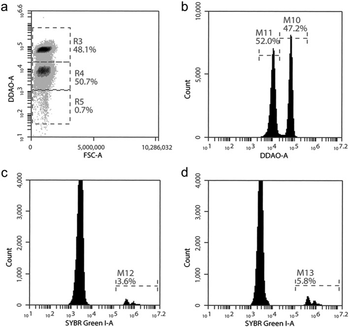
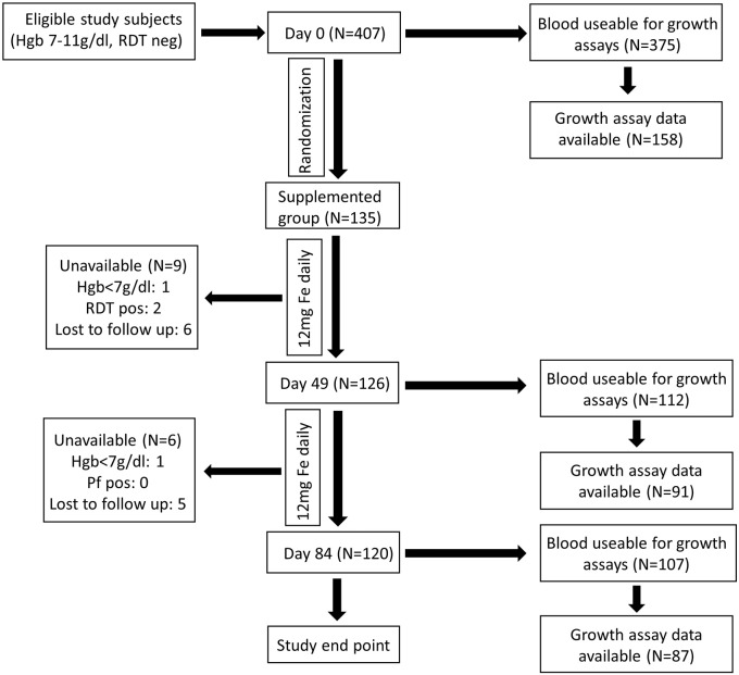
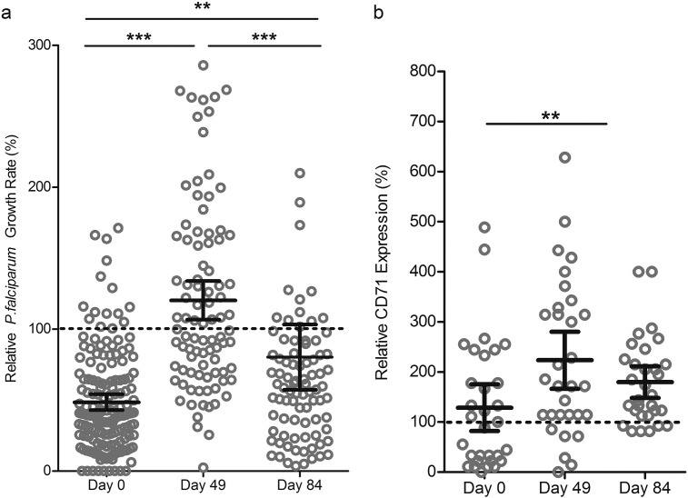
Methods: This observational cohort study occurred in a malaria-endemic region where sickle-cell trait is also common. We studied fresh RBCs from anemic children (135 children; age 6-24months; hemoglobin <11g/dl) participating in an iron supplementation trial (ISRCTN registry, number ISRCTN07210906) in which they received iron (12mg/day) as part of a micronutrient powder for 84days. Children donated RBCs at baseline, Day 49, and Day 84 for use in flow cytometry-based in vitro growth and invasion assays with P. falciparum laboratory and field strains. In vitro parasite growth in subject RBCs was the primary endpoint.

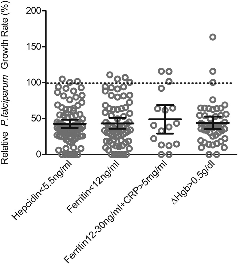
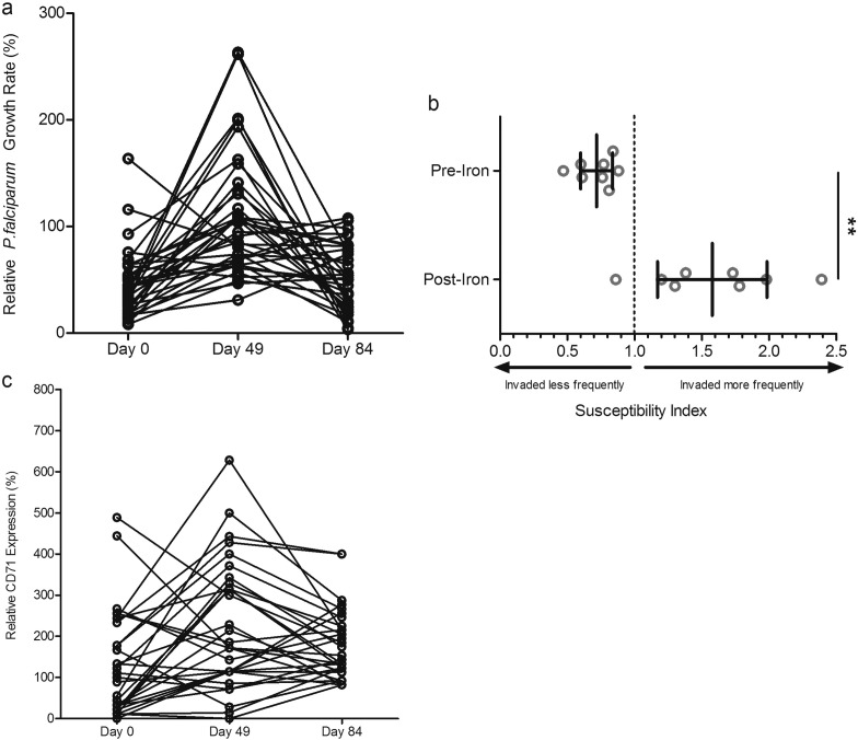
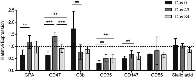
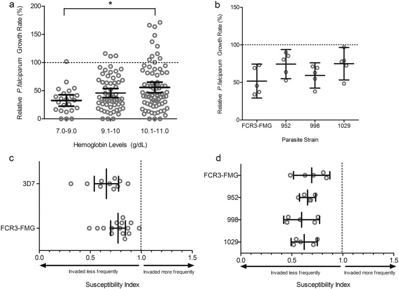
Findings: Anemia substantially reduced the invasion and growth of both laboratory and field strains of P. falciparum in vitro (~10% growth reduction per standard deviation shift in hemoglobin). The population level impact against erythrocytic stage malaria was 15.9% from anemia compared to 3.5% for sickle-cell trait. Parasite growth was 2.4 fold higher after 49days of iron supplementation relative to baseline (p<0.001), paralleling increases in erythropoiesis.

Interpretation: These results confirm and quantify a plausible mechanism by which anemia protects African children against falciparum malaria, an effect that is substantially greater than the protection offered by sickle-cell trait. Iron supplementation completely reversed the observed protection and hence should be accompanied by malaria prophylaxis. Lower hemoglobin levels typically seen in populations of African descent may reflect past genetic selection by malaria.

Funding: National Institute of Child Health and Development, Bill and Melinda Gates Foundation, UK Medical Research Council (MRC) and Department for International Development (DFID) under the MRC/DFID Concordat.

Keywords: Anemia; Hemoglobin; Iron; Iron supplementation; Malaria; Sickle cell trait.


Figures

Supplemental Fig. 1
Supplemental Fig. 1
Parasite growth rates in RBCs from children categorized by different definitions of anemia at baseline. In analysis of parasite growth rates in RBCs from children at Day 0, we stratified participants (all anemic) using four different definitions to categorize the severity and type of iron deficiency in the presence or absence of inflammation: those with 1) hepcidin < 5.5 ng/ml (n = 82); 2) ferritin < 12 ng/ml (n = 69); 3) ferritin12-30 ng/ml with CRP > 5 mg/ml (n = 17); 4) hemoglobin increase of > 0.5 g/dl from baseline after 49d or 84d of daily iron supplementation (n = 46); definitions 1–4 are not necessarily mutually exclusive. Of note, everyone in our population had a raised serum transferrin receptor (sTfR):log ferritin index > 2 which is highly suggestive of iron deficiency. Growth rate values are presented relative to growth in RBCs from non-anemic donors. Each dot represents the mean result of triplicate growth assays from each donor and the error bars represent 95% CI. Mean growth rate results (with 95%CI) are: hepcidin < 5.5 ng/ml = 42.89% (37.11–48.67%); ferritin < 12 ng/ml = 43.34% (36.01–50.68%); ferritin 12-30 ng/ml with CRP > 5 mg/ml = 49.08% (29.16–69.00%), ΔHgb > 0.5 g/dl = 44.04% (35.14–52.93%). There are no significant differences between the means.
Supplemental Fig. 2
Supplemental Fig. 2
Gating strategy to highlight adaptation of RBC barcoding assay to the field setting using basic two-color flow cytometry. a) RBCs from different blood donors are differentially labelled with CellTrace Far Red DDAO (1 μM (R3) or 0.1 μM (R4)) to distinguish between donor populations. Late stage purified parasites grown in unlabeled RBCs (R5) are seeded into the differentially labelled RBCs which have been combined in equal proportion. b) M10 represents the 1 μM Far Red DDAO labelled RBCs from a non-anemic donor and M11 represents the 0.1 μM Far Red DDAO labelled RBCs from an anemic donor. Gating cells on M11 (c) or M10 (d) allows for Sybr Green I DNA dye detection of parasite infected RBCs in the RBCs from anemic donors (M12, c) or from non-anemic donors (M13, d). Parasitemia in each cell population is compared to calculate the invasion SI. The percentages in the flow plots represent the percent of total cells within the indicated gate.
Supplemental Fig. 3
Supplemental Fig. 3
Changes in parasite growth, invasion, and reticulocytosis in RBCs from anemic children before and after daily iron supplementation. a) Levels of parasite growth rates increase over time in anemic children undergoing iron supplementation, as depicted by line graph in order to highlight changes for each individual that had data available at all timepoints (n = 35 children with complete repeat growth measures at Day 0, 49, and 84, with 86% having increased growth rate at Day 49) One-way repeated measures ANOVA of growth rate values indicates the means are significantly different between Days (p < 0.0001); post-hoc analysis with Tukey's test indicates significant differences between Day 0 and Day 49 means (p < 0.001) and Day 49 and Day 84 means (p < 0.001), but no significance between Day 0 and Day 84 for those children with repeat measures. b) Direct comparison of invasion into RBCs from non-anemic donors to RBCs from 8 anemic children either before or during 12 mg daily iron supplementation. Each experiment was performed in triplicate for each blood donor. The marker represents the SI point estimate and the bar represents the 95% CI. An SI of 1.0 indicates no difference in parasite invasion of the two RBC populations. Student's t-test indicates significant differences between pre- and post-iron SI values (**p < 0.01). c) Line graph of CD71 repeated measures (n = 31 children with complete repeat CD71 measures at Day 0, 49, and 84). In 21 of these children, the relative percent CD71 positive cells increased from Day 0 to Day 49. See Fig. 3B for repeated measures ANOVA statistics.
Supplemental Fig. 4
Supplemental Fig. 4
Surface markers of RBC age and integrity change in a pattern consistent with an increase in erythropoiesis in anemic children undergoing iron supplementation (12 mg daily). We measured GPA (an abundant sialoglycoprotein which contributes to RBC surface charge and is found at higher levels on younger RBCs (Beeson et al., 2016)), CD47 (an anti-phagocytic marker which influences RBC senescence and is found in lower levels in RBCs that have been in circulation longer or are less healthy (Lutz, 2004)), surface deposition of complement factor C3b (higher levels of which would correlate with increased RBC time in circulation, or less healthy RBC membranes (Gwamaka et al., 2012)), and levels of P. falciparum merozoite receptors (CD35, CD147, CD55, and sialic acid residues). Note that GPA is also a merozoite receptor, and CD35 and CD55 involved in the complement system have also been described as reflecting RBC age (more abundant on younger/healthier RBCs (Gwamaka et al., 2012)), as has sialic acid abundance (reduced on older RBCs) (Lutz, 2004). CD147, known as basigin, is the only known essential P. falciparum invasion receptor (Crosnier et al., 2011). Data represent relative expression based on anemic donor RBC MFI values (GPA, CD47, CD35, CD147, CD55, and sialic acid residues) or percent positive population values (C3b), compared to RBCs from a non-anemic donor not receiving iron supplementation (relative expression = 1.0). RBCs from the same 8 donors were examined over time. Error bars represent the 95% CIs. If indicated, one-way repeated measures ANOVA with post-hoc Tukey's test analysis indicates significant difference between expression levels (*p < 0.05, **p < 0.01, ***p < 0.001).
Fig. 1
Fig. 1
Description of subjects and flow chart of sample collection and assays performed. Blood samples for hematological, biochemical, and parasite growth analyses were drawn at Day 0, as well as Day 49 and Day 84 for those taking iron. A full hematology panel was measured in EDTA-stabilized blood (Medonic M20M GP). We also assayed plasma ferritin, soluble transferrin receptor (sTfR), serum iron, transferrin saturation (TSAT), C-reactive protein (CRP), alpha 1-acid glycoprotein (AGP) (Cobas Integra 400 plus); and hepcidin (Hepcidin-25 (human) EIA Kit (Bachem)). Genotyping for hemoglobinopathies was performed using hemoglobin electrophoresis. Glucose-6-phosphate dehydrogenase (G6PD) enzyme activity was measured by commercial kit (R&D Diagnostics Ltd). For malaria assays, 2.5 ml of venous blood was drawn directly into microvette tubes containing CPDA-1 (Sarstedt, Germany). Unavailable donors include safety exclusion (Hgb < 7 g/dl or positive malaria test, RDT pos) or general loss to follow up (withdrawal and travel). Failure to collect blood from subjects (e.g. from phlebotomy failure, subject moved or withdrew, or became significantly ill) was 7.8% (32/407) at Day 0, 17.0% (23/135) at Day 49, and 20.7% (28/135) at Day 84. RBCs from study subjects were evaluated with in vitro P. falciparum growth assays (using strain FCR3-FMG) as a proxy measure for malaria susceptibility. In order to standardize the growth assays, control for inter-assay variability and variability between parasite preparations, assays on clinical samples were run in parallel with and reported relative to growth assays done using RBCs from non-anemic donors. Each available blood sample at every time point was subjected to growth assays but not all produced growth data, as some blood was unusable (e.g. clotted, hemolysed, contaminated). Further growth data exclusions (e.g. parasites died or control blood did not provide a readable output for comparison) do not represent population sampling bias, as subject characteristics are the same between those with and without corresponding growth data (Supplemental Table 2).
Fig. 2
Fig. 2
Parasite growth and invasion in RBCs from anemic children (Hgb < 11 g/dl) at baseline. A) P. falciparum (strain FCR3-FMG) growth rates are proportional to hemoglobin concentration. Growth assays were performed in RBCs drawn from anemic children at baseline (Day 0) and values are presented relative to growth in RBCs from non-anemic donors. Each dot represents the mean result of triplicate growth assays from each donor and the error bars represent 95% CI. One-way ANOVA indicates the means are significantly different between Days (p < 0.05); specifically, post-hoc analysis with Tukey's test indicates significant differences between Hgb levels 7–9 g/dl and 10.1–11 g/dl (*p < 0.05). B) P. falciparum clinical isolates from The Gambia exhibit decreased growth in RBCs from anemic children at Day 0. Growth of 3 different clinical strains (952, 998, 1029) was compared to growth of a laboratory strain (FCR3-FMG) in RBCs from five anemic children. Each dot represents the mean result of triplicate growth assays from each donor relative to growth in non-anemic RBCs and error bars represent the 95% CI. The mean relative growth rate in anemic RBCs for each strain is decreased compared to 100% growth in non-anemic RBCs. C) Direct comparison of invasion into RBCs from anemic and non-anemic donors using P. falciparum laboratory strains. Invasion experiments for RBCs from all anemic donors (drawn at Day 0) were performed independently and each experiment was performed in triplicate. Data show the mean SI using RBCs from 10 anemic donors for strain 3D7 and 15 for FCR3-FMG. The SI defines the relative susceptibility to invasion of two different types of RBCs. The marker represents the SI point estimate and the bar represents the 95% CI. An SI of 1.0 indicates no difference in parasite invasion of two RBC populations. Both strains 3D7 and FCR3-FMG give SI values significantly decreased from the control value of 1.0. D) Direct comparison of invasion into RBCs from either anemic or non-anemic donors using clinical strains of P. falciparum. Invasion experiments for RBCs from all anemic donors (drawn at Day 0) were performed independently and each experiment was performed in triplicate. Data show the mean SI using RBCs from 5 anemic donors for all strains (FCR3-FMG, 952, 998, 1029). The marker represents the SI point estimate and the bar represents the 95% CI. An SI of 1.0 indicates no difference in parasite invasion of two RBC populations.
Fig. 3:
Fig. 3
Malaria susceptibility increases transiently during iron supplementation and anemic children receiving iron supplements have increased numbers of young RBCs. A) P. falciparum in vitro growth rates in RBCs from anemic children increase over time with iron supplementation (12 mg iron daily for 84 d). Parasite growth assays were conducted in RBCs from children at Day 0, Day 49, and Day 84 using strain FCR3-FMG. Growth rates are reported relative to growth in RBCs from non-anemic donors. Each dot represents the mean of triplicate assays and error bars represent the 95% CI. Differences between growth rates at the different timepoints were significant (p < 0.0001 by one-way ANOVA); specifically, post-hoc analysis with Tukey's test indicates significant differences between Day 0 and Day 49 (***p < 0.001) and Day 49 and Day 84 (***p < 0.001), as well as Day 0 and Day 84 (**p < 0.01). n = 158 children at Day 0, n = 91 children at Day 49, and n = 87 children at Day 84. B) Levels of CD71 positive RBCs increase over time in anemic children undergoing iron supplementation. Percent CD71-positive RBCs was measured by flow cytometry analysis of CD71 surface expression. Error bars represent the 95% CI; one-way repeated measures ANOVA indicates the means are significantly different between Days (p < 0.01, n = 31); post-hoc analysis with Tukey's test indicates significant differences between Day 0 and Day 49 (**p < 0.001) but not between Day 49 and Day 84, nor Day 0 and Day 84.

Similar articles

Cited by

References

    1. Beeson J.G., Drew D.R., Boyle M.J., Feng G., Fowkes F.J.I., Richards J.S. Merozoite surface proteins in red blood cell invasion, immunity and vaccines against malaria. FEMS Microbiol. Rev. 2016;40:343–372. - PMC - PubMed
    1. Brandão M.M., Castro M.d.L.R.B., Fontes A., Cesar C.L., Costa F.F., Saad S.T.O. Impaired red cell deformability in iron deficient subjects. Clin. Hemorheol. Microcirc. 2009;43:217–221. - PubMed
    1. Clark M.A., Goheen M.M., Fulford A., Prentice A.M., Elnagheeb M.A., Patel J., Fisher N., Taylor S.M., Kasthuri R.S., Cerami C. Host iron status and iron supplementation mediate susceptibility to erythrocytic stage Plasmodium falciparum. Nat. Commun. 2014;5:4446. - PMC - PubMed
    1. Clark M.A., Goheen M.M., Spidale N.A., Kasthuri R.S., Fulford A., Cerami C. RBC barcoding allows for the study of erythrocyte population dynamics and P. falciparum merozoite invasion. PloS One. 2014;9 - PMC - PubMed
    1. Cox S.E., Doherty C.P., Atkinson S.H., Nweneka C.V., Fulford A.J.C., Sirugo G., Rockett K.A., Kwiatkowski D.P., Prentice A.M. Haptoglobin genotype, anaemia and malaria in Gambian children. Tropical Med. Int. Health. 2008;13:76–82. - PubMed

MeSH terms