

Predictors of malignancy in patients with pheochromocytomas/paragangliomas: Asian Indian experience
- PMID: 27852633
- PMCID: PMC5314950
- DOI: 10.1530/EC-16-0086
Predictors of malignancy in patients with pheochromocytomas/paragangliomas: Asian Indian experience
Abstract
Background and aims: Malignant transformation of pheochromocytomas/paragangliomas (PCC/PGL) is a rare occurrence, and predictive factors for the same are not well understood. This study aims to identify the predictors of malignancy in patients with PCC/PGL.
Materials and methods: We performed a retrospective analysis of 142 patients with either PCC or PGL registered at our institute between 2000 and 2015. Records were evaluated for clinical parameters like age, gender, familial/syndromic presentation, symptomatic presentation, biochemistry, size, number and location of tumours and presence of metastases and mode of its diagnosis.
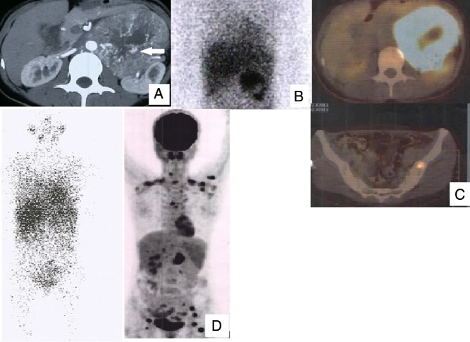
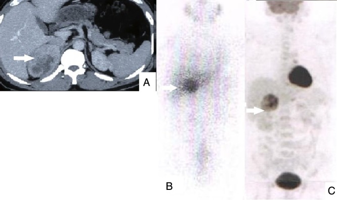
Results: Twenty patients were found to have metastases; 13 had metastases at diagnosis and seven during follow-up. Metastases were detected by radiology (CT-neck to pelvis) in 11/20 patients (5/13 synchronous and 6/7 metachronous), 131I-metaiodobenzylguanidine in five (2/12 synchronous and 3/6 metachronous) patients and 18F-flurodeoxyglucose PET/CT in 15 (12/12 synchronous and 3/3 metachronous) patients. Malignant tumours were significantly larger than benign tumours (8.3 ± 4.1 cm, range: 3-22 cm vs 5.7 ± 2.3 cm, range: 2-14 cm, P = 0.0001) and less frequently metanephrine secreting. On linear regression analysis, tumour size and lack of metanephrine secretion were the independent predictors of malignancy.
Conclusions: Patients with primary tumour size >5.7 cm and lack of metanephrine secretory status should be evaluated for possible malignancy not only at diagnosis but also in the postoperative period. As compared to CT and 131I-MIBG scan, 18F-flurodeoxyglucose PET/CT analyses are better (sensitivity: 100%) for the diagnosis of metastases in our study.
Keywords: 18F-flurodeoxyglucose PET/CT; clinical predictors; malignant paraganglioma; malignant pheochromocytoma.
© 2016 The authors.
Figures
References
-
- DeLellis RA, Lloyd RV, Heitz PU, Eng C. Pathology and Genetics of Tumours of Endocrine Organs. World Health Organization Classification of Tumours. Lyon, France: IARC Press, 2004.
-
- Lenders JW, Duh QY, Eisenhofer G, Gimenez-Roqueplo AP, Grebe SK, Murad MH, Naruse M, Pacak K, Young WF, Jr, Endocrine Society Pheochromocytoma and paraganglioma: an endocrine society clinical practice guideline. Journal of Clinical Endocrinology and Metabolism 2014. 99 1915–1942. ( 10.1210/jc.2014-1498) - DOI - PubMed
-
- Timmers HJ, Chen CC, Carrasquillo JA, Whatley M, Ling A, Havekes B, Eisenhofer G, Martiniova L, Adams KT, Pacak K. Comparison of 18F-fluoro-L-DOPA, 18F-fluoro-deoxyglucose, and 18F-fluorodopamine PET and 123I-MIBG scintigraphy in the localization of pheochromocytoma and paraganglioma. Journal of Clinical Endocrinology and Metabolism 2009. 12 4757–4767. ( 10.1210/jc.2009-1248) - DOI - PMC - PubMed
LinkOut - more resources
Full Text Sources
Other Literature Sources
Research Materials
