. 2016 Nov 16;36(46):11693-11703.
doi: 10.1523/JNEUROSCI.1081-16.2016.

Amyloid-β Peptide Nitrotyrosination Stabilizes Oligomers and Enhances NMDAR-Mediated Toxicity

Affiliations

Amyloid-β Peptide Nitrotyrosination Stabilizes Oligomers and Enhances NMDAR-Mediated Toxicity

Biuse Guivernau et al. J Neurosci. .

Abstract

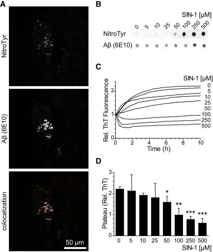
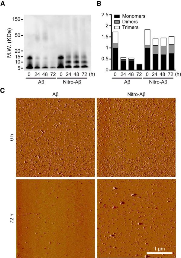
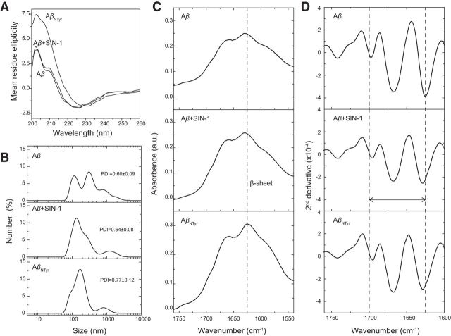
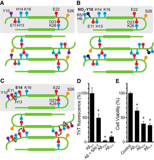
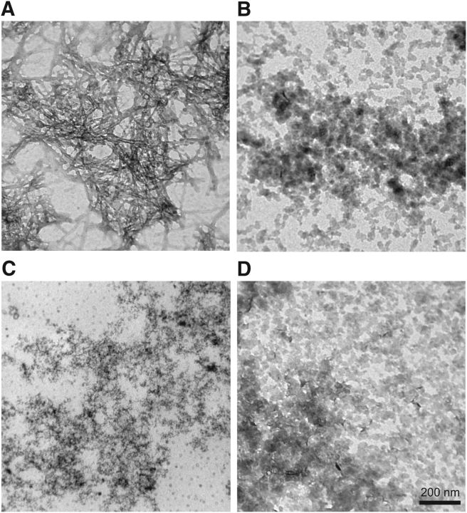
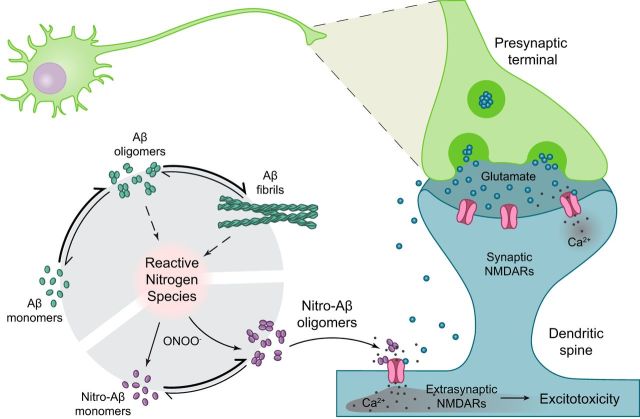
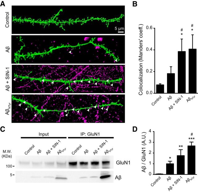
Alzheimer's disease (AD) is a neurodegenerative disorder characterized by the pathological aggregation of the amyloid-β peptide (Aβ). Monomeric soluble Aβ can switch from helicoidal to β-sheet conformation, promoting its assembly into oligomers and subsequently to amyloid fibrils. Oligomers are highly toxic to neurons and have been reported to induce synaptic transmission impairments. The progression from oligomers to fibrils forming senile plaques is currently considered a protective mechanism to avoid the presence of the highly toxic oligomers. Protein nitration is a frequent post-translational modification under AD nitrative stress conditions. Aβ can be nitrated at tyrosine 10 (Y10) by peroxynitrite. Based on our analysis of ThT binding, Western blot and electron and atomic force microscopy, we report that Aβ nitration stabilizes soluble, highly toxic oligomers and impairs the formation of fibrils. We propose a mechanism by which fibril elongation is interrupted upon Y10 nitration: Nitration disrupts fibril-forming folds by preventing H14-mediated bridging, as shown with an Aβ analog containing a single residue (H to E) replacement that mimics the behavior of nitrated Aβ related to fibril formation and neuronal toxicity. The pathophysiological role of our findings in AD was highlighted by the study of these nitrated oligomers on mouse hippocampal neurons, where an increased NMDAR-dependent toxicity of nitrated Aβ oligomers was observed. Our results show that Aβ nitrotyrosination is a post-translational modification that increases Aβ synaptotoxicity.

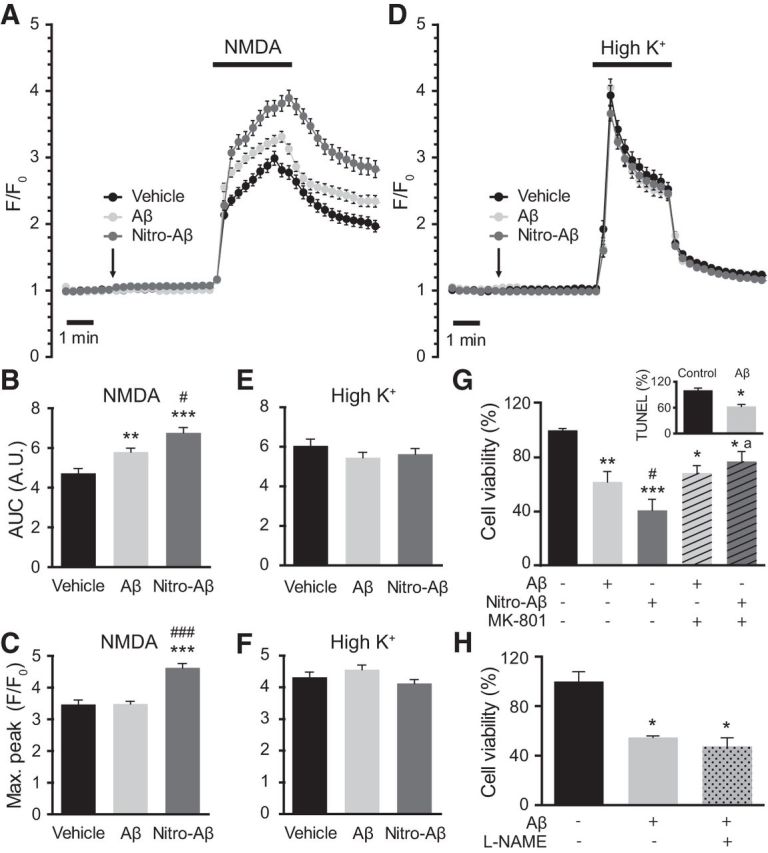
Significance statement: We report that nitration (i.e., the irreversible addition of a nitro group) of the Alzheimer-related peptide amyloid-β (Aβ) favors the stabilization of highly toxic oligomers and inhibits the formation of Aβ fibrils. The nitrated Aβ oligomers are more toxic to neurons due to increased cytosolic calcium levels throughout their action on NMDA receptors. Sustained elevated calcium levels trigger excitotoxicity, a characteristic event in Alzheimer's disease.

Keywords: Alzheimer; NMDA Rc; amyloid; nitrotyrosination; oligomers; peroxynitrite.


Figures

Figure 1.
Figure 1.
Aβ nitrotyrosination impairs fibril formation. A, Presence of nitrotyrosinated Aβ within amyloid plaques of the hippocampus from an AD patient. Colocalized pixels appear in white. B, Dot blot of synthetic Aβ42 treated with increasing concentrations of SIN-1 probed with an anti-nitrotyrosine and 6E10 anti-Aβ antibodies. C, ThT aggregation assay with synthetic Aβ42 treated with increasing concentrations of SIN-1. Graph represents fluorescence values relative to time 0. Mean of four independent experiments. D, Plateau phase ThT values of the data shown in C. Mean ± SEM of 3–6 independent experiments. *p < 0.05 (Student's t test). **p < 0.01 (Student's t test). ***p < 0.001 (Student's t test).
Figure 2.
Figure 2.
Aβ nitrotyrosination stabilizes low molecular weight oligomers. A, Representative Western blot of Aβ42 control and Aβ42 treated with 100 μm SIN-1 (nitro-Aβ) at different times of aggregation probed with 6E10 anti-Aβ antibody. B, Relative amounts of oligomeric species obtained by Western blot. Mean of three independent experiments. C, Oligomer visualization using AFM of Aβ42 and nitro-Aβ at 0 and 72 h of aggregation on a mica surface.
Figure 3.
Figure 3.
Aβ nitrotyrosination generates low molecular weight oligomers with β-sheet structure. A, DLS. Size distribution measured by DLS as a function of number of particles. All the Aβ oligomers tested show a high and very similar polydispersity index (>0.6). B, CD spectroscopy. CD measurements of Aβ42 and nitrotyrosinated samples showing a minimum at ∼225 nm and a maximum at ∼200 nm, indicative of β-sheet structure. C, ATR-FTIR spectroscopy of the amide I region for different Aβ42 oligomers in a thin hydrated film. All spectra show the characteristic β-sheet secondary structure peak at ∼1630 cm, following the band assignments of Byler and Susi (1986). D, Second derivative of the ATR-FTIR is used here to illustrate better the different secondary structures due to band narrowing (Wahlstrom et al., 2008).
Figure 4.
Figure 4.
Disruption of Y10–S26 interaction impairs fibril formation. A, Model of interprotofibrillar Aβ42. The interaction between Y10 and S26 stabilizes the fibril and contributes to its progression. B, Upon the nitration of Y10, because the nitro group is negatively charged, it is arrested by the side-chains of either R5 or H6, impeding its interaction with S26 and destabilizing the fibril. C, Our mutant H14 to E14 also would trigger a rearrangement of the side-chains of the N terminus, disrupting the interaction between Y10 and S26 as it happens upon nitration. D, ThT fluorescence after 72 h of aggregation of Aβ42, Aβ42 treated with 100 μm SIN-1, synthetically nitrated Aβ42 (AβNTyr), and AβE14 mutant. Values are expressed as a percentage compared with Aβ42. Mean ± SEM of four independent experiments. *p < 0.001 (one-way ANOVA using Dunnett's post test). E, Cell viability assay performed by the MTT method. Hippocampal neurons were treated with 10 μm of AβWT, AβNTyr, and the mutant AβE14 for 24 h. Values are expressed as the percentage of the control. Mean ± SEM of 13–16 independent experiments. *p < 0.001 (one-way ANOVA using Dunnett's post test).
Figure 5.
Figure 5.
Nitrotyrosination stabilizes oligomers. Transmission electron microscopy of the following: A, AβWT; B, AβWT + 100 μm SIN-1; C, AβNTyr; D, AβE14 negatively stained with uranyl acetate after 72 h of aggregation.
Figure 6.
Figure 6.
Nitro-Aβ oligomers affect calcium homeostasis and exert NMDA-dependent toxicity. Cells were treated for 5 min with 10 μm Aβ or nitro-Aβ and stimulated with bath application of either 100 μm NMDA (A) or 52.5 μm KCl (D). Mean ± SEM of 82–87 cells for NMDA and 98–104 for KCl. The area under the curve (AUC) (B, C) and the maximum response peak (E, F) were calculated for each cell response. G, Cell viability assay performed by the MTT method. Hippocampal neurons were treated with 10 μm Aβ and nitro-Aβ with or without 10 μm MK-801 for 5 min, and cell viability was assessed after 24 h. Untreated neurons were taken as 100% of viability. Inset, A control of Aβ toxicity studied by TUNEL assay. Mean ± SEM of three or four independent experiments. H, Cell viability assayed with the MTT method in hippocampal neurons treated with Aβ in the presence or absence of the NOS inhibitor l-NAME for 5 min. Cell viability was assessed after 24 h. Mean ± SEM of three independent experiments. *p < 0.05, compared with control (one-way ANOVA using Newman–Keuls post test). **p < 0.01, compared with control (one-way ANOVA using Newman–Keuls post test). ***p < 0.001, compared with control (one-way ANOVA using Newman–Keuls post test). #p < 0.05, compared with Aβ (one-way ANOVA using Newman–Keuls post test). ###p < 0.001, compared with Aβ (one-way ANOVA using Newman–Keuls post test). ap < 0.05 compared with nitro-Aβ by (one-way ANOVA using Newman–Keuls post test). Student's t test for the TUNEL study.
Figure 7.
Figure 7.
Aβ oligomers and fibrils trigger the formation of nitrogen reactive species. In this context, peroxynitrite reacts with proteins, affecting their function. One of these modifications is protein nitrotyrosination, which is highly promoted in AD brains. Aβ has a potential nitration site in Y10 and is nitrated in the disease. Nitrotyrosination of Aβ impairs amyloid fibril formation and stabilizes soluble, highly toxic oligomers. Glutamatergic synapses are the most abundant within the hippocampus and the first to be affected in AD. Glutamate is released from the presynaptic neuron into the synaptic cleft; and when it is released in a large quantity, it spills to the perisynaptic space, partially activating extrasynaptic NMDARs. When glutamatergic stimulation is particularly high, and especially when extrasynaptic NMDARs are activated, the excitotoxic apoptotic pathway is triggered. Nitro-Aβ oligomers make the neurons more susceptible to excitotoxic insults.
Figure 8.
Figure 8.
Increased nitro-Aβ oligomers binding to the synapses. Cells were treated for 5 min with 10 μm Aβ or nitro-Aβ (SIN-1-treated Aβ and AβNTyr). A, Three-dimensional rendering of dendritic spines. Green represents the neuron volume (GFP). Magenta represents Aβ and nitro-Aβ oligomers. Nitro-Aβ oligomers contact the dendritic spines (arrows). B, Colocalization analysis of A. M1 Manders' coefficient refers to the fraction of Aβ overlapping spines. Mean ± SEM of 5–8 neurons from three different experiments. C, Representative Western blot of Aβ and nitro-Aβ coimmunoprecipitated with the NMDAR subunit GluN1 from hippocampal neurons. D, Quantification of C. Mean ± SEM of 5–7 independent experiments. *p < 0.05, compared with control (one-way ANOVA using Newman–Keuls post test). **p < 0.01, compared with control (one-way ANOVA using Newman–Keuls post test). ***p < 0.001, compared with control (one-way ANOVA using Newman–Keuls post test). #p < 0.01, compared with Aβ (one-way ANOVA using Newman–Keuls post test).

References

    1. Arimon M, Díez-Pérez I, Kogan MJ, Durany N, Giralt E, Sanz F, Fernàndez-Busquets X. Fine structure study of Aβ1–42 fibrillogenesis with atomic force microscopy. FASEB J. 2005;19:1344–1346. doi: 10.1096/fj.04-3137fje. - DOI - PubMed
    1. Beckman JS, Koppenol WH. Nitric oxide, superoxide, and peroxynitrite: the good, the bad, and ugly. Am J Physiol. 1996;271:C1424–C1437. - PubMed
    1. Berman HM, Westbrook J, Feng Z, Gilliland G, Bhat TN, Weissig H, Shindyalov IN, Bourne PE. The Protein Data Bank. Nucleic Acids Res. 2000;28:235–242. doi: 10.1093/nar/28.1.235. - DOI - PMC - PubMed
    1. Berridge MJ. Calcium hypothesis of Alzheimer's disease. Pflugers Arch. 2010;459:441–449. doi: 10.1007/s00424-009-0736-1. - DOI - PubMed
    1. Bitan G, Teplow DB. Preparation of aggregate-free, low molecular weight amyloid-beta for assembly and toxicity assays. Methods Mol Biol. 2005;299:3–9. - PubMed

Publication types

MeSH terms

LinkOut - more resources