. 2016 Dec 30;291(53):27147-27159.
doi: 10.1074/jbc.M116.754887. Epub 2016 Nov 16.

Targeted Elimination of G Proteins and Arrestins Defines Their Specific Contributions to Both Intensity and Duration of G Protein-coupled Receptor Signaling

Affiliations

Targeted Elimination of G Proteins and Arrestins Defines Their Specific Contributions to Both Intensity and Duration of G Protein-coupled Receptor Signaling

Elisa Alvarez-Curto et al. J Biol Chem. .

Abstract

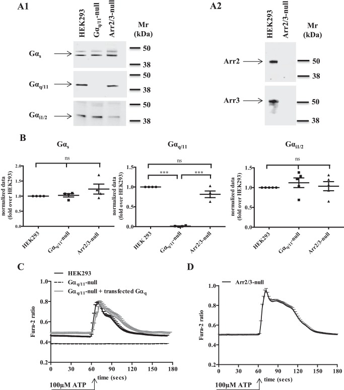
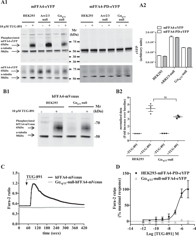
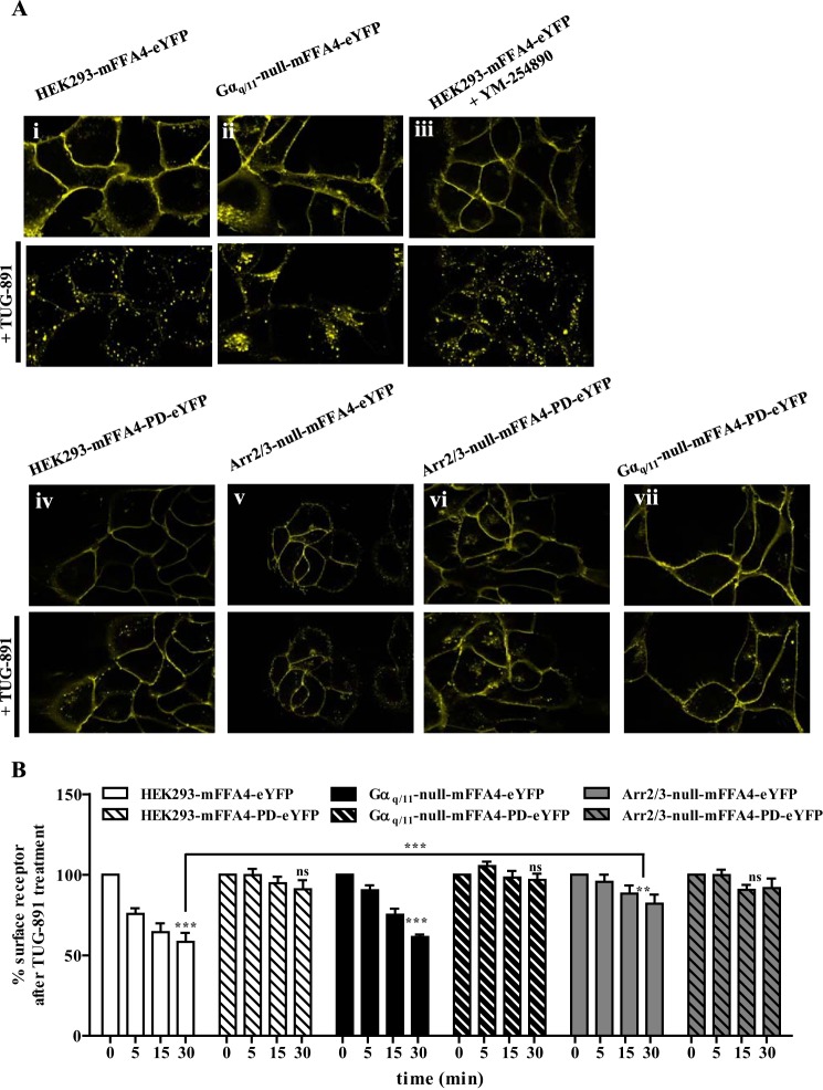
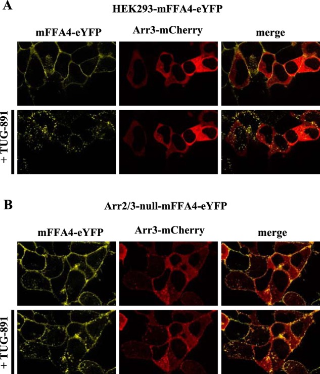
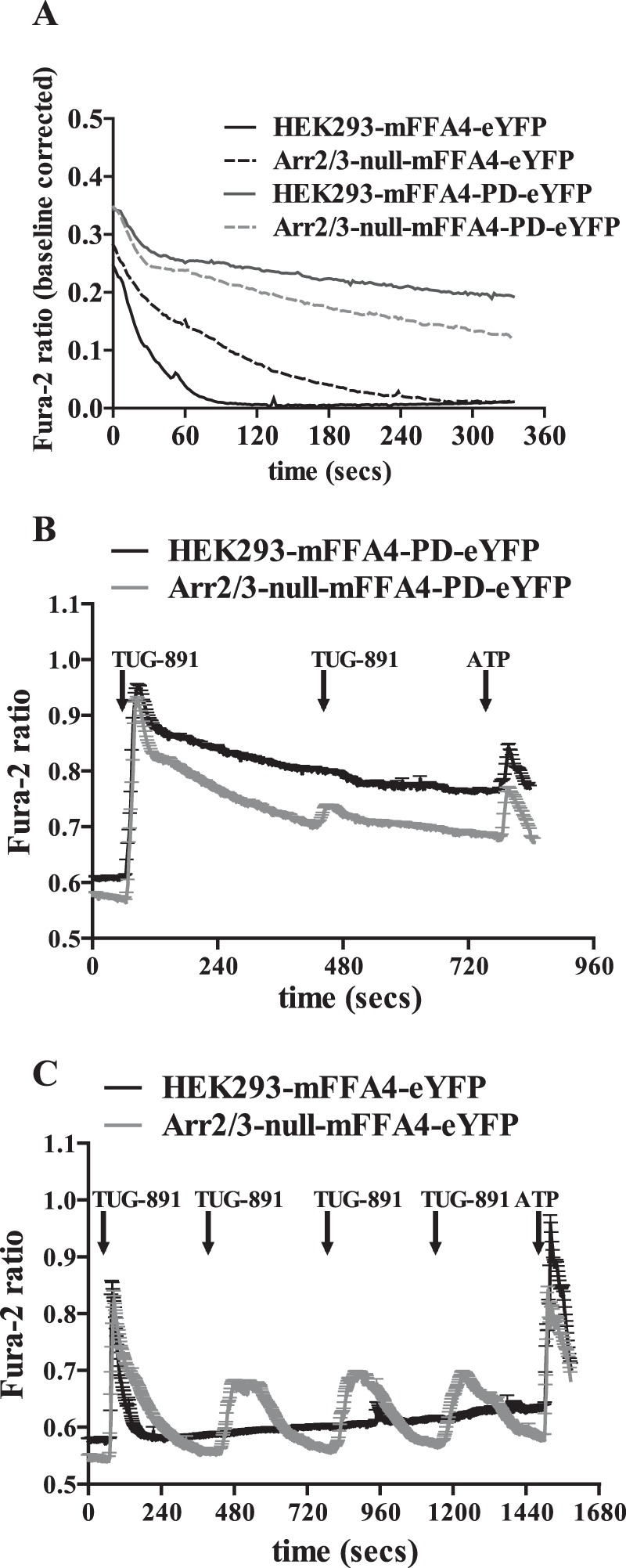
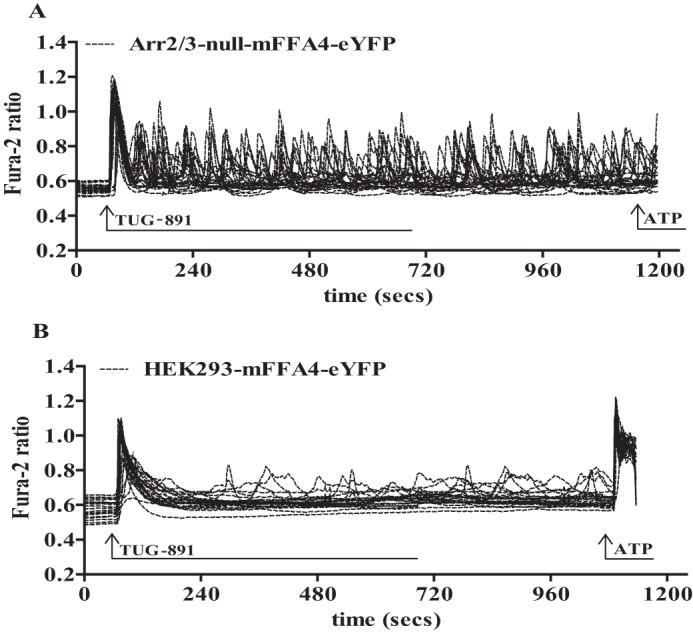
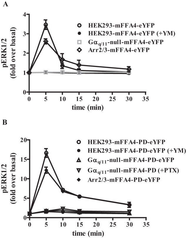
G protein-coupled receptors (GPCRs) can initiate intracellular signaling cascades by coupling to an array of heterotrimeric G proteins and arrestin adaptor proteins. Understanding the contribution of each of these coupling options to GPCR signaling has been hampered by a paucity of tools to selectively perturb receptor function. Here we employ CRISPR/Cas9 genome editing to eliminate selected G proteins (Gαq and Gα11) or arrestin2 and arrestin3 from HEK293 cells together with the elimination of receptor phosphorylation sites to define the relative contribution of G proteins, arrestins, and receptor phosphorylation to the signaling outcomes of the free fatty acid receptor 4 (FFA4). A lack of FFA4-mediated elevation of intracellular Ca2+ in Gαq/Gα11-null cells and agonist-mediated receptor internalization in arrestin2/3-null cells confirmed previously reported canonical signaling features of this receptor, thereby validating the genome-edited HEK293 cells. FFA4-mediated ERK1/2 activation was totally dependent on Gq/11 but intriguingly was substantially enhanced for FFA4 receptors lacking sites of regulated phosphorylation. This was not due to a simple lack of desensitization of Gq/11 signaling because the Gq/11-dependent calcium response was desensitized by both receptor phosphorylation and arrestin-dependent mechanisms, whereas a substantially enhanced ERK1/2 response was only observed for receptors lacking phosphorylation sites and not in arrestin2/3-null cells. In conclusion, we validate CRISPR/Cas9 engineered HEK293 cells lacking Gq/11 or arrestin2/3 as systems for GPCR signaling research and employ these cells to reveal a previously unappreciated interplay of signaling pathways where receptor phosphorylation can impact on ERK1/2 signaling through a mechanism that is likely independent of arrestins.

Keywords: CRISPR/Cas; G protein; G protein-coupled receptor (GPCR); arrestin; calcium intracellular release; extracellular-signal-regulated kinase (ERK); fatty acid.


Figures

FIGURE 1.
FIGURE 1.
Characteristics of Gαq/Gα11 and arrestin2/3-null HEK293 cells. Immunoblotting studies were performed on (A1) membrane preparations of parental HEK293 cells and clones from genome-edited cells that eliminate expression of Gαq + Gα11 or arrestin2 + arrestin3. Such samples were probed to detect expression of the long and short isoforms of Gαs, Gαq + Gα11, and Gαi1/2. A2, cytosolic preparations from wild type HEK293 cells and arrestin2/3-null cells were immunoblotted to detect expression of arrestin2 (upper panel) or arrestin3 (lower panel). Representative immunoblots are displayed in both A1 and A2. B, a series of such immunoblots was quantified by densitometric analysis. Individual results of immunoblots taken from at least four separate cell preparations are shown relative to levels detected in parental HEK293 cells. Values from individual experiments are shown as the group means ± S.E. ns = not significantly different; ***, different at p < 0.001. C, single cell Ca2+ imaging was performed on parental HEK293 cells and on both Gαq/Gα11-null cells and such cells into which Gαq had been re-introduced. ATP (100 μm) was added at the indicated time. D, studies akin to those in C were performed in arrestin2/3-null cells. ATP (100 μm) was added at the indicated time.
FIGURE 2.
FIGURE 2.
Wild type but not PD forms of FFA4 became phosphorylated on amino acids Thr347 and Ser350 upon the addition of agonist. Parental, Gαq/Gα11-null and arrestin2/3-null HEK293 cells stably expressing either mFFA4-eYFP (left-hand side) or mFFA4-PD-eYFP (right-hand side) were stimulated with vehicle or TUG-891 (10 μm, 5 min). Subsequently, cell lysates were resolved by SDS-PAGE and immunoblotted with a mixture of (mouse FFA4 specific) anti-phospho Thr347-Ser350 (22) and anti-α-tubulin antibodies, detecting the specific 65-kDa and 50-kDa polypeptides corresponding to the receptor and α-tubulin bands respectively (A1, upper panels). Parallel immunoblots were probed with a mixture of anti-GFP (recognizes also eYFP) and anti-α-tubulin antibodies (A1, lower panels). The higher level of TUG-891 mediated mFFA4-eYFP phosphorylation in the arrestin2/3-null HEK293 cells reflects the higher level expression of mFFA4 receptor protein in this clone (A1, lower panel). This was confirmed by assessment of eYFP fluorescence in cells in each clone (A2) and was also the case for mFFA4-PD-eYFP expressed in the arrestin2/3-null background (A2). B, parental and Gαq/Gα11-null HEK293 cells expressing hFFA4-mVenus were also stimulated with vehicle or TUG-891 (10 μm, 5 min). Phosphorylation of the receptor was detected with a human FFA4 specific anti-phospho Thr347-Ser350 antiserum (21), and the co-addition of anti-α-tubulin antibodies provided loading controls (B1). B2, quantification of the extent of agonist-induced phosphorylation was produced by densitometric analysis of three experiments, each performed on samples derived from separate cell treatments: ns = not significantly different. C, HEK293 and Gαq/Gα11-null cells expressing hFFA4- mVenus were challenged with 3 μm TUG-891 in single-cell imaging Ca2+ experiments. D, calcium mobilization experiments were performed on populations of parental HEK293 and Gαq/Gα11-null cells expressing the mFFA4-eYFP construct. These demonstrated that mFFA4-mediated Ca2+ release was lost in the absence of Gαq/Gα11.
FIGURE 3.
FIGURE 3.
FFA4-mediated inositol phosphate generation is lacking in Gαq/Gα11-null HEK293 cells. Parental and Gαq/Gα11-null HEK293 cells stably expressing human (A) or mouse (B) FFA4 were used to measure basal and TUG-891-mediated levels of inositol monophosphates.
FIGURE 4.
FIGURE 4.
Effects of elimination of Gαq + Gα11 or arrestin2 + arrestin3 on agonist-induced internalization of FFA4. A, parental (i, iii, and iv), Gαq/Gα11-null (ii and vii), or arrestin2/3-null (v and vi) HEK293 cells stably expressing mFFA4-eYFP (i–iii and v) or mFFA4-PD-eYFP (iv and vi–vii) were exposed to TUG-891 (10 μm) (upper panels, t = 0; lower panels, t = 30 min). In iii, the Gαq/Gα11 inhibitor YM-254890 (100 nm) was added 30 min before the addition of TUG-891. Representative images of experiments performed at least three times are shown. B, cell surface ELISA experiments were performed to detect the FLAG-epitope tag introduced into the extracellular N-terminal domain on each form of FFA4 at various times after the addition of TUG-891. 100% represents receptor at the cell surface before the addition of TUG-891. Agonist-induced internalization was observed at the 30-min time point (**, p < 0.01; ***, p < 0.001). The extent of internalization of mFFA4-eYFP was greater (p < 0.001) in parental than in arrestin2/3-null HEK293 cells. ns = not significantly different from t = 0.
FIGURE 5.
FIGURE 5.
Reintroduction of arrestin3 into arrestin2/3-null HEK293 cells restored agonist-mediated internalization of FFA4. Parental (A) or arrestin2/3-null (B) HEK293 cells stably expressing mFFA4-eYFP were transfected transiently to express arrestin3-mCherry and treated with TUG-891 (10 μm) (upper panels: t = 0; lower panels: t = 30 min). Representative images of the location of mFFA4-eYFP (yellow channel) and arrestin3-mCherry (red channel) are shown. In the right-hand panels these images are merged to provide color overlap.
FIGURE 6.
FIGURE 6.
Lack of arrestin2/3 and resistance to agonist-induced receptor phosphorylation extended the kinetics of Ca2+ elevation in HEK293 cells. Intracellular Ca2+ levels and how these were affected over time by TUG-891-induced activation of FFA4 were assessed in a series of single cell Ca2+ imaging studies performed in both parental and arrestin2/3-null HEK293 cells expressing either wild type or mFFA4-PD as indicated. A, representative traces in parental HEK293 cells expressing wild type mFFA4 (HEK293-mFFA4-eYFP), parental cells expressing mFFA4-PD (HEK293-mFFA4-PD-eYFP), arrestin2/3-null cells expressing wild type mFFA4 (Arr2/3-null-mFFA4-eYFP), and arrestin2/3-null cells expressing mFFA4-PD (Arr2/3-null-mFFA4-PD-eYFP). B, shows data from studies on parental and arrestin2/3-null HEK293 cells expressing mFFA4-PD in which after the addition and washout of two treatments with TUG-891 ATP was added at 760 s to activate endogenously expressed P2Y receptors. C, a single exposure to TUG-891 was sufficient to fully desensitize mFFA4 in parental HEK293 cells but not in arrestin2/3-null cells. Time(s) of addition of TUG-891 is shown as are subsequent additions of ATP.
FIGURE 7.
FIGURE 7.
Repetitive Ca2+ spiking in arrestin2/3-null HEK293 cells reflected a lack of FFA4 desensitization. Arrestin2/3-null (A) and parental (B) HEK293 cells expressing mFFA4-eYFP were exposed to a single, maintained concentration of TUG-891 (3 μm) at the indicated point. Studies across 25 cells analyzed in single-cell Ca2+ imaging studies were overlaid. At the indicated time ATP (100 μm) was added.
FIGURE 8.
FIGURE 8.
FFA4-PD generated enhanced activation of ERK1/2 MAP kinase phosphorylation largely via Gαq/Gα11-mediated signals. ERK1/2 phosphorylation studies were performed using wild type (A) or mFFA4-PD-eYFP (B) stably expressed in each of parental, Gαq/Gα11-null, and arrestin2/3-null HEK293 cells. In certain experiments the Gαq + Gα11 inhibitor YM-254890 (YM, 100 nm) was added 30 min before the agonist TUG-891. In other studies cells were pretreated with pertussis toxin (PTX) overnight before the addition of TUG-891. ERK1/2 phosphorylation was measured at the indicated time points and is presented as -fold over basal. NOTE: the different scale of the y axis in A and B.

Similar articles

Cited by

References

    1. Lefkowitz RJ. (2013) A brief history of G-protein-coupled receptors (Nobel Lecture). Angew. Chem. Int. Ed. Engl. 52, 6366–6378 - PubMed
    1. Luttrell L. M., and Gesty-Palmer D. (2010) Beyond desensitization: physiological relevance of arrestin-dependent signaling. Pharmacol. Rev. 62, 305–330 - PMC - PubMed
    1. Smith J. S., and Rajagopal S. (2016) The β-arrestins: multifunctional regulators of G protein-coupled receptors. J. Biol. Chem. 291, 8969–8977 - PMC - PubMed
    1. Gurevich V. V., and Gurevich E. V. (2015) Arrestins: critical players in trafficking of many GPCRs. Prog. Mol. Biol. Transl. Sci. 132, 1–14 - PMC - PubMed
    1. Schmid C. L., and Bohn L. M. (2009) Physiological and pharmacological implications of β-arrestin regulation. Pharmacol. Ther. 121, 285–293 - PMC - PubMed

Publication types

MeSH terms

Substances