. 2017 Jan 27;292(4):1490-1509.
doi: 10.1074/jbc.M116.758185. Epub 2016 Nov 16.

Phosphorylation of Human Retinoid X Receptor α at Serine 260 Impairs Its Subcellular Localization, Receptor Interaction, Nuclear Mobility, and 1α,25-Dihydroxyvitamin D3-dependent DNA Binding in Ras-transformed Keratinocytes

Affiliations

Phosphorylation of Human Retinoid X Receptor α at Serine 260 Impairs Its Subcellular Localization, Receptor Interaction, Nuclear Mobility, and 1α,25-Dihydroxyvitamin D3-dependent DNA Binding in Ras-transformed Keratinocytes

Sylvester Jusu et al. J Biol Chem. .

Abstract

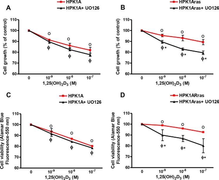
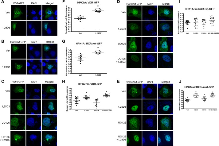
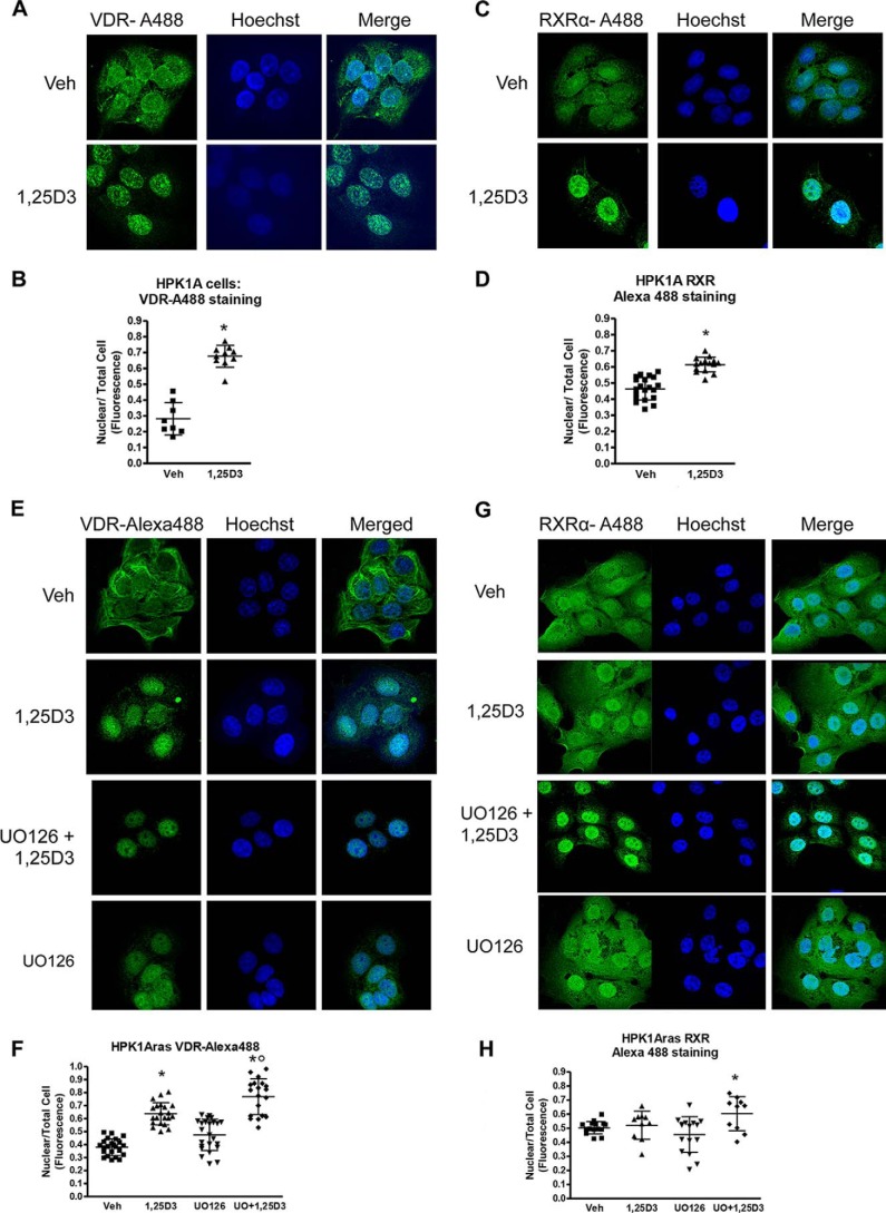
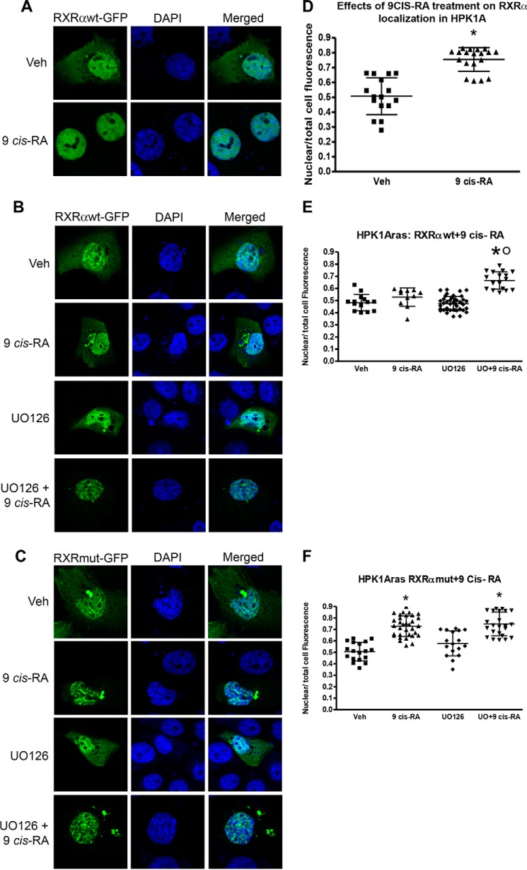
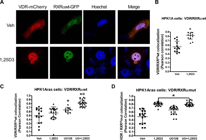
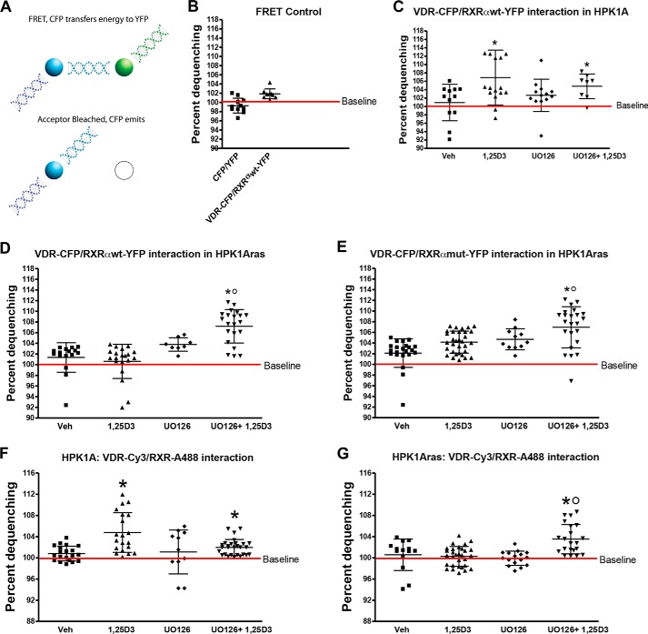
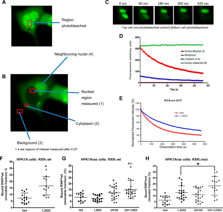
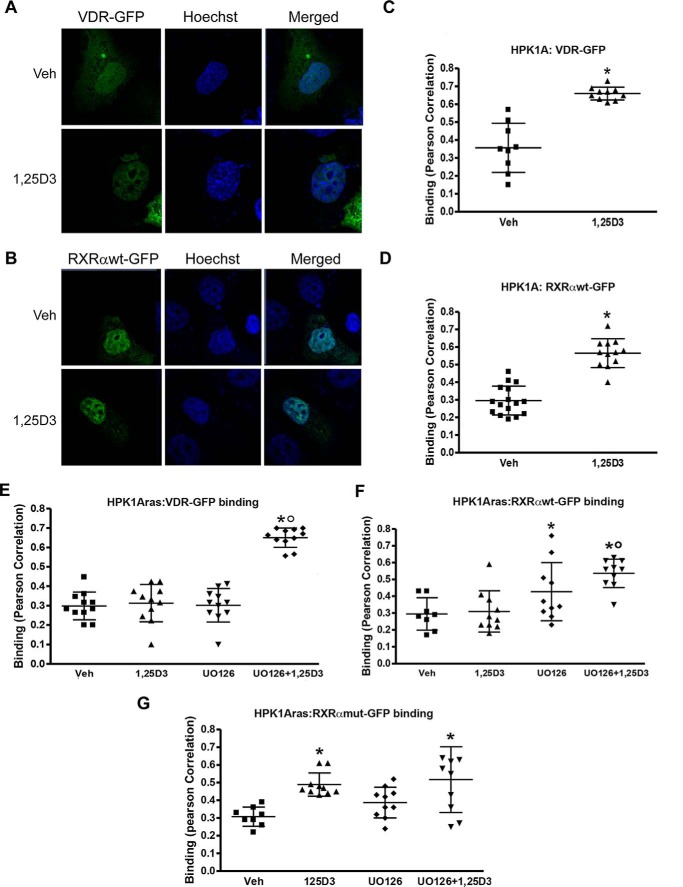
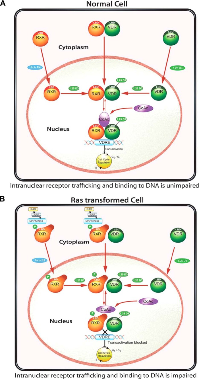
Human retinoid X receptor α (hRXRα) plays a critical role in DNA binding and transcriptional activity through heterodimeric association with several members of the nuclear receptor superfamily, including the human vitamin D receptor (hVDR). We previously showed that hRXRα phosphorylation at serine 260 through the Ras-Raf-MAPK ERK1/2 activation is responsible for resistance to the growth inhibitory effects of 1α,25-dihydroxyvitamin D3 (1α,25(OH)2D3), the biologically active metabolite of vitamin D3 To further investigate the mechanism of this resistance, we studied intranuclear dynamics of hVDR and hRXRα-tagged constructs in living cells together with endogenous and tagged protein in fixed cells. We find that hVDR-, hRXRα-, and hVDR-hRXRα complex accumulate in the nucleus in 1α,25(OH)2D3-treated HPK1A cells but to a lesser extent in HPK1ARas-treated cells. Also, by using fluorescence resonance energy transfer (FRET), we demonstrate increased interaction of the hVDR-hRXRα complex in 1α,25(OH)2D3-treated HPK1A but not HPK1ARas cells. In HPK1ARas cells, 1α,25(OH)2D3-induced nuclear localization and interaction of hRXRα are restored when cells are treated with the MEK1/2 inhibitor UO126 or following transfection of the non-phosphorylatable hRXRα Ala-260 mutant. Finally, we demonstrate using fluorescence loss in photobleaching and quantitative co-localization with chromatin that RXR immobilization and co-localization with chromatin are significantly increased in 1α,25(OH)2D3-treated HPK1ARas cells transfected with the non-phosphorylatable hRXRα Ala-260 mutant. This suggests that hRXRα phosphorylation significantly disrupts its nuclear localization, interaction with VDR, intra-nuclear trafficking, and binding to chromatin of the hVDR-hRXR complex.

Keywords: fluorescence loss in photobleaching (FLIP); fluorescence resonance energy transfer (FRET); mitogen-activated protein kinase (MAPK); phosphorylation; retinoid; retinoid X receptor; vitamin D; vitamin D receptor.


Figures

FIGURE 1.
FIGURE 1.
Effects of 1,25(OH)2D3 and UO126 on cell growth (A and B) and viability (C and D). HPK1A (A) and HPK1ARas (B) cells were grown in DMEM containing 10% FBS. At 40% confluency the medium was changed, and cells were treated with increasing concentrations of 1,25(OH)2D3 in the absence or presence of UO126 (10−6 m). After 72 h, cells were collected, and cells were counted using a Coulter counter. HPK1A (C) and HPK1ARas (D) cells were seeded at a density of 5 × 10³ cells/well. Twenty four hours later, cells were treated with vehicle, 1,25(OH)2D3 alone, or a combination of UO126 and 1,25(OH)2D3 for 72 h. Alamar blue metabolism was used to assess cell viability. Values are expressed as a percentage of vehicle-treated cells. All values represent means ± S.D. (bars) of at least three separate experiments conducted in triplicate. In the cell viability experiment (C and D) HPK1A and HPK1ARas showed very little variation in the absence of UO126, and error bars are less than the width of the plotted points. Closed circles indicate a significant growth inhibition in either HPK1A + UO126 or HPK1ARas + UO126 cells as compared with vehicle-treated cells. Open circles indicate a significant growth inhibition in either HPK1A or HPK1ARas cells as compared with vehicle-treated cells. Asterisks indicate a significant difference between vehicle-treated and drug-treated cells. A p value of p < 0.05 was considered significant.
FIGURE 2.
FIGURE 2.
Effects of 1,25(OH)2D3 with or without UO126 treatment on nuclear localization of VDR and RXRα in HPK1A and HPK1ARas cells. HPK1A cells were transfected with VDR-GFP (A) or RXRα-WT-GFP (B) followed by treatment with either vehicle (veh) or 1,25(OH)2D3 (1,25D3). Similarly, HPK1ARas cells were transfected with either VDR-GFP (C), RXRα-WT-GFP (D), or RXRα-mut-GFP (E) followed by treatment with either vehicle, 1,25(OH)2D3, UO126 alone, or a combination of UO126 and 1,25(OH)2D3. Nuclear localization was assessed as under “Experimental Procedures.” Scatter plots show the quantitation of fluorescence of nuclear receptors normalized to total cell fluorescence (F–J). Values represent mean ± S.D. of at least 10 different cells. Asterisks indicate a significant difference in nuclear localization between 1,25(OH)2D3 treatment alone compared with vehicle-treated control. Open circles indicate a significant difference in nuclear localization of receptors in 1,25(OH)2D3-treated cells alone compared with combined UO126 and 1,25(OH)2D3 treatment. A p value of p < 0.05 was considered significant.
FIGURE 3.
FIGURE 3.
Effects of 1,25(OH)2D3 with or without UO126 treatment on nuclear localization of endogenous VDR and RXRα in HPK1A and HPK1ARas cells. HPK1A cells were grown and treated with either vehicle (Veh) or 1,25(OH)2D3 (1,25D3). The cells were also fixed and stained with VDR-Alexa-488 (A) or RXRα-Alexa-488 (C) antibodies to detect VDR or RXRα. Alexa-488 (C) followed by treatment with either vehicle (veh) or 1,25(OH)2D3. Hoechst dye was used as a DNA marker as discussed under “Experimental Procedures.” Similarly, HPK1ARas cells were grown and treated with either vehicle or 1,25(OH)2D3. The cells were then fixed and stained with VDR-Alexa-488 (E) or RXRα-Alexa-488 (G) antibodies to detect VDR or RXRα. Hoechst dye was also used as a DNA marker as discussed under “Experimental Procedures.” Nuclear localization was assessed as under “Experimental Procedures.” Scatter plots show the quantitation of fluorescence of nuclear receptors normalized to total cell fluorescence (B, D, F, and H). Values represent mean ± S.D. of at least 10 different cells. Asterisks indicate a significant difference in nuclear localization between 1,25(OH)2D3 treatment alone compared with vehicle-treated control. Open circles indicate a significant difference in nuclear localization of receptors in 1,25(OH)2D3-treated cells alone compared with combined UO126 and 1,25(OH)2D3 treatment. A p value of p < 0.05 was considered significant.
FIGURE 4.
FIGURE 4.
Effects of 9-cis-retinoic acid on RXRα subcellular localization in HPK1A and HPK1ARas cells. HPK1A cells were transfected with RXRα-WT-GFP (A and D) followed by treatment with either vehicle (Veh) or 9-cis-RA for 4 h. Similarly, HPK1ARas cells were transfected with either RXRα-WT-GFP (B and E) or RXRα-mut-GFP (C and F) followed by treatment with either vehicle, 9-cis-RA, UO126 alone, or a combination of UO126 and 9-cis-RA. Nuclear localization was assessed as under “Experimental Procedures.” Scatter plots show the quantitation of fluorescence of nuclear receptors normalized to total cell fluorescence (D–F). Values represent mean ± S.D. of at least 10 different cells. Asterisks indicate a significant difference in nuclear localization between 9-cis-RA treatment alone compared with vehicle-treated control. Open circles indicate a significant difference in nuclear localization of receptors in 9-cis-RA-treated cells alone compared with combined UO126 and 9-cis-RA treatment. A p value of p < 0.05 was considered significant.
FIGURE 5.
FIGURE 5.
Effects of RXRα phosphorylation on VDR/hRXRα co-trafficking in HPK1A and HPK1ARas cells. A, cells were co-transfected with either VDR-mCherry/RXRαWT-GFP or VDR-mCherry/RXRαmut-GFP. Following transfection, cells were treated with either vehicle, 1,25(OH)2D3 (1,25D3), UO126 alone, or a combination of UO126 and 1,25(OH)2D3. Scatter plots show co-localization measurement using Pearson correlation coefficient of HPK1A cells co-transfected with VDR-mCherry/RXRαWT-GFP (B) or HPK1ARas cells co-transfected with either VDR-mCherry/RXRαWT-GFP (C) or with or VDR-mCherry/RXRαmut-GFP (D). Values are mean ± S.D. of at least 10 cells per treatment condition. Asterisks indicate a significant difference in interaction between 1,25(OH)2D3 treatment alone compared with vehicle-treated control. Open circles indicate a significant difference in interaction between 1,25(OH)2D3-treated cells alone compared with combined UO126 and 1,25(OH)2D3 treatment. A p value of p < 0.05 was considered significant.
FIGURE 6.
FIGURE 6.
Effects of RXRα phosphorylation on VDR and RXR interaction in HPK1A and HPK1ARas cells. A, FRET was measured by acceptor photobleaching as described under “Experimental Procedures.” Cells were co-transfected with either VDR-CFP/RXRαWT-YFP or VDR-CFP/RXRαmut-YFP. Following transfection, cells were treated with either vehicle (Veh), 1,25(OH)2D3 (1,25D3), UO126 alone, or a combination of UO126 and 1,25(OH)2D3 (C–E). B, HPK1A cells were co-transfected with either CFP/YFP (negative control) or VDR-CFP/RXRαWT-YFP plasmids. Similarly, both HPK1A (F) and HPK1ARas (G) cells were grown and treated with either vehicle, 1,25(OH)2D3, UO126 alone, or a combination of UO126 and 1,25(OH)2D3. The cells were then fixed and co-stained with VDR-Cy3 and RXRα-Alexa-488 antibodies to detect both endogenous VDR and RXRα. FRET was measured by acceptor photobleaching as described under “Experimental Procedures.” Scatter plots show FRET measurement of HPK1A cells co-transfected with VDR-CFP/RXRαWT-YFP (C) or co-stained with VDR-cy3 and RXRα-Alexa-488 antibodies (F). Similarly, in HPK1ARas cells scatter plots show cells co-transfected with either VDR-CFP/RXRαWT-YFP (D), VDR-CFP/RXRαmut-YFP (E), or co-stained with VDR-cy3 and RXRα-Alexa-488 antibodies (G). Values are mean percentage dequenching ± S.D. of at least 10 cells per treatment condition. FRET baseline was set at 100%. Asterisks indicate a significant difference in interaction between 1,25(OH)2D3 treatment alone compared with vehicle-treated control. Open circles indicate a significant difference in interaction between 1,25(OH)2D3-treated cells alone compared with combined UO126 and 1,25(OH)2D3 treatment. A p value of p < 0.05 was considered significant.
FIGURE 7.
FIGURE 7.
Effects of RXRα phosphorylation on nucleocytoplasmic kinetics of RXRα in HPK1A and HPK1ARas cells. FLIP methodology was used to assess nucleocytoplasmic kinetics. HPK1A cells were transfected with RXRαWT-GFP (A–E). Following transfection, live cells were treated with either vehicle (Veh) or 1,25(OH)2D3 (1,25D3) and nucleocytoplasmic trafficking was measured using confocal microscopy (see “Experimental Procedures”). Nuclear area was selected and photobleached (A), and other regions of interests measured but not photobleached (B, 1–3). C, time course showing (i) an unbleached nucleus of a neighboring cell and (ii) a cell with a bleached nucleus. The normalized fluorescent intensity of the unbleached and bleached nuclei above is shown in D. The dissociation curve of vehicle and 1,25(OH)2D3-treated cells are shown in E. The bound fractions of HPK1A cells transfected with RXRαWT-GFP (F) or HPK1ARas cells transfected with either RXRαWT-GFP (G) or RXRαmut-GFP (H) are similarly shown. Values are mean ± S.D. of at least 10 cells per treatment. Asterisks indicate a significant difference in bound fraction between vehicle (Veh) and 1,25(OH)2D3 (1,25D3) cells. A p value < 0.05 was considered significant.
FIGURE 8.
FIGURE 8.
Determination of receptor/DNA interaction. HPK1A cells transfected with VDR-GFP (green) and treated with either vehicle or 1,25(OH)2D3 (1,25D3) post-transfection were stained with Hoechst dye (blue, A). Quantitation of binding between DNA (Hoechst) and VDR (GFP) was assessed using confocal microscopy and Pearson correlation (B). Similarly, cells were transfected with RXRαWT-GFP (C), and binding was assessed following treatment as above (D). Next, HPK1ARas cells were transfected with either VDR-GFP (E), RXRαWT-GFP (F), or RXRαmut-GFP (G), and binding was assessed following treatment with either vehicle (veh), 1,25(OH)2D3, UO126 alone, or a combination of UO126 and 1,25(OH)2D3. Values are mean ± S.D. of at least 10 cells per treatment. Asterisks indicate a significant increase in DNA/receptor interaction in vehicle compared with 1,25(OH)2D3-treated cells. Open circles indicate a significant difference in interaction between 1,25(OH)2D3-treated cells alone compared with combined UO126 and 1,25(OH)2D3 treatment. A p value of p < 0.05 was considered significant.
FIGURE 9.
FIGURE 9.
Proposed model for nuclear import of VDR, RXR, and VDR/RXR interaction and DNA binding in non-transformed and Ras-transformed cells. In normal cells (A), the nuclear import of VDR and RXR is mediated by their respective ligands. Once in the nucleus, 1,25(OH)2D3 binding to VDR is critical for the VDR-RXR heterodimer interaction and binding to the hormone-response elements, recruitment of co-factors (CoAc), and effect on 1,25(OH)2D3 signaling. In the Ras-transformed keratinocyte (B), phosphorylation of RXR prevents the nuclear translocation of RXR and binding of the VDR-RXR complex to the hormone-response element. The recruitment of co-factors was impaired thus preventing 1,25(OH)2D3 signaling. Using either the MEK inhibitor UO126 or a non-phosphorylable RXR mutant, we can restore the cells nuclear import of RXR, VDR/RXR, as well as interaction with DNA and 1,25(OH)2D3 and VDR signaling.

Similar articles

Cited by

References

    1. Bikle D. D. (2012) Vitamin and bone. Curr. Osteoporos. Rep. 10, 151–159 - PMC - PubMed
    1. van Leeuwen J. P., van Driel M., van den Bemd G. J., and Pols H. A. (2001) Vitamin D control of osteoblast function and bone extracellular matrix mineralization. Crit. Rev. Eukaryot. Gene Expr. 11, 199–226 - PubMed
    1. Chen J., Dosier C. R., Park J. H., De S., Guldberg R. E., Boyan B. D., and Schwartz Z. (2016) Mineralization of three-dimensional osteoblast cultures is enhanced by the interaction of 1α,25-dihydroxyvitamin D3 and BMP2 via two specific vitamin D receptors. J. Tissue Eng. Regen. Med. 10, 40–51 - PubMed
    1. Haussler M. R., Whitfield G. K., Kaneko I., Haussler C. A., Hsieh D., Hsieh J. C., and Jurutka P. W. (2013) Molecular mechanisms of vitamin D action. Calcif. Tissue Int. 92, 77–98 - PubMed
    1. Shevde N. K., Plum L. A., Clagett-Dame M., Yamamoto H., Pike J. W., and DeLuca H. F. (2002) A novel potent analog of 1α,25-dihydroxyvitamin D3 selectively induces bone formation. Proc. Natl. Acad. Sci. U.S.A. 99, 13487–13491 - PMC - PubMed

Publication types

MeSH terms

Grants and funding