. 2016 Dec 30;291(53):27265-27278.
doi: 10.1074/jbc.M116.736801. Epub 2016 Nov 16.

Differential Receptor Binding and Regulatory Mechanisms for the Lymphangiogenic Growth Factors Vascular Endothelial Growth Factor (VEGF)-C and -D

Affiliations

Differential Receptor Binding and Regulatory Mechanisms for the Lymphangiogenic Growth Factors Vascular Endothelial Growth Factor (VEGF)-C and -D

Natalia Davydova et al. J Biol Chem. .

Abstract

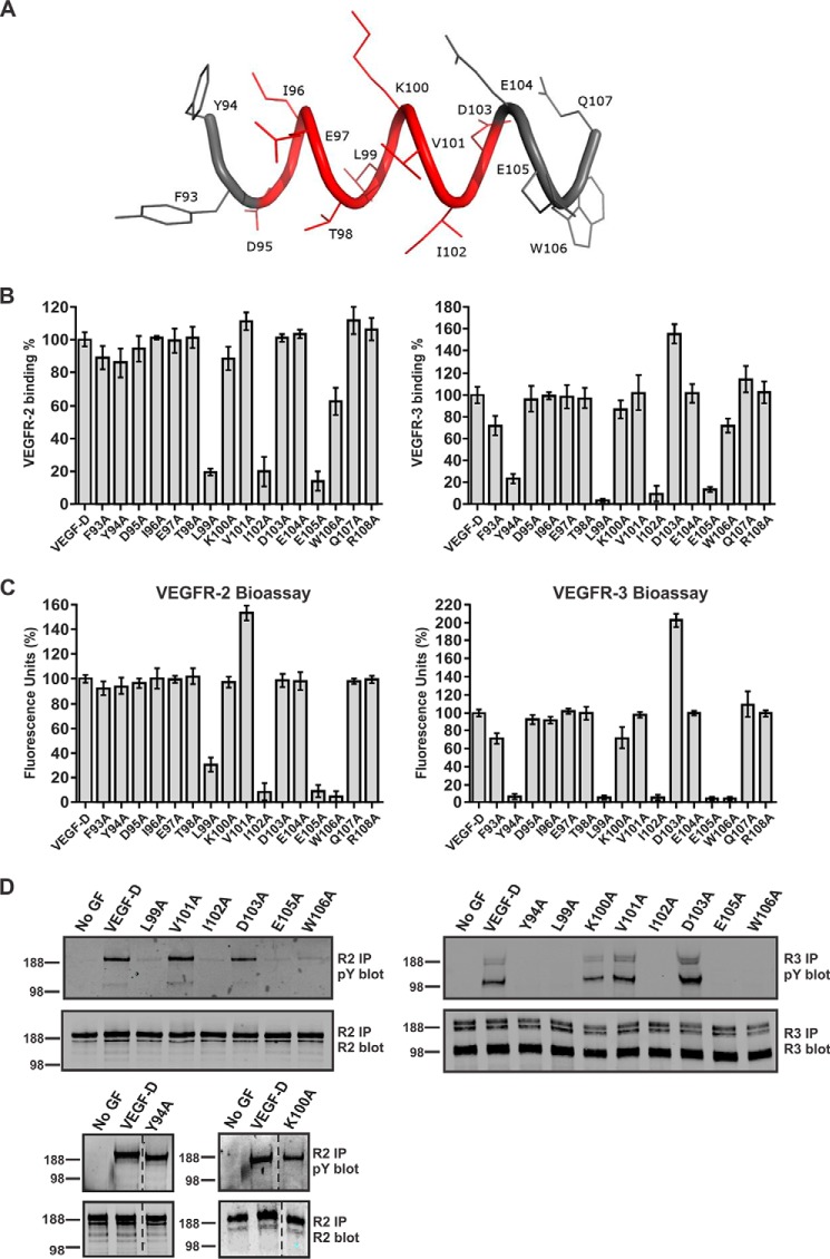
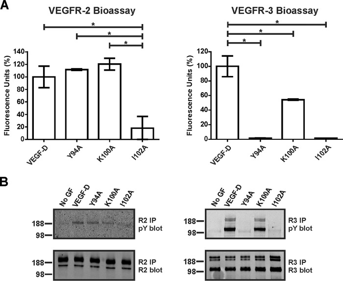
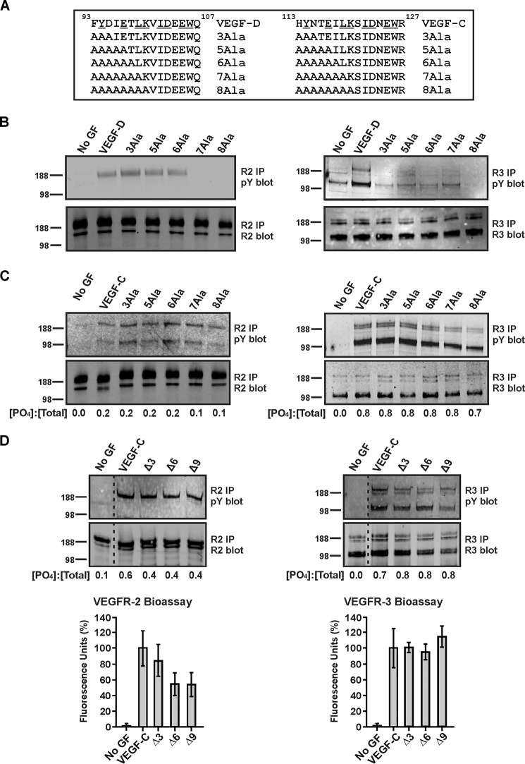
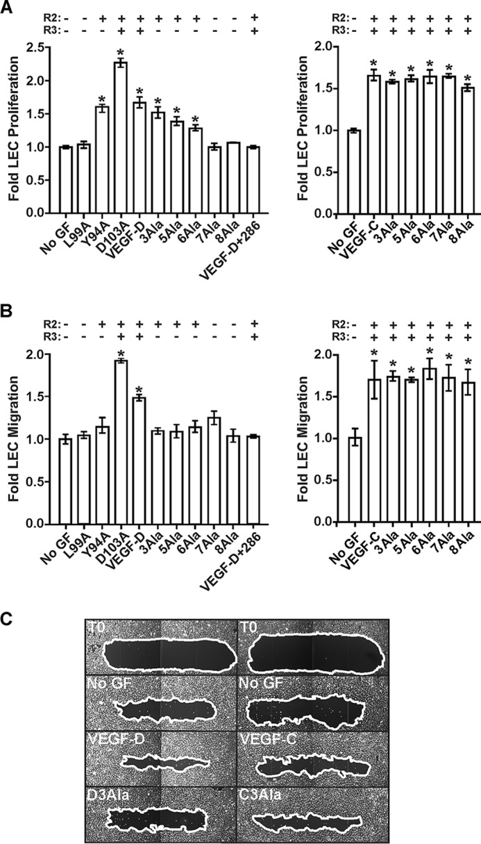
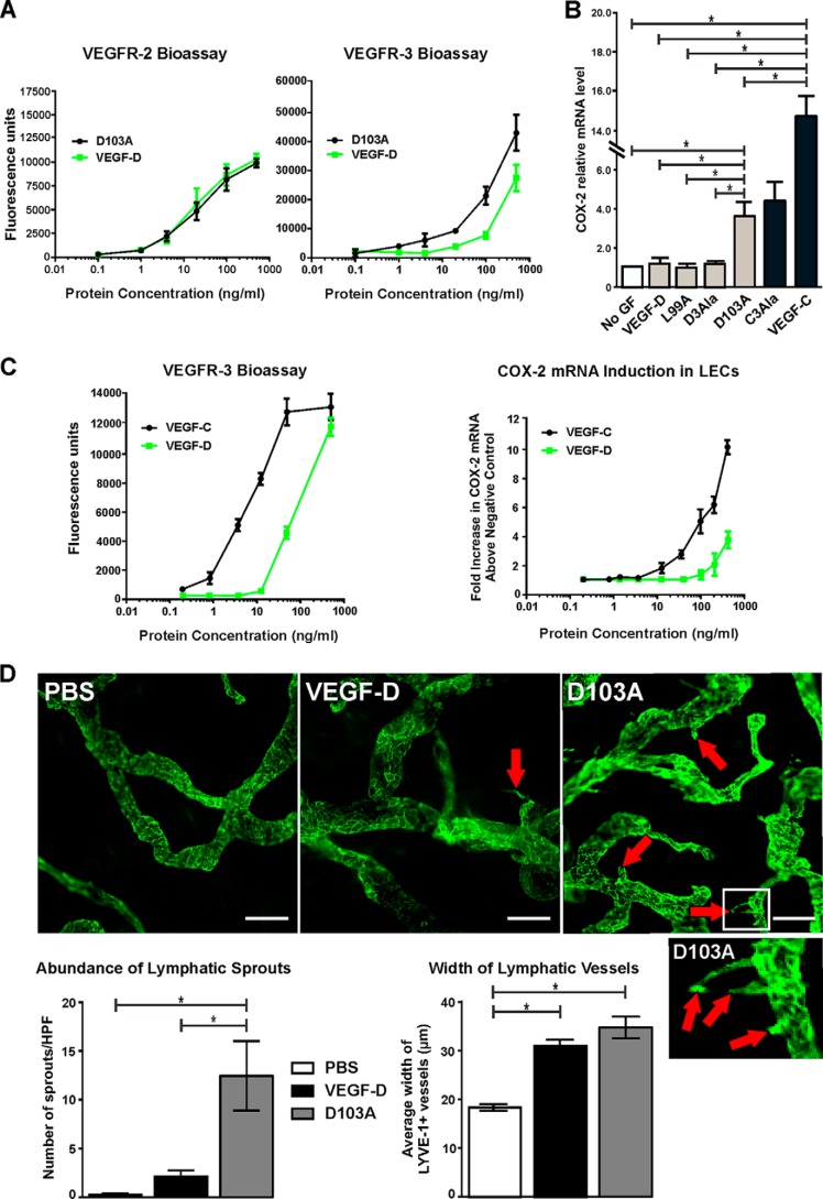
VEGF-C and VEGF-D are secreted glycoproteins that induce angiogenesis and lymphangiogenesis in cancer, thereby promoting tumor growth and spread. They exhibit structural homology and activate VEGFR-2 and VEGFR-3, receptors on endothelial cells that signal for growth of blood vessels and lymphatics. VEGF-C and VEGF-D were thought to exhibit similar bioactivities, yet recent studies indicated distinct signaling mechanisms (e.g. tumor-derived VEGF-C promoted expression of the prostaglandin biosynthetic enzyme COX-2 in lymphatics, a response thought to facilitate metastasis via the lymphatic vasculature, whereas VEGF-D did not). Here we explore the basis of the distinct bioactivities of VEGF-D using a neutralizing antibody, peptide mapping, and mutagenesis to demonstrate that the N-terminal α-helix of mature VEGF-D (Phe93-Arg108) is critical for binding VEGFR-2 and VEGFR-3. Importantly, the N-terminal part of this α-helix, from Phe93 to Thr98, is required for binding VEGFR-3 but not VEGFR-2. Surprisingly, the corresponding part of the α-helix in mature VEGF-C did not influence binding to either VEGFR-2 or VEGFR-3, indicating distinct determinants of receptor binding by these growth factors. A variant of mature VEGF-D harboring a mutation in the N-terminal α-helix, D103A, exhibited enhanced potency for activating VEGFR-3, was able to promote increased COX-2 mRNA levels in lymphatic endothelial cells, and had enhanced capacity to induce lymphatic sprouting in vivo This mutant may be useful for developing protein-based therapeutics to drive lymphangiogenesis in clinical settings, such as lymphedema. Our studies shed light on the VEGF-D structure/function relationship and provide a basis for understanding functional differences compared with VEGF-C.

Keywords: angiogenesis; endothelial cell; lymphangiogenesis; mutagenesis in vitro; receptor; vascular endothelial growth factor (VEGF).


Figures

FIGURE 1.
FIGURE 1.
Neutralizing effect of mAb 286, mapping of its binding site, and analysis of binding to VEGF-D variants with mutated residues in N-terminal α-helix. A, the capacity of mAb 286 to block binding and cross-linking, by VEGF-DΔNΔC, of chimeric receptors containing VEGFR-2 (left) or VEGFR-3 (right) extracellular domains was assessed in bioassays (see “Experimental Procedures”). Also included were neutralizing mAb VD1, which binds loop 2 of VEGF-DΔNΔC, and mAb VD4, which binds, but does not neutralize, VEGF-DΔNΔC (39). B, peptide-based mapping of the mAb 286 binding site in VEGF-DΔNΔC by ELISA (see “Experimental Procedures”). The ratio of signal to background for the interaction of mAb 286 with immobilized peptides is shown on the y axis of the graph, and the x axis indicates the identifier numbers of peptides. Top box above the graph, amino acid sequence for the VEGF homology domain of human VEGF-D; N-terminal residue (phenylalanine) is number 89, and the C-terminal residue (arginine) is 205. Bottom box above the graph, examples of peptides used in mapping (mAb 286 binding site is in a rectangle). The FLAG sequence is shown in boldface type in peptide 36, which lacks the VEGF-D-derived sequence, and was the negative control. C, detection of VEGF-DΔNΔC variants by Western blotting under reducing and denaturing conditions using mAb 286 (top) or M2 anti-FLAG mAb as a positive control (bottom). Each well contained 30 ng of purified protein. VEGF-D, VEGF-DΔNΔC; variants of this protein each have one residue mutated to alanine, as indicated. Positions of molecular mass markers (in kDa) are shown to the left. The histogram under the blots shows intensities of bands for VEGF-D variants (mean ± S.D.) relative to the intensity of the band for VEGF-DΔNΔC, as determined from Western blots with mAb 286. D, analysis of mAb 286 binding to VEGF-DΔNΔC variants by ELISA. M2 was used for capture and mAb 286 for detection; the y axis shows binding of variant proteins compared with VEGF-DΔNΔC (the latter defined as 100% binding), and the x axis lists VEGF-D variants. Equal amounts of VEGF-DΔNΔC and variants were used. For A, B, and D, assays were conducted three times. Columns, mean; error bars, S.D.
FIGURE 2.
FIGURE 2.
Interaction of VEGFR-2 and VEGFR-3 with VEGF-DΔNΔC variants. A, representation of structure for part of the N-terminal α-helix (93FYDIETLKVIDEEWQ107) in human mature VEGF-D with the mAb 286 binding site shown in red. B, analysis of binding of VEGF-DΔNΔC variants to VEGFR-2 (left) and VEGFR-3 (right) by ELISA (see “Experimental Procedures”). y axes show binding of variant proteins compared with VEGF-DΔNΔC (the latter defined as 100%), and x axes define the mutated amino acid in each variant. The same amount of each VEGF-DΔNΔC variant was used. VEGF-D, VEGF-DΔNΔC. Assays were conducted three times. Columns, mean; error bars, S.D. C, bioassays for binding and cross-linking of the extracellular domains of VEGFR-2 (left) and VEGFR-3 (right) by VEGF-DΔNΔC variants. The same amount of each VEGF-DΔNΔC variant was used in each assay. Results are expressed as a percentage of fluorescence units generated by VEGF-DΔNΔC variants relative to VEGF-DΔNΔC (y axes). x axes define the mutated amino acid in each variant. Assays were conducted five times. Columns, mean; error bars, S.D. D, receptor phosphorylation induced by selected VEGF-DΔNΔC variants. Adult LECs were stimulated with matched quantities of VEGF-DΔNΔC or its variants or left unstimulated (No GF). Lysates were immunoprecipitated (IP) with an antibody against VEGFR-2 (left) or VEGFR-3 (right) and analyzed by reducing SDS-PAGE and Western blotting with an antibody against phosphotyrosine (pY) to assess activation of receptors (top blot in each pair) or with an antibody against VEGFR-2 (bottom blot in each pair on the left) or VEGFR-3 (right bottom blot) to confirm the presence of each receptor. VEGFR-2 migrated predominantly at ∼230 kDa, whereas VEGFR-3 migrated as three bands, a ∼125-kDa cleaved form and two uncleaved forms of ∼175 and ∼195 kDa that differed in degree of glycosylation. Sizes of molecular mass markers (in kDa) are shown to the left of the panels. Dotted lines indicate where irrelevant tracks have been excised from images.
FIGURE 3.
FIGURE 3.
Receptor binding and activation by untagged VEGF-D variants. A, bioassays for binding and cross-linking of extracellular domains of VEGFR-2 (left) and VEGFR-3 (right) with altered versions of VEGF-DΔNΔC, Y94A, K100A, and I102A lacking FLAG tag. The same amount of each VEGF-DΔNΔC variant was used. Results are expressed as a percentage of fluorescence units generated relative to untagged VEGF-DΔNΔC (y axis). VEGF-D, untagged form of VEGF-DΔNΔC. Assays were conducted three times. Columns, mean; error bars, S.D. *, statistically significant differences as assessed by one-way analysis of variance with Tukey's post hoc test. B, adult LECs were stimulated with matched quantities of untagged variants or left unstimulated (No GF). Lysates were immunoprecipitated (IP) with antibody against VEGFR-2 (left) or VEGFR-3 (right) and analyzed by reducing SDS-PAGE and Western blotting with antibody against phosphotyrosine (pY) to assess receptor activation (top blots) or with antibody against VEGFR-2 (bottom left blot) or VEGFR-3 (bottom right blot) to confirm the presence of each receptor. Sizes of molecular mass markers (in kDa) are shown to the left of the panels.
FIGURE 4.
FIGURE 4.
Effects of mutating residues in N-terminal α-helices of VEGF-DΔNΔC or VEGF-CΔNΔC. A, sequences within the N-terminal α-helices of human VEGF-DΔNΔC (VEGF-D) and VEGF-CΔNΔC (VEGF-C) (top, with identical residues underlined) with variants in which multiple residues were altered to alanine shown below. B and C, blots show analyses of receptor phosphorylation by variants of VEGF-DΔNΔC and VEGF-CΔNΔC, respectively. D, blots show analyses of receptor phosphorylation induced by VEGF-CΔNΔC and mutants of VEGF-CΔNΔC lacking residues 113–115 (designated Δ3), 113–118 (Δ6), and 113–121 (Δ9). Graphs below blots show the results of bioassays of binding and cross-linking of VEGFR-2 and VEGFR-3 extracellular domains by VEGF-C variants (data are mean percentage of fluorescence relative to VEGF-CΔNΔC ± S.D.). For blots in B–D, adult LECs were stimulated with VEGF-DΔNΔC, VEGF-CΔNΔC or their variants or left unstimulated (No GF). Lysates were immunoprecipitated with antibody against VEGFR-2 (left-hand blots) or VEGFR-3 (right-hand blots) and analyzed by reducing SDS-PAGE and Western blotting with antibody against phosphotyrosine to assess receptor activation (top blots) or with antibody against VEGFR-2 (bottom left blots) or VEGFR-3 (bottom right blots) to confirm the presence of each receptor. Sizes of molecular mass markers (in kDa) are shown to the left of the blots. The amounts of VEGF-D or VEGF-C variants were matched in each experiment. Dotted lines indicate where irrelevant tracks have been excised from the images. In C and D, numbers under the lanes of blots represent the ratios of the intensities of phosphorylated receptor signals to intensities of total receptor signals ([PO4]/[total]) for each ligand treatment as determined by calculating the mean ratios from two independent experiments. The ratios for VEGFR-2 were derived by combining the intensities of the signals for bands in the size range of 188–230 kDa (note that the lower band of ∼125 kDa in the top left blot of C was not used because it probably represents co-immunoprecipitated VEGFR-3 arising from receptor heterodimers, as reported previously (68)), whereas those for VEGFR-3 are based on combining the intensities of the ∼125-, ∼175-, and ∼195-kDa forms of this receptor. pY, phosphotyrosine; IP, immunoprecipitation.
FIGURE 5.
FIGURE 5.
Analyses of the role of N-terminal α-helices of mature VEGF-D and VEGF-C for proliferation and migration by LECs. A, LEC proliferation assays. Adult LECs were treated with VEGF-DΔNΔC (VEGF-D), VEGF-CΔNΔC (VEGF-C), or their variants or left untreated (No GF). VEGF-D+286, combination of VEGF-DΔNΔC and a 10-fold molar excess of mAb 286. y axes represent proliferation by LECs stimulated with growth factor relative to that of unstimulated cells. x axes denote VEGF-D variants (left) and VEGF-C variants (right) used in assays. B, LEC migration assay. The capacity of variant proteins to induce cell migration was assessed in a scratch wound assay. Neonatal LECs were wounded, and the amount of wound closure was calculated for each variant as described under “Experimental Procedures.” y axes show migration of cells stimulated with growth factor relative to that of unstimulated cells. x axes denote VEGF-D variants (left) and VEGF-C variants (right) used in assays. C, images of selected scratch wounds. Wounds were imaged immediately post-wounding (T0, two examples) and after 24-h treatment with VEGF-DΔNΔC, VEGF-CΔNΔC, or the 3Ala variant of each (D3Ala and C3Ala, respectively). No GF, two results after 24 h with no growth factor. White lines, edges of the wounds. In A and B, the capacity of variants to activate VEGFR-2 (R2) or VEGFR-3 (R3) is indicated above the graphs, and asterisks indicate that results differ from No GF in a statistically significant fashion, as assessed by one-way analysis of variance with Tukey's post hoc test. The amounts of VEGF-D or VEGF-C variants were matched in each assay.
FIGURE 6.
FIGURE 6.
Assessment of the D103A variant and VEGF-DΔNΔC for receptor interactions, stimulation of COX-2 expression, and sprouting lymphangiogenesis. A, bioassays for binding and cross-linking of extracellular domains of VEGFR-2 (left) and VEGFR-3 (right) with VEGF-DΔNΔC (VEGF-D) and the D103A variant of VEGF-DΔNΔC. Data points, mean; error bars, S.D. B, effect of VEGF-DΔNΔC, the D103A variant, and other selected variants of VEGF-DΔNΔC (gray bars) and VEGF-CΔNΔC (VEGF-C) and the 3Ala variant of VEGF-C (C3Ala) (black bars) on the level of COX-2 mRNA in adult LECs as assessed by quantitative RT-PCR (D3Ala denotes the 3Ala variant of VEGF-DΔNΔC). Cells were exposed to 100 ng/ml ligands for 30 min before lysis for RNA preparation, as described under “Experimental Procedures.” COX-2 mRNA levels were normalized to β-actin and are expressed relative to the level in cells that were not treated with ligand (No GF). Columns, mean; error bars, S.D. C, titrations of VEGF-DΔNΔC and VEGF-CΔNΔC in the VEGFR-3 bioassay (left) and for the capacity to increase COX-2 mRNA levels in LECs (right). -Fold increases of COX-2 mRNA are relative to cells that were not treated with growth factor. In both graphs, data points indicate the mean, and error bars denote S.D. D, VEGF-DΔNΔC and the D103A variant (1 μg) were subcutaneously injected into ears of mice every 24 h for 3 days, as described under “Experimental Procedures”; PBS was the negative control. Ears were harvested and stained for lymphatics using antibody to LYVE-1 (green); the vessels shown are predominantly initial lymphatics. A high power image of the region within the white rectangle in the D103A image, showing three lymphatic sprouts, is shown below the lower power D103A image. Red arrows indicate lymphatic sprouts, which are quantitated in the left-hand graph; scale bars, 50 μm. HPF, high powered field. The width of LYVE-1-positive lymphatics is quantified in the right-hand graph. In both graphs, columns show mean and error bars denote S.E. In B and D, asterisks indicate statistically significant differences, as assessed by one-way analysis of variance with Tukey's post hoc test.

Similar articles

Cited by

References

    1. Stacker S. A., Williams S. P., Karnezis T., Shayan R., Fox S. B., and Achen M. G. (2014) Lymphangiogenesis and lymphatic vessel remodelling in cancer. Nat. Rev. Cancer 14, 159–172 - PubMed
    1. Zheng W., Aspelund A., and Alitalo K. (2014) Lymphangiogenic factors, mechanisms, and applications. J. Clin. Invest. 124, 878–887 - PMC - PubMed
    1. Duong T., Koltowska K., Pichol-Thievend C., Le Guen L., Fontaine F., Smith K. A., Truong V., Skoczylas R., Stacker S. A., Achen M. G., Koopman P., Hogan B. M., and Francois M. (2014) VEGFD regulates blood vascular development by modulating SOX18 activity. Blood 123, 1102–1112 - PubMed
    1. Ober E. A., Olofsson B., Mäkinen T., Jin S. W., Shoji W., Koh G. Y., Alitalo K., and Stainier D. Y. (2004) Vegfc is required for vascular development and endoderm morphogenesis in zebrafish. EMBO Rep. 5, 78–84 - PMC - PubMed
    1. Karnezis T., Shayan R., Caesar C., Roufail S., Harris N. C., Ardipradja K., Zhang Y. F., Williams S. P., Farnsworth R. H., Chai M. G., Rupasinghe T. W., Tull D. L., Baldwin M. E., Sloan E. K., Fox S. B., Achen M. G., and Stacker S. A. (2012) VEGF-D promotes tumor metastasis by regulating prostaglandins produced by the collecting lymphatic endothelium. Cancer Cell 21, 181–195 - PubMed

Publication types

MeSH terms

Substances

Associated data