. 2017 Jan 3;91(2):e01795-16.
doi: 10.1128/JVI.01795-16. Print 2017 Jan 15.

Honey Bee Deformed Wing Virus Structures Reveal that Conformational Changes Accompany Genome Release

Affiliations

Honey Bee Deformed Wing Virus Structures Reveal that Conformational Changes Accompany Genome Release

Lindsey J Organtini et al. J Virol. .

Abstract

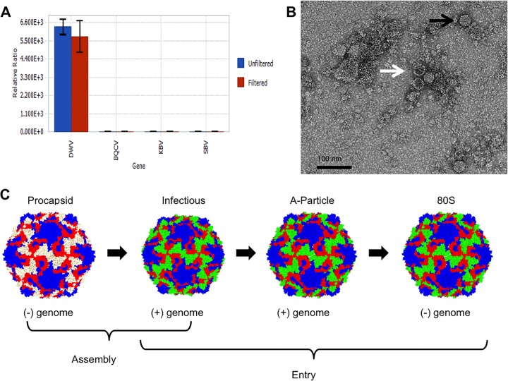
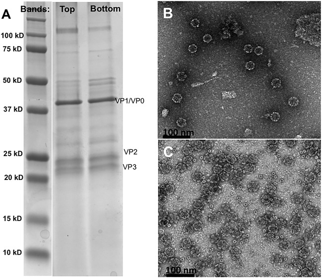
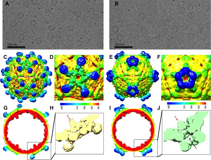
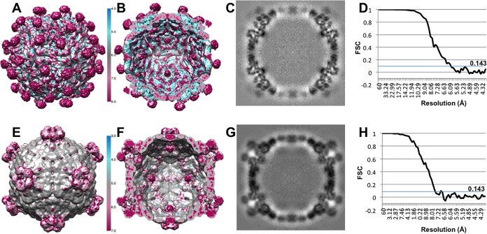
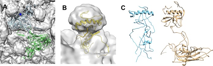
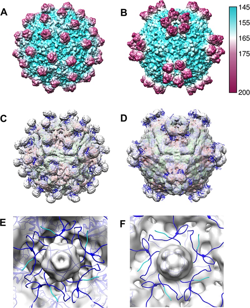
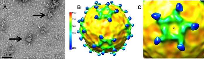
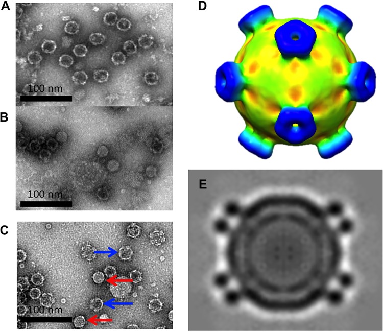
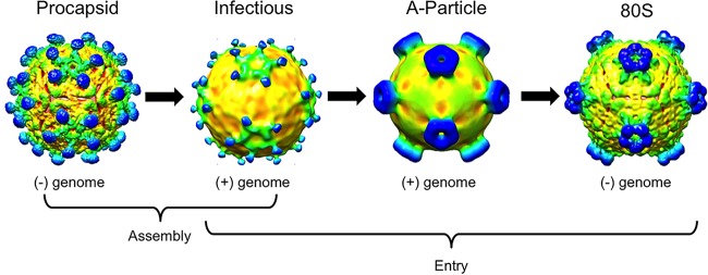
The picornavirus-like deformed wing virus (DWV) has been directly linked to colony collapse; however, little is known about the mechanisms of host attachment or entry for DWV or its molecular and structural details. Here we report the three-dimensional (3-D) structures of DWV capsids isolated from infected honey bees, including the immature procapsid, the genome-filled virion, the putative entry intermediate (A-particle), and the empty capsid that remains after genome release. The capsids are decorated by large spikes around the 5-fold vertices. The 5-fold spikes had an open flower-like conformation for the procapsid and genome-filled capsids, whereas the putative A-particle and empty capsids that had released the genome had a closed tube-like spike conformation. Between the two conformations, the spikes undergo a significant hinge-like movement that we predicted using a Robetta model of the structure comprising the spike. We conclude that the spike structures likely serve a function during host entry, changing conformation to release the genome, and that the genome may escape from a 5-fold vertex to initiate infection. Finally, the structures illustrate that, similarly to picornaviruses, DWV forms alternate particle conformations implicated in assembly, host attachment, and RNA release.

Importance: Honey bees are critical for global agriculture, but dramatic losses of entire hives have been reported in numerous countries since 2006. Deformed wing virus (DWV) and infestation with the ectoparasitic mite Varroa destructor have been linked to colony collapse disorder. DWV was purified from infected adult worker bees to pursue biochemical and structural studies that allowed the first glimpse into the conformational changes that may be required during transmission and genome release for DWV.

Keywords: 5-fold spikes; 80S; DWV; conformation change; deformed wing virus; honey bee; insect; life cycle; picornavirus; procapsid.


Figures

FIG 1
FIG 1
Detection of DWV in honey bee lysate. (A) Quantitative real-time PCR of DWV-infected honey bee homogenates. Unfiltered and filtered honey bee extracts were subjected to qRT-PCR using primers corresponding to four different common honey bee viruses, including DWV, black queen cell virus (BQCV), Kashmir bee virus (KBV), and sacbrood virus (SBV). The ratio of RNA of each virus to an internal control gene is shown. DWV was the only virus detected, and the amount of virus detected was unaffected by the filtration process. (B) Visualization of DWV capsids. Negatively stained TEM of DWV-infected bee homogenate shows a mixture of full capsids (black arrow) and empty capsids (white arrow). (C) A schematic showing that for picornaviruses, two types of capsids, procapsid and genome-filled virus capsid, are assembled. During host attachment and entry, an entry intermediate, the A-particle, is formed and is then triggered to release genome, leaving an empty capsid, referred to as an 80S capsid.
FIG 2
FIG 2
(A) SDS-PAGE staining with Coomassie blue shows that proteins corresponding to VP1 and VP0 (VP1/4), VP2, and VP3 can be detected in the top and lower bands. A protein standard (lane 1) is used as a sizing ladder. (B and C) Material from each of the two bands that formed during sucrose gradient purification were negatively stained and imaged by TEM. The top (B) and lower (C) bands both contain empty capsids.
FIG 3
FIG 3
Cryo-EM reconstructions of two different types of empty DWV capsids. (A and B) Micrographs of (A) DWV1 and (B) DWV2 show empty capsids. (C to J) The DWV1 (C, D, G, and H) (6.1-Å) and DWV2 (E, F, I, and J) (7.6-Å) cryo-EM reconstructions are visualized at a contour level of 1σ, surface rendered, and colored radially according to the scale bar. (G to J) Cutaway and closeup views of the virus 5-fold vertices show the different conformations of the density spikes (blue) and the 5-fold density plug (red arrows) that may block access to the capsid interior. (J) Tenuous connections (black arrow) seem to hold a 5-fold density plug (red arrow) at the inner capsid surface, although there is a clear opening through the capsid shell at each 5-fold vertex which is better visualized with a continuous color scheme for DWV1 (yellow) and DVV2 (green) in the zoomed views.
FIG 4
FIG 4
Quality and resolution of the DWV cryo-EM reconstructions. DWV1 (A and B) and DWV2 (E and F) are colored according to local-resolution estimations (see scale bars). The capsid shells reach higher resolutions than the flexible 5-fold decorations, with DWV1 spikes at lower resolution than the DWV2 spikes. (C and G) The map central sections (protein is indicated in black) of DWV1 (C) and DWV2 (G) show the quality of the maps. (D and H) Because the data sets were split initially and the halves reconstructed separately, the resolution for each reconstruction was assessed using the gold standard Fourier shell correlation (FSC) cutoff value of 0.143, yielding 6.1-Å and 7.6-Å resolutions for DWV1 (D) and DWV2 (H), respectively.
FIG 5
FIG 5
Alignment of DWV and Ljungan virus VP1 residues shows C-terminal extension of DWV VP1. The two viruses share 49% sequence identity. *, fully conserved residue; :, residues that share similar properties; ., residues that share weakly similar properties.
FIG 6
FIG 6
DWV capsid composition and spike movement. (A) The Ljungan virus structure (PDB ID 3JB4) (blue and green ribbon) fitted into the DWV1 map illustrates that the last C-terminal residue of VP1 maps to the base of the 5-fold spike density (blue sphere). Symmetry axes are indicated, and the strand-swapping mechanism can be seen where VP2 ribbons cross the 2-fold density bridge. (B) The last 171 C-terminal VP1 residues of DWV were predicted to form a helix-loop structure (yellow) that was fitted into the spike density. (C) The predicted model of DWV VP1 C-terminal extension has a structure similar to the VP1 crystal structure of slow bee paralysis virus (PDB ID 5J98) (22).
FIG 7
FIG 7
Comparison of DWV1 and DWV2 to SBPV. (A and B) Two structures of slow bee paralysis virus (PDB ID 5J98 and 5J96) (22) were used to calculate ∼7-Å surface-rendered radially colored (see key) maps to compare gross surface topologies to those of DWV1 and DWV2, respectively. (C and D) The SBPV structures (VP1, -2, and -3 are color coded blue, green, and red according to the gene order presented in Fig. 8) were fitted into the DWV1 and DWV2 cryo-EM maps (transparent gray), and correlation coefficients were obtained that signified a moderately poor fit despite the obvious overall similarities. (E and F) The zoomed view shows the internal surface of DWV1 and DWV2, respectively (gray), at the 5-fold vertex, with SBPV structures fitted to show that the VP1 N termini (blue) surround the 5-fold vertex. If VP0 were not cleaved, the VP4 portion would map to this region, which is similar to the location of VP4 in picornaviruses. The large gray plug of unfilled density was equal in magnitude to the capsid density.
FIG 8
FIG 8
The gene order for SBPV, DWV, and picornaviruses is shown with boxes color coded to represent VP1, VP2, VP3, and VP4 (blue, green, red, and yellow, respectively). The alignment of the P1 region encoding the structural proteins for DWV and SBPV (33% sequence similarity) has been similarly color coded with lines to indicate viral proteins. For consistency, the DWV published gene order and the picornavirus color code were used throughout this work. *, fully conserved residue; :, residues that share similar properties; ., residues that share weakly similar properties.
FIG 9
FIG 9
The structure of the RNA-filled DWV capsid from infectious bee lysates. (A) Negatively stained micrographs of infected bee homogenate were used to select 102 genome-filled infectious virus capsids for a low resolution (∼25-Å resolution) negative-stain reconstruction to reveal the spike conformation. (B and C) The RNA-filled capsids have spikes in the open conformation, similarly to the DWV1 reconstruction.
FIG 10
FIG 10
Results from the second purification of infectious honey bee lysate. (A and B) Negatively stained TEM images of the (A) top and (B) lower bands. The top band remained empty, as before, whereas the lower band contained approximately 50% genome-filled virus. (C) A negatively stained micrograph of an aliquot from the lower band after incubation at 37°C for 24 h shows different distributions, as most of the filled capsids had lost the genome (blue arrows). The filled viruses that retain the genome appear to have less dense centers than those previous observed (red arrows), consistent with the presence of A-particles. The low-resolution (18-Å) cryo-EM reconstruction (D) and central section (E) of putative A-particle (C) reveal that the spikes had undergone a conformational change into the closed tube-like form after incubation.
FIG 11
FIG 11
Similarly to picornavirus, DWV assembles an empty capsid in addition to RNA-filled capsids. This procapsid structure resembles the infectious virus, whereas the putative A-particle resembles the 80S empty capsid. The putative A-particle and 80S-like empty capsid have different conformations for the 5-fold spikes.

References

    1. Evans JD, Schwarz RS. 2011. Bees brought to their knees: microbes affecting honey bee health. Trends Microbiol 19:614–620. doi: 10.1016/j.tim.2011.09.003. - DOI - PubMed
    1. Potts SG, Biesmeijer JC, Kremen C, Neumann P, Schweiger O, Kunin WE. 2010. Global pollinator declines: trends, impacts and drivers. Trends Ecol Evol 25:345–353. doi: 10.1016/j.tree.2010.01.007. - DOI - PubMed
    1. van Engelsdorp D, Hayes J, Underwood RM, Pettis J. 2008. A survey of honey bee colony losses in the U.S., fall 2007 to spring 2008. PLoS One 3:e4071. doi: 10.1371/journal.pone.0004071. - DOI - PMC - PubMed
    1. Genersch E, Aubert M. 2010. Emerging and re-emerging viruses of the honey bee (Apis mellifera L.). Vet Res 41:54. doi: 10.1051/vetres/2010027. - DOI - PMC - PubMed
    1. Mondet F, de Miranda JR, Kretzschmar A, Le Conte Y, Mercer AR. 2014. On the front line: quantitative virus dynamics in honeybee (Apis mellifera L.) colonies along a new expansion front of the parasite Varroa destructor. PLoS Pathog 10:e1004323. doi: 10.1371/journal.ppat.1004323. - DOI - PMC - PubMed