. 2017 Jan 3;91(2):e02095-16.
doi: 10.1128/JVI.02095-16. Print 2017 Jan 15.

Robust Central Nervous System Pathology in Transgenic Mice following Peripheral Injection of α-Synuclein Fibrils

Affiliations

Robust Central Nervous System Pathology in Transgenic Mice following Peripheral Injection of α-Synuclein Fibrils

Jacob I Ayers et al. J Virol. .

Abstract

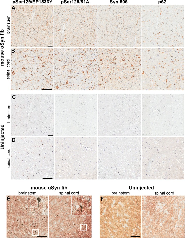
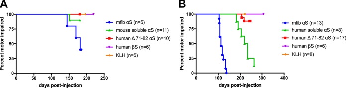
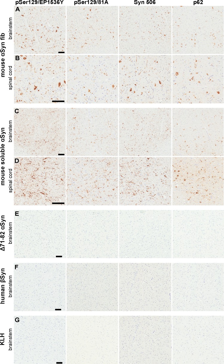
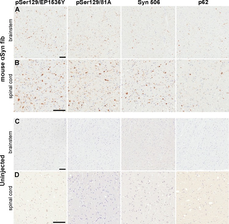
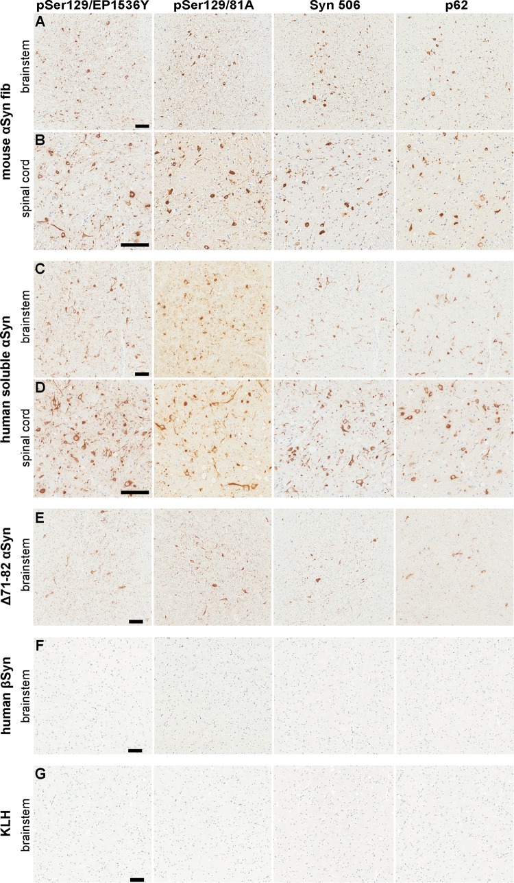
Misfolded α-synuclein (αS) is hypothesized to spread throughout the central nervous system (CNS) by neuronal connectivity leading to widespread pathology. Increasing evidence indicates that it also has the potential to invade the CNS via peripheral nerves in a prion-like manner. On the basis of the effectiveness following peripheral routes of prion administration, we extend our previous studies of CNS neuroinvasion in M83 αS transgenic mice following hind limb muscle (intramuscular [i.m.]) injection of αS fibrils by comparing various peripheral sites of inoculations with different αS protein preparations. Following intravenous injection in the tail veins of homozygous M83 transgenic (M83+/+) mice, robust αS pathology was observed in the CNS without the development of motor impairments within the time frame examined. Intraperitoneal (i.p.) injections of αS fibrils in hemizygous M83 transgenic (M83+/-) mice resulted in CNS αS pathology associated with paralysis. Interestingly, injection with soluble, nonaggregated αS resulted in paralysis and pathology in only a subset of mice, whereas soluble Δ71-82 αS, human βS, and keyhole limpet hemocyanin (KLH) control proteins induced no symptoms or pathology. Intraperitoneal injection of αS fibrils also induced CNS αS pathology in another αS transgenic mouse line (M20), albeit less robustly in these mice. In comparison, i.m. injection of αS fibrils was more efficient in inducing CNS αS pathology in M83 mice than i.p. or tail vein injections. Furthermore, i.m. injection of soluble, nonaggregated αS in M83+/- mice also induced paralysis and CNS αS pathology, although less efficiently. These results further demonstrate the prion-like characteristics of αS and reveal its efficiency to invade the CNS via multiple routes of peripheral administration.

Importance: The misfolding and accumulation of α-synuclein (αS) inclusions are found in a number of neurodegenerative disorders and is a hallmark feature of Parkinson's disease (PD) and PD-related diseases. Similar characteristics have been observed between the infectious prion protein and αS, including its ability to spread from the peripheral nervous system and along neuroanatomical tracts within the central nervous system. In this study, we extend our previous results and investigate the efficiency of intravenous (i.v.), intraperitoneal (i.p.), and intramuscular (i.m.) routes of injection of αS fibrils and other protein controls. Our data reveal that injection of αS fibrils via these peripheral routes in αS-overexpressing mice are capable of inducing a robust αS pathology and in some cases cause paralysis. Furthermore, soluble, nonaggregated αS also induced αS pathology, albeit with much less efficiency. These findings further support and extend the idea of αS neuroinvasion from peripheral exposures.

Keywords: Lewy pathology; alpha-synuclein; peripheral; prions; transgenic mice.


Figures

FIG 1
FIG 1
Induction of CNS αS inclusion pathology in M83+/+ mice following tail vein injection of αS fibs. (A and B) Representative images showing αS inclusion pathology in the brain stem (A) and spinal cord (B) of M83+/+ mice injected in the tail vein with 20 μg mouse αS fibs. (C and D) Representative images show the lack of αS inclusion pathology in the brain stem (C) and spinal cord (D) of uninjected age-matched M83+/+ mice. Tissue sections were stained with antibodies to αS phosphorylated at Ser129 (EP1536Y and 81A), anti-αS (Syn 506) or anti-p62/sequestosome (a general marker of inclusion pathology) as indicated in each panel. Tissue sections were counterstained with hematoxylin. (E and F) Representative images revealing the presence of argyrophilic deposits in the brain stems and spinal cords (as indicated by insets [×3 magnification]) of M83+/+ mice injected in the tail vein with 20 μg mouse αS fibs (E), and a lack of deposits in age-matched uninjected M83+/+ mice (F). Bars = 100 μm.
FIG 2
FIG 2
Induction of motor impairments and paralysis in M83+/− mice peripherally challenged with various forms of αS. (A) Kaplan-Meier survival plot shows a decreased time to end stage (due to motor impairments) for M83+/− Tg mice injected i.p. with mouse αS fibs compared to M83+/− mice injected i.p. with Δ71-82 αS, soluble nonaggregated mouse αS, βS, or KLH. A mouse euthanized at an earlier time point due to a non-disease-related complication (i.e., dermatitis) was excluded from the data. mfib, mouse αS fib. (B) Bilateral i.m. injection in the hind limb muscle of M83+/− Tg mice with mouse αS fibs resulted in paralysis in all of the mice by 134 dpi. i.m. injections with soluble, nonaggregated human αS also resulted in the majority of animals developing paralysis, whereas only 2 of the 17 mice injected with Δ71-82 αS developed disease, and none of the mice injected with βS, or KLH ever displayed symptoms. Mice euthanized at earlier time points due to non-disease-related complications (i.e., fighting wounds) were excluded from these data (Table 1).
FIG 3
FIG 3
Induction of CNS αS inclusion pathology in M83+/− mice following i.p. injection of αS. (A and B) Representative images showing αS inclusion pathology in the brain stem (A) and spinal cord (B) of M83+/− mice i.p. injected with 50 μg mouse αS fibs. (C and D) Representative images showing αS inclusion pathology in the brain stem (C) and spinal cord (D) of M83+/− mice i.p. injected with 50 μg soluble mouse αS that developed motor impairments. (E to G) Representative images showing a paucity of αS inclusion pathology in the brain stems of M83+/− mice i.p. injected with 50 μg of Δ71-82 αS (E), βS (F), or KLH (G). Tissue sections were stained with antibodies to αS phosphorylated at Ser129 (EP1536Y and 81A), anti-αS (Syn 506), or anti-p62/sequestosome (a general marker of inclusion pathology) as indicated in each panel. Tissue sections were counterstained with hematoxylin. Bars = 100 μm.
FIG 4
FIG 4
Induction of CNS αS inclusion pathology in M20+/− mice following i.p. injection of αS fibs. (A and B) Representative images showing αS inclusion pathology in the brain stem (A) and spinal cord (B) of M20+/− mice that were i.p. injected with 50 μg mouse αS fibs and developed a moribund state. (C and D) Representative images depicting a paucity of αS inclusion pathology in the brain stem (C) and spinal cord (D) of uninjected older (450 days) M20+/− mice. Tissue sections were stained with antibodies to αS phosphorylated at Ser129 (EP1536Y and 81A), anti-αS (Syn 506) or anti-p62/sequestosome (a general marker of inclusion pathology) as indicated in each panel. Tissue sections were counterstained with hematoxylin. Bars = 100 μm.
FIG 5
FIG 5
Induction of CNS αS inclusion pathology in M83+/− mice following i.m. injection of αS fibs and soluble αS. (A and B) Representative images showing αS inclusion pathology in the brain stem (A) and spinal cord (B) of M83+/− mice i.m. injected with 20 μg mouse αS fibs. (C and D) Representative images showing αS inclusion pathology in the brain stem (C) and spinal cord (D) of an M83+/− mouse that was i.m. injected with 20 μg soluble human αS that developed motor impairments. (E) Representative images showing αS inclusion pathology in the brain stem of one of the M83+/− mice i.m. injected with 20 μg Δ71-82 αS that developed motor impairments. Paucity of αS inclusion pathology as shown in the brain stems of M83+/− mice i.m. injected with 20 μg βS (F) or KLH (G). Tissue sections were stained with antibodies to αS phosphorylated at Ser129 (EP1536Y and 81A), anti-αS (Syn 506), or anti-p62/sequestosome (a general marker of inclusion pathology) as indicated in each panel. Tissue sections were counterstained with hematoxylin. Bars = 100 μm.
FIG 6
FIG 6
Negative staining electron microscopy of mouse αS fibs after water bath sonication. Representative images showing αS fibs that have been broken up into an array of polymers by water bath sonication. Grids were stained with 1% uranyl acetate. Bar = 200 nm.

Similar articles

Cited by

References

    1. Waxman EA, Giasson BI. 2009. Molecular mechanisms of alpha-synuclein neurodegeneration. Biochim Biophys Acta 1792:616–624. doi:10.1016/j.bbadis.2008.09.013. - DOI - PMC - PubMed
    1. Goedert M. 1997. Familial Parkinson's disease. The awakening of alpha-synuclein. Nature 388:232–233. - PubMed
    1. Goedert M. 2001. Alpha-synuclein and neurodegenerative diseases. Nat Rev Neurosci 2:492–501. doi:10.1038/35081564. - DOI - PubMed
    1. Cookson MR. 2005. The biochemistry of Parkinson's disease. Annu Rev Biochem 74:29–52. doi:10.1146/annurev.biochem.74.082803.133400. - DOI - PubMed
    1. Goedert M, Spillantini MG, Del Tredici K, Braak H. 2013. 100 years of Lewy pathology. Nat Rev Neurol 9:13–24. doi:10.1038/nrneurol.2012.242. - DOI - PubMed

MeSH terms

LinkOut - more resources