. 2017 Jan 18;91(3):e01956-16.
doi: 10.1128/JVI.01956-16. Print 2017 Feb 1.

A Novel Rabies Vaccine Expressing CXCL13 Enhances Humoral Immunity by Recruiting both T Follicular Helper and Germinal Center B Cells

Affiliations

A Novel Rabies Vaccine Expressing CXCL13 Enhances Humoral Immunity by Recruiting both T Follicular Helper and Germinal Center B Cells

Zhao Wang et al. J Virol. .

Abstract

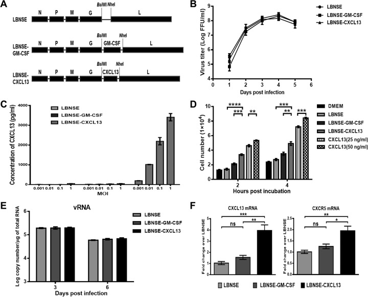
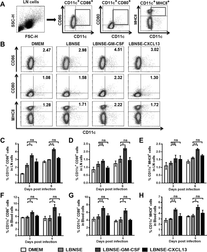
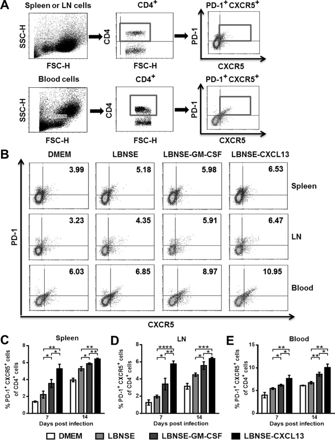
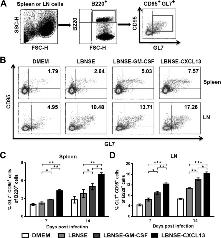
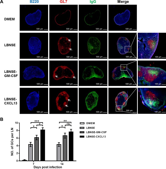
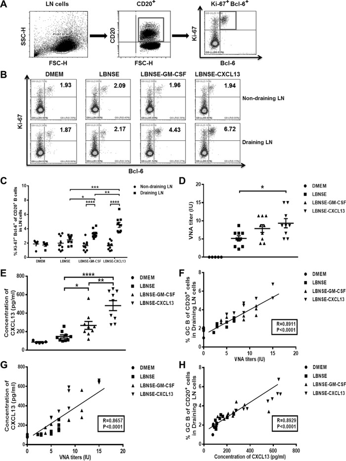
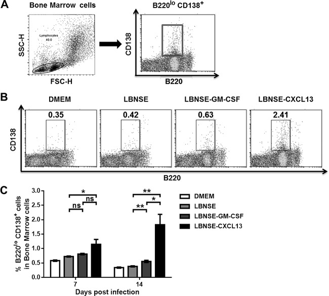
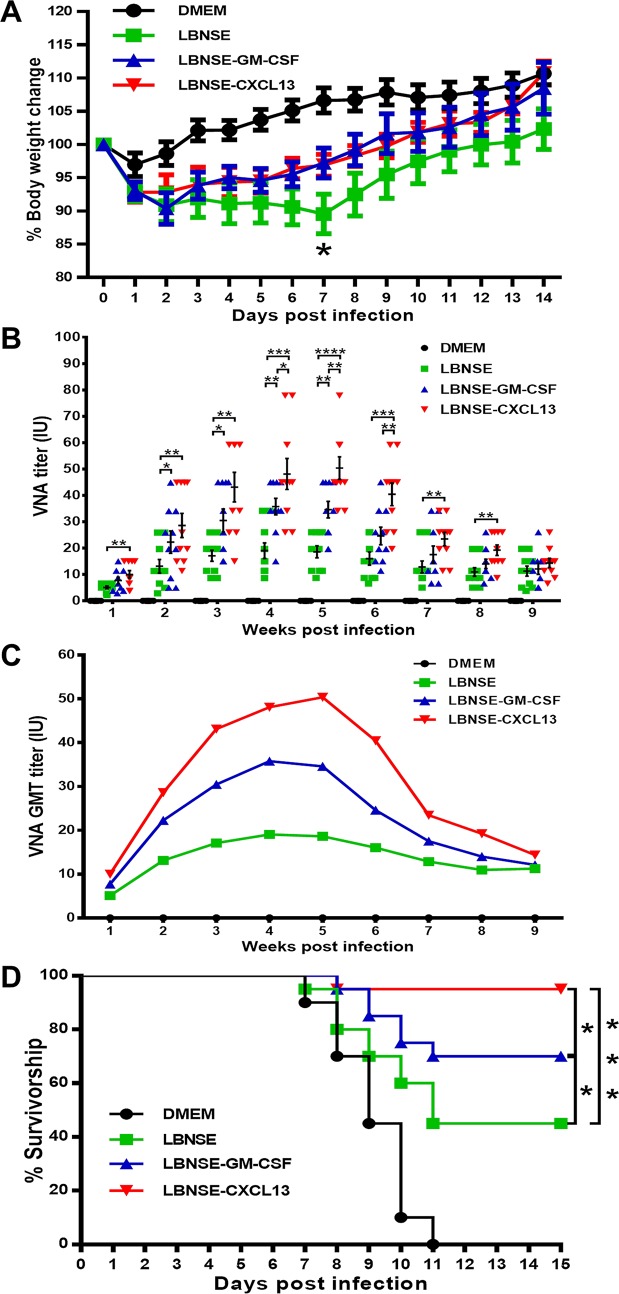
Rabies remains a public health threat in most parts of the world, and approximately 99% of the cases are transmitted by dogs. There is an urgent need to develop an efficacious and affordable vaccine to control canine-transmitted rabies in developing countries. Our previous studies demonstrate that overexpression of chemokines/cytokines such as CCL-3 (MIP-1α) and granulocyte-macrophage colony-stimulating factor (GM-CSF) can enhance the immunogenicity of rabies vaccines. In the present study, the chemokine CXCL13 was inserted into the genome of the recombinant rabies virus (rRABV) strain LBNSE, and the effect of the chemokine CXCL13 on the immunogenicity of RABV was investigated. It was found that LBNSE-CXCL13 recruited follicular helper T (Tfh) and germinal center (GC) B cells, promoted the formation of GCs, and increased the population of plasma cells in immunized mice. Further studies showed that mice immunized with LBNSE-CXCL13 produced more rabies virus-neutralizing antibodies (VNAs) and developed better protection than those immunized with the parent virus LBNSE or the GM-CSF-expressing RABV (LBNSE-GM-CSF). Collectively, these findings provide a better understanding of the role of CXCL13 expression in the immunogenicity of the RABV, which may help in designing more-efficacious rabies vaccines.

Importance: Rabies is endemic in most parts of the world, and more effort is needed to develop affordable and effective vaccines to control or eliminate this disease. The chemokine CXCL13 recruits both Tfh and B cells, which is essential for the homing of Tfh cells and the development of B cell follicles. In this study, the effect of the overexpression of CXCL13 on the immunogenicity of the RABV was evaluated in a mouse model. We found that CXCL13 expression promoted humoral immunity by recruiting Tfh and GC B cells, facilitating the formation of GCs, and increasing the number of plasma cells. As expected, the overexpression of CXCL13 resulted in enhanced virus-neutralizing antibody (VNA) production and protection against a virulent RABV challenge. These findings provide a better understanding of the role of CXCL13 in RABV-induced immune responses, which will help in designing more efficacious rabies vaccines.

Keywords: CXCL13; T follicular helper cells; germinal center B cells; humoral immunity; rabies virus.


Figures

FIG 1
FIG 1
Construction and characterization of the rRABV expressing CXCL13. (A) Schematic diagram for the construction of LBNSE, LBNSE-GM-CSF, and LBNSE-CXCL13. The mouse CXCL13 gene was cloned and inserted into the RABV genome in place of the deleted long noncoding region, and rRABVs were rescued according to the method described in Materials and Methods. (B) A multistep growth curve was generated in BSR cells. Cells were infected with LBNSE, LBNSE-GM-CSF, or LBNSE-CXCL13 at a multiplicity of infection (MOI) of 0.01 FFU and incubated at 37°C. Viruses were harvested at 1, 2, 3, 4, and 5 dpi, and viral titers were determined. All titrations were carried out in quadruplicate, and the data are presented as the means ± standard deviations (SD). (C) Production of CXCL13 in BSR cells. Cells were infected with different viruses at MOIs of 0.001, 0.01, 0.1, and 1. After incubation at 37°C for 24 h, the culture supernatants were collected, and the CXCL13 concentrations produced by the indicated rRABVs were determined with a commercial ELISA kit. (D) Chemotactic effects of cultured medium from BSR cells infected with rRABVs at an MOI of 1 on mouse splenocytes. Splenocytes (5 × 10⁵) were applied to the upper wells of chemotaxis chambers. Two and 4 h later, the cells migrating to the bottom chamber were counted. (E and F) BALB/c mice were inoculated via i.m. injection of 1 × 10⁶ FFU of rRABVs. The muscles from the hind legs of mice (n = 3) were harvested at 3 and 6 dpi. Total RNA was extracted, and viral genomic RNA (vRNA) (E) and CXCL13 mRNA and CXCR5 mRNA (F) were analyzed via qRT-PCR. All the data are expressed as the means ± SD. Asterisks indicate significant differences between the indicated experimental groups.
FIG 2
FIG 2
Recruitment and/or activation of DCs in mice postinfection with rRABVs. BALB/c mice (n = 3) were infected via i.m. injection of 1 × 10⁶ FFU of different rRABVs, and the draining (inguinal) lymph nodes (LNs) and blood were harvested at 3 and 6 dpi. Single-cell suspensions were prepared, stained with antibodies against DCs and DC activation markers, and analyzed via flow cytometry. (A and B) Representative gating strategies for the detection of DCs (A) and representative flow cytometric plots of DCs (B) from the draining LNs are shown. (C to H) Analyses for activated DCs (CD11c+ CD86+, CD11c+ CD80+, or CD11c+ MHCII+) from the draining LNs (C, D, E) and the blood (F, G, H) at 3 and 6 dpi are presented. All the data are expressed as the means ± standard errors of the means (SEM) (n = 3). Asterisks indicate significant differences between the indicated experimental groups.
FIG 3
FIG 3
Recruitment of Tfh cells by CXCL13. BALB/c mice (n = 3) were infected via i.m. injection of 1 × 10⁶ FFU of different rRABVs, and the spleens, draining LNs, and blood were harvested at 7 and 14 dpi. Single-cell suspensions were prepared, stained with antibodies against Tfh cells and Tfh cell activation markers, and analyzed via flow cytometry. (A and B) Representative gating strategies for the detection of Tfh cells (A) and representative flow cytometric plots of Tfh cells (B) are shown. (C to E) The results of a detailed analysis for activated Tfh cells (CD4+ CXCR5+ PD-1+) at 7 and 14 dpi are presented for the spleen (C), the draining LNs (D), and the blood (E). Data are expressed as the means ± SEM (n = 3). Asterisks indicate significant differences between the indicated experimental groups.
FIG 4
FIG 4
Recruitment of GC B cells by CXCL13. BALB/c mice (n = 3) were infected via i.m. injection of 1 × 10⁶ FFU of different rRABVs, and the spleens and draining LNs were harvested at 7 and 14 dpi. Single-cell suspensions were prepared, stained with antibodies against GC B cells and GC B cell activation markers, and analyzed via flow cytometry. (A and B) Representative gating strategies for the detection of GC B cells (A) and representative flow cytometric plots of GC B cells (B) are shown. (C and D) The results of a detailed analysis for activated GC B cells (B220+ CD95+ GL7+) at 7 and 14 dpi are presented for the spleen (C) and the draining LNs (D). Data are presented as the means ± SEM (n = 3). Asterisks indicate significant differences between the indicated experimental groups.
FIG 5
FIG 5
Expression of CXCL13 facilitates the formation of GCs. BALB/c mice (n = 3) were infected via i.m. injection of 1 × 10⁶ FFU of different rRABVs, and the draining LNs were collected at 7 and 14 dpi. Then, the draining LNs were excised, and tissue sections were prepared and stained for germinal centers (GL7, red; B220, blue; and IgG, green). Scale bars, 500 μm or 100 μm (rightmost column only). (A) Representative results are shown. (B) The numbers of GCs formed at 7 and 14 dpi were calculated. All the data are expressed as the means ± SEM (n = 3). Asterisks indicate significant differences between the indicated experimental groups; ns, not significant.
FIG 6
FIG 6
Plasma CXCL13 levels correlate with GC activity and VNA titers in mice. BALB/c mice were infected via i.m. injection of 1 × 10⁶ FFU of DMEM (n = 5), LBNSE (n = 9), LBNSE-GM-CSF (n = 9), or LBNSE-CXCL13 (n = 9), and then draining (inguinal) and nondraining (cervical) LNs were collected at 7 dpi. Single-cell suspensions were prepared, stained with antibodies against GC B cells and GC B cell activation markers, and analyzed via flow cytometry. (A and B) Representative gating strategies for the detection of GC B cells (A) and representative flow cytometric plots of plasma B cells (B) are shown. (C) The results of a detailed analysis of activated GC B cells (CD20+ Ki-67+ Bcl-6+) at 7 dpi are presented for the draining and nondraining LNs. (D) Serum samples were harvested at 7 dpi, and RABV VNA titers were measured via FAVN tests as described in Materials and Methods. (E) The concentration of plasma CXCL13 was determined using a commercial ELISA kit. (F) Correlations of GC B cell activity in the draining LNs and RABV VNA titers in mice 7 days after immunization were determined. (G) Correlations of plasma CXCL13 concentrations and RABV VNA titers in mice 7 days after immunization were determined. (H) Correlations of GC B cell activity in the draining LNs and plasma CXCL13 concentrations in mice 7 days after immunization were determined. All the data are expressed as the means ± SEM (n = 3). Asterisks indicate significant differences between the indicated experimental groups.
FIG 7
FIG 7
Expression of CXCL13 promotes an increase in the quantity of plasma cells. BALB/c mice (n = 3) were infected via i.m. injection of 1 × 10⁶ FFU of different rRABVs, and the bone marrow samples were harvested at 7 and 14 dpi. Single-cell suspensions were prepared, stained with antibodies against plasma B cells and plasma B cell activation markers, and analyzed via flow cytometry. (A and B) Representative gating strategies for the detection of plasma B cells (A) and representative flow cytometric plots of plasma B cells (B) are shown. (C) The results of a detailed analysis of activated plasma B cells (B220low CD138+) at 7 and 14 dpi are presented for the bone marrow samples. All the data are expressed as the means ± SEM (n = 3). Asterisks indicate significant differences between the indicated experimental groups.
FIG 8
FIG 8
Pathogenicity and immunogenicity of rRABVs in mice. (A) ICR mice were infected i.c. with 1 × 10⁶ FFU of different rRABVs or DMEM (mock infection), and their body weights were monitored daily for 2 weeks. Data are presented as the mean values ± SEM (n = 9 or 10). (B and C) Groups of ICR mice (n = 10) were immunized via i.m. injection of 1 × 10⁶ FFU of rRABVs. At indicated time points postimmunization, blood samples were collected and VNA titers were measured via FAVN tests. Titers were normalized to international units based on the WHO standard and are expressed as mathematic mean titers (B) and geometric mean titers (C). (D) Groups of ICR mice (n = 20) were immunized i.m. with 1 × 10⁶ FFU of rRABVs. Two weeks after immunization, the mice were challenged i.c. with 50 LD50 of pathogenic RABV strain CVS-24 and observed for another 2 weeks, and survivorship was recorded. Asterisks indicate significant differences between the indicated experimental groups as determined from one-way analysis of variance (ANOVA) (A and B) or Fisher's exact tests (× 2).

Similar articles

Cited by

References

    1. Davis BM, Rall GF, Schnell MJ. 2015. Everything you always wanted to know about rabies virus (but were afraid to ask). Annu Rev Virol 2:451–471. doi:10.1146/annurev-virology-100114-055157. - DOI - PMC - PubMed
    1. Albertini AA, Ruigrok RW, Blondel D. 2011. Rabies virus transcription and replication. Adv Virus Res 79:1–22. doi:10.1016/B978-0-12-387040-7.00001-9. - DOI - PubMed
    1. Lankester F, Hampson K, Lembo T, Palmer G, Taylor L, Cleaveland S. 2014. Infectious disease. Implementing Pasteur's vision for rabies elimination. Science 345:1562–1564. doi:10.1126/science.1256306. - DOI - PMC - PubMed
    1. Wunner WH, Briggs DJ. 2010. Rabies in the 21 century. PLoS Negl Trop Dis 4:e591. doi:10.1371/journal.pntd.0000591. - DOI - PMC - PubMed
    1. Fooks AR, Banyard AC, Horton DL, Johnson N, McElhinney LM, Jackson AC. 2014. Current status of rabies and prospects for elimination. Lancet 384:1389–1399. doi:10.1016/S0140-6736(13)62707-5. - DOI - PMC - PubMed

MeSH terms

LinkOut - more resources