. 2017 Jan 18;91(3):e01785-16.
doi: 10.1128/JVI.01785-16. Print 2017 Feb 1.

Mapping of Functional Subdomains in the Terminal Protein Domain of Hepatitis B Virus Polymerase

Affiliations

Mapping of Functional Subdomains in the Terminal Protein Domain of Hepatitis B Virus Polymerase

Daniel N Clark et al. J Virol. .

Abstract

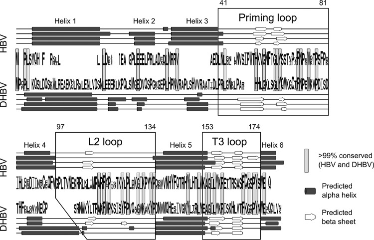
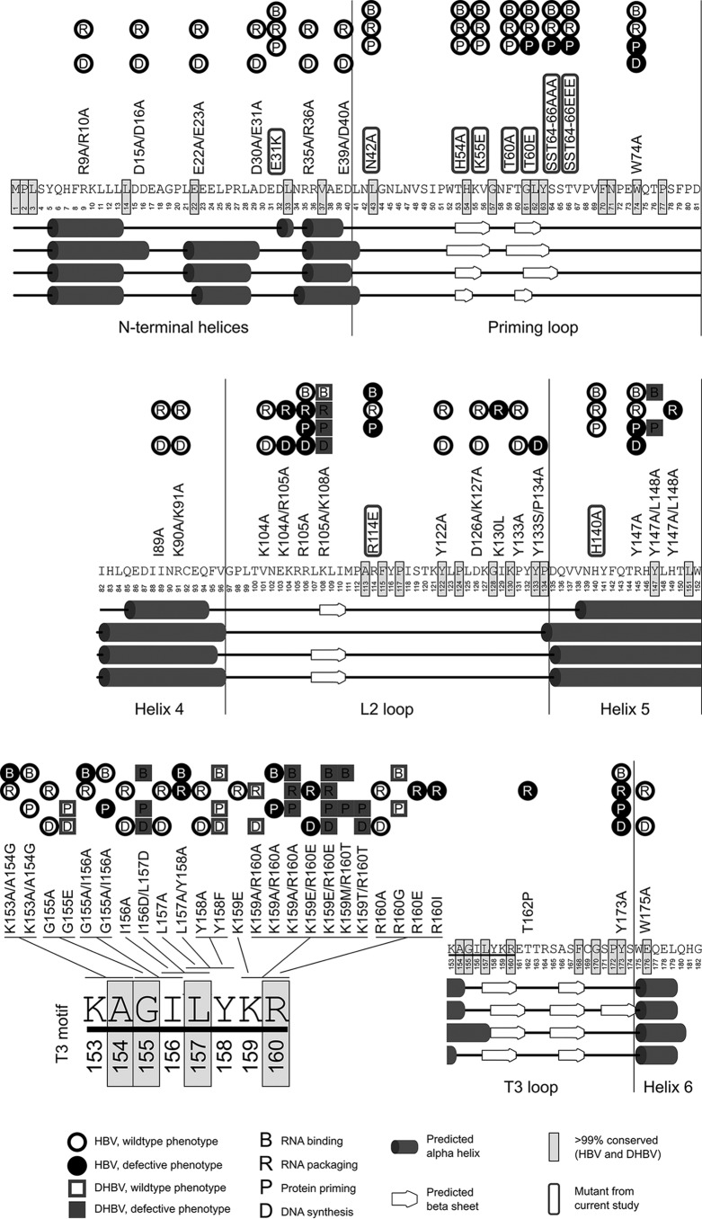
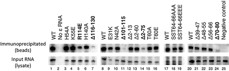
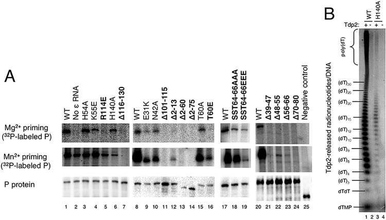
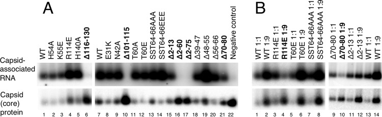
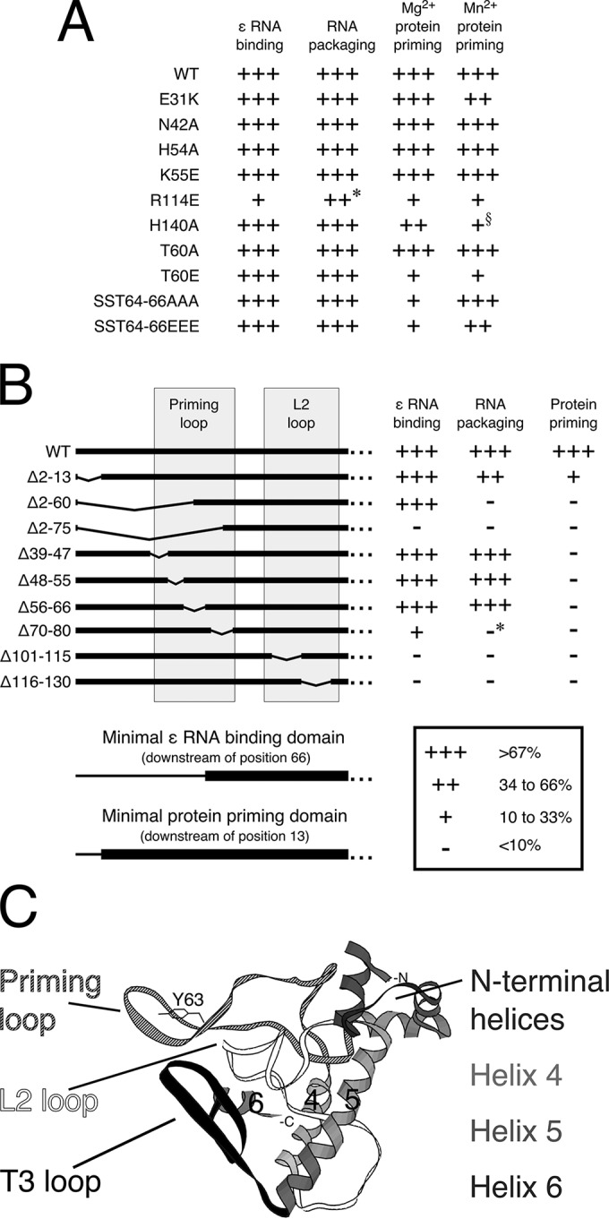
Hepatitis B virus (HBV) encodes a multifunction reverse transcriptase or polymerase (P), which is composed of several domains. The terminal protein (TP) domain is unique to HBV and related hepadnaviruses and is required for specifically binding to the viral pregenomic RNA (pgRNA). Subsequently, the TP domain is necessary for pgRNA packaging into viral nucleocapsids and the initiation of viral reverse transcription for conversion of the pgRNA to viral DNA. Uniquely, the HBV P protein initiates reverse transcription via a protein priming mechanism using the TP domain as a primer. No structural homologs or high-resolution structure exists for the TP domain. Secondary structure prediction identified three disordered loops in TP with highly conserved sequences. A meta-analysis of mutagenesis studies indicated these predicted loops are almost exclusively where functionally important residues are located. Newly constructed TP mutations revealed a priming loop in TP which plays a specific role in protein-primed DNA synthesis beyond simply harboring the site of priming. Substitutions of potential sites of phosphorylation surrounding the priming site demonstrated that these residues are involved in interactions critical for priming but are unlikely to be phosphorylated during viral replication. Furthermore, the first 13 and 66 TP residues were shown to be dispensable for protein priming and pgRNA binding, respectively. Combining current and previous mutagenesis work with sequence analysis has increased our understanding of TP structure and functions by mapping specific functions to distinct predicted secondary structures and will facilitate antiviral targeting of this unique domain.

Importance: HBV is a major cause of viral hepatitis, liver cirrhosis, and hepatocellular carcinoma. One important feature of this virus is its polymerase, the enzyme used to create the DNA genome from a specific viral RNA by reverse transcription. One region of this polymerase, the TP domain, is required for association with the viral RNA and production of the DNA genome. Targeting the TP domain for antiviral development is difficult due to the lack of homology to other proteins and high-resolution structure. This study mapped the TP functions according to predicted secondary structure, where it folds into alpha helices or unstructured loops. Three predicted loops were found to be the most important regions functionally and the most conserved evolutionarily. Identification of these functional subdomains in TP will facilitate its targeting for antiviral development.

Keywords: DNA polymerase; RNA binding; hepadnavirus; hepatitis B virus; protein priming; reverse transcriptase; terminal protein.


Figures

FIG 1
FIG 1
Conservation in HBV and DHBV in the primary and predicted secondary structure of the TP domain of the P protein. For comparison between HBV and DHBV, the sequence for the TP domain is shown, divided into two rows. HBV (n = 583) and DHBV (n = 45) TP domain sequences were aligned and used to create a WebLogo, where the top letter is the most common, the height of the stack indicates relative sequence conservation at that position, and the height of individual letters indicates the proportion of sequences which use that amino acid. Several software programs (from top to bottom: PsiPred, SABLE, I-TASSER, and QUARK) were used to analyze the consensus sequence and predict secondary structure; predicted alpha helices and beta sheets are shown as cylinders and arrows, respectively. Between the two species, the majority of predicted secondary structure elements overlapped. Many of the conserved sequences (light gray boxes) are in loops between helices. Three disordered conserved loops are highlighted in boxes. Numbering for HBV is according to genotype D, and boundary numbers near the boxes indicate HBV amino acid positions.
FIG 2
FIG 2
Meta-analysis of the phenotypes of known mutants within the TP domain of the HBV P protein. The consensus sequence for the TP domain is shown, divided into three rows, and numbered according to genotype D. Residues with homology to DHBV are highlighted (light gray boxes). Predicted alpha helices and beta sheets are shown as cylinders and arrows, respectively, as in Fig. 1. The TP domain is grouped (vertical lines) into seven subdomains according to predicted secondary structures. Known mutants in each of these subdomains are shown above the TP sequence. The filled shapes represent a defective phenotype (significant decrease or loss of function due to a mutation), and empty shapes represent WT phenotype (activity not significantly impaired by the mutation); circles are from studies with the HBV P, and squares are from DHBV studies. The novel mutants tested in the current study are circled with a gray line. The four commonly tested functions are shown by letter: B for RNA binding, R for RNA packaging, P for protein priming, and D for DNA synthesis. If a shape is absent, the mutant was not tested in that assay. The T3 motif, underlined on the right side of the 3rd row, is expanded on the left side of the 3rd row to show the many mutations that have been constructed in this subdomain. Except for helix 5, all defective mutants are found within the predicted loop subdomains.
FIG 3
FIG 3
ε RNA binding activities of HBV TP mutants. The HBV P, either WT or mutated, was expressed in HEK293T cells together with the ε RNA and purified by immunoprecipitation. RNA was extracted from both the immunopurified P (top) and cytoplasmic lysates (bottom) and resolved by urea-PAGE gel, followed by detection using an ε RNA-specific radiolabeled riboprobe. A control with no ε RNA expression was included (lane 2). The negative control (lane 25) expressed a mutant P containing only amino acids 1 to 175 and 300 to 775, known to be inactive in all P functions. This experiment was repeated at least three times; representative images are shown. Mutants with a defective phenotype are highlighted in boldface.
FIG 4
FIG 4
In vitro protein priming activities of HBV TP mutants. (A) The HBV P, either WT or mutated, was expressed in HEK293T cells together with the ε RNA and purified by immunoprecipitation. Radiolabeled dGTP and either Mg2+ (top) or Mn2+ (middle) were added to allow protein priming, during which the nucleotide(s) become covalently attached to the P protein. Levels of radiolabeled P proteins were detected by autoradiography (top and middle) following SDS-PAGE resolution of the priming reactions. Levels of total P proteins were detected by Western blotting (bottom) using an antibody against the triple-FLAG tag at the N terminus of each P construct. A control with no ε RNA expression was included (lane 2). The negative control (lane 25) expressed a P mutant containing only amino acids 1 to 175 and 300 to 775, known to be inactive in all P functions. This experiment was repeated at least three times; representative images are shown. Mutants with a defective phenotype are highlighted in boldface. (B) Analysis of DNA synthesis products by the WT HBV P protein and the H140A mutant in the protein-primed terminal transferase reaction. WT or H140A mutant P was expressed in HEK293T cells and purified by immunoprecipitation. Priming assays were performed with Mn2+ and radiolabeled TTP. Priming products were then mock treated (lanes 2 and 4) or treated with Tdp2 (lanes 1 and 3) in order to release the nucleotides/DNA strands attached to the P protein. Samples were resolved by urea-PAGE and detected by autoradiography. The lengths of the nucleotides/DNA strands are indicated.
FIG 5
FIG 5
RNA packaging activities of the HBV TP mutants. (A) Nucleocapsids were purified from HEK293T cells which were cotransfected with the indicated HBV P construct and pCMVHBV-Pol, which expresses the pgRNA and all HBV proteins except P. (Top) Levels of RNA packaging activity were measured by probing a nitrocellulose membrane with a plus-strand-specific probe following resolution of nucleocapsids on an agarose gel. (Bottom) Levels of capsid proteins were measured on the same membrane by Western blotting with an anti-HBV core antibody. The negative control (lane 22) expressed a P mutant containing only amino acids 1 to 175 and 300 to 775, known to be inactive in all P functions. Mutants with a defective phenotype are highlighted in boldface. (B) Effect on pgRNA packaging by expressing various levels of the P and capsid proteins in the trans-complementation assay. HBV nucleocapsids were purified from HEK293T cells that were cotransfected with the indicated P:P ratios and analyzed as described for panel A. Samples with less RNA packaging at the 1:9 ratio of P:P are highlighted in boldface. These experiments were repeated at least three times; representative images are shown.
FIG 6
FIG 6
Summary of TP mutant phenotypes. The activity levels of the substitution mutants (A) and N-terminal truncation and internal deletion mutants (B) are represented by symbols (>67%, +++; 34 to 66%, ++; 10 to 33%, +; <10%, −). Results represent averages from at least three independent experiments. *, RNA packaging levels of these mutants were based on comparison with the WT at a 1:9 ratio of P:P construct in transfection, which were significantly lower relative to the WT than those measured at the 1:1 ratio (Fig. 5B). All other mutants showed similar levels of RNA packaging relative to the WT at either 1:1 or 1:9 P:P. §, More severe defect in priming activity with Mn2+ than Mg2+ (Fig. 4). (B) The priming loop and L2 loop (gray boxes) are shown. For the mutant constructs, solid bars represent the sequences that are retained, and thin dipped lines represent deleted sequences. The protein priming results were the same using both Mn2+ and Mg2+ and are shown in a single column for clarity. At the bottom, the minimal necessary domains (thick bars) for ε RNA binding (downstream of position 66) and protein priming (downstream of position 13) are shown. Only the first 140 positions are shown (the remainder of TP is indicated by “…”). The image is drawn to scale. (C) The three-dimensional structure of TP as predicted using QUARK. The seven putative secondary structural elements of TP are indicated and shown in different shades. The N and C termini of TP are labeled, as is the Y63 priming site. The three loops, extending outward, are predicted to interact with the viral RNA or other factors during the various steps of viral DNA synthesis (see the text for details).

Similar articles

Cited by

References

    1. Summers J, Mason WS. 1982. Replication of the genome of a hepatitis B-like virus by reverse transcription of an RNA intermediate. Cell 29:403–415. doi:10.1016/0092-8674(82)90157-X. - DOI - PubMed
    1. Hu J, Seeger C. 2015. Hepadnavirus genome replication and persistence. Cold Spring Harb Perspect Med 5:a021386. doi:10.1101/cshperspect.a021386. - DOI - PMC - PubMed
    1. Hu J. 2016. Hepatitis B virus virology and replication, p 1–34. In Liaw Y-F, Zoulim F (ed), Hepatitis B virus in human diseases. Humana Press, Springer, New York, New York.
    1. Hu J, Seeger C. 1996. Expression and characterization of hepadnavirus reverse transcriptases. Methods Enzymol 275:195–208. doi:10.1016/S0076-6879(96)75013-9. - DOI - PubMed
    1. Clark DN, Hu J. 2015. Unveiling the roles of HBV polymerase for new antiviral strategies. Future Virol 10:283–295. doi:10.2217/fvl.14.113. - DOI - PMC - PubMed