. 2017 Jan 18;91(3):e02045-16.
doi: 10.1128/JVI.02045-16. Print 2017 Feb 1.

Kaposi's Sarcoma-Associated Herpesvirus MicroRNAs Target GADD45B To Protect Infected Cells from Cell Cycle Arrest and Apoptosis

Affiliations

Kaposi's Sarcoma-Associated Herpesvirus MicroRNAs Target GADD45B To Protect Infected Cells from Cell Cycle Arrest and Apoptosis

Xiaoyan Liu et al. J Virol. .

Abstract

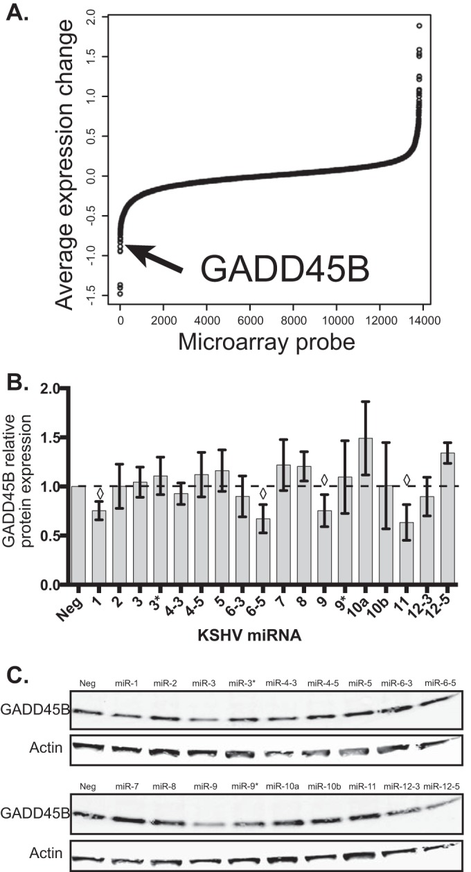
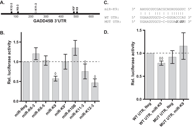
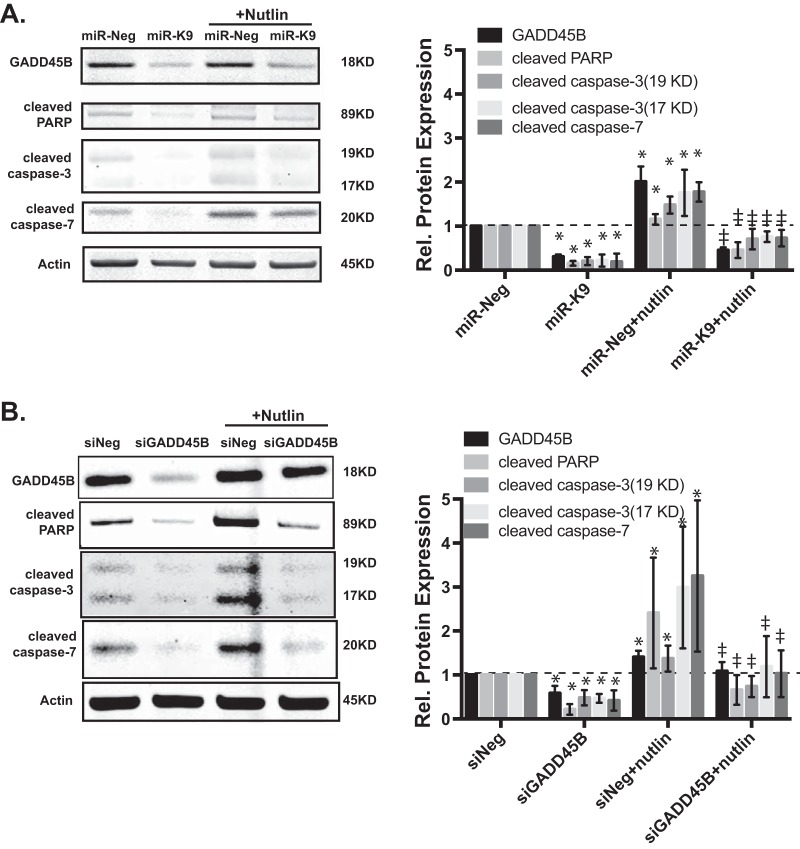
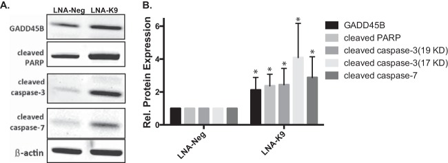
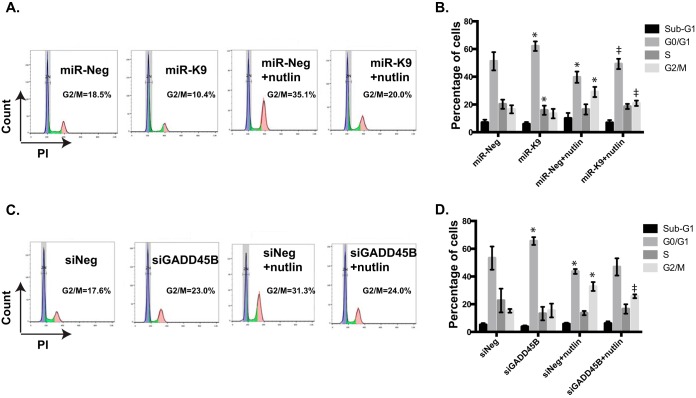
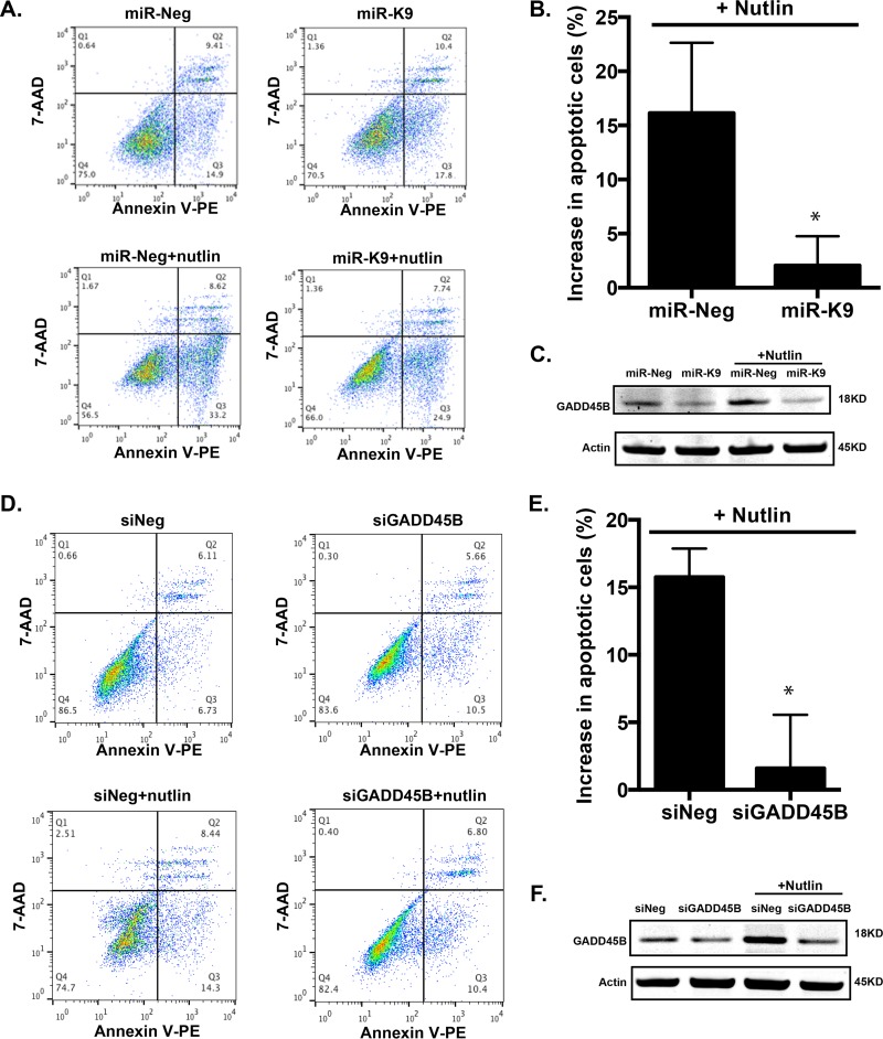
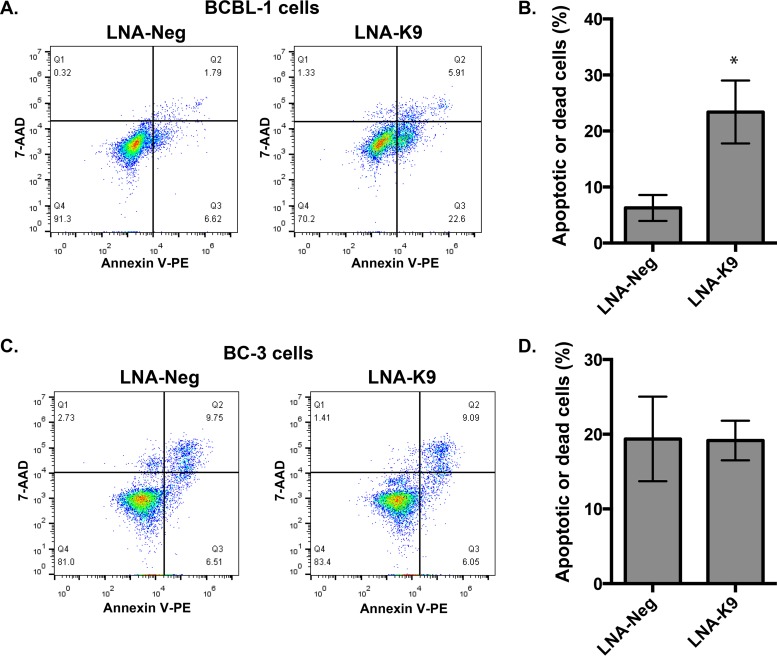
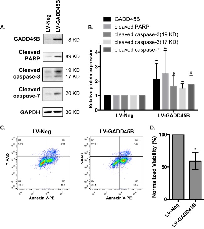
Kaposi's sarcoma is one of the most common malignancies in HIV-infected individuals. The responsible agent, Kaposi's sarcoma-associated herpesvirus (KSHV; HHV8), expresses multiple microRNAs (miRNAs), but the targets and functions of these miRNAs are not completely understood. After infection in primary endothelial cells with KSHV, growth arrest DNA damage-inducible gene 45 beta (GADD45B) is one of the most repressed genes using genomic expression profiling. GADD45B was also repressed in mRNA expression profiling experiments when KSHV miRNAs were introduced to uninfected cells. We hypothesized that KSHV miRNAs target human GADD45B to protect cells from consequences of DNA damage, which can be triggered by viral infection. Expression of GADD45B protein is induced by the p53 activator, Nutlin-3, and KSHV miRNA-K9 inhibits this induction. In addition, Nutlin-3 increased apoptosis and cell cycle arrest based on flow cytometry assays. KSHV miR-K9 protected primary endothelial cells from apoptosis and cell cycle arrest following Nutlin-3 treatment. Similar protective phenotypes were seen for targeting GADD45B with short interfering RNAs (siRNAs), as with miR-K9. KSHV miR-K9 also decreased the protein levels of cleaved caspase-3, cleaved caspase-7, and cleaved poly(ADP-ribose) polymerase (PARP). In B lymphocytes latently infected with KSHV, specific inhibitors of KSHV miR-K9 led to increased GADD45B expression and apoptosis, indicating that miR-K9 is important for reducing apoptosis in infected cells. Furthermore, ectopic expression of GADD45B in KSHV-infected cells promoted apoptosis. Together, these results identify a new miRNA target and demonstrate that KSHV miRNAs are important for protecting infected cells from DNA damage responses.

Importance: Kaposi's sarcoma-associated herpesvirus is a leading cause of cancers in individuals with AIDS. Promoting survival of infected cells is essential for maintaining viral infections. A virus needs to combat various cellular defense mechanisms designed to eradicate the viral infection. One such response can include DNA damage response factors, which can promote an arrest in cell growth and trigger cell death. We used a new approach to search for human genes repressed by small nucleic acids (microRNAs) expressed by a gammaherpesvirus (KSHV), which identified a gene called GADD45B as a target of microRNAs. Repression of GADD45B, which is expressed in response to DNA damage, benefited survival of infected cells in response to a DNA damage response. This information could be used to design new treatments for herpesvirus infections.

Keywords: DNA damage; Kaposi's sarcoma-associated herpesvirus; cell cycle; microRNA.


Figures

FIG 1
FIG 1
GADD45B expression is repressed by KSHV infection and specific KSHV miRNAs. (A) Microarray data were analyzed for changes after KSHV infection (48 h) or transfection of KSHV miRNAs (30 h). Average expression changes are shown from the two conditions and sorted by expression change. The arrow shows the location of the probe corresponding to GADD45B. (B) Primary endothelial cells were transfected with individual miRNA mimics and harvested 48 h after transfection. Protein expression changes of GADD45B (normalized to the loading control beta-actin) were obtained by immunoblotting using fluorescently labeled secondary antibodies and normalized to a nontargeting negative-control miRNA (miR-Neg). ♢, P value of <0.05 compared to miR-Neg using the Student t test. (C) A representative Western blot is shown for GADD45B (18 kDa) and beta-actin (45 kDa). The loading control was beta-actin.
FIG 2
FIG 2
KSHV miRNAs target the 3′UTR of GADD45B. (A) Sequences show the predicted target site for miRNAs. Filled circles are for perfect seed matches, and the open circle denotes an imperfect seed-matching site. (B) Cells were transfected with a GADD45B 3′UTR reporter and various KSHV miRNAs. Data are presented compared to an internal control luciferase reporter and normalized to the miR-Neg control. (C) Point mutations to disrupt miR-K9 interaction with the 3′UTR are shown. (D) Luciferase assay results are shown with the wild-type GADD45B 3′UTR (WT UTR) on the left and the mutant 3′UTR (MUT UTR) on the right with either the control miRNA (Neg) or miR-K9. P values of <0.05 (♢) and <0.01 (♢♢) were determined compared to miR-Neg using the Student t test.
FIG 3
FIG 3
Repression of GADD45B results in lower levels of apoptosis markers in response to Nutlin-3 treatment. (A) Primary endothelial cells were transfected with miRNAs (24 h) and then treated with Nutlin-3 for 24 h. The protein expression of GADD45B and apoptosis markers was analyzed by Western blotting (n = 3). (B) Assays similar to those performed for panel A, but cells were transfected with siRNAs targeting GADD45B (n = 5). Graphs from biological replicates are shown on the right. *, P value of <0.05 compared to miNeg/siNeg without Nutlin-3; ‡, P value of <0.05 compared to miNeg/siNeg with Nutlin.
FIG 4
FIG 4
Repression of miR-K9 in KSHV-infected cells. (A) Infected B lymphocytes (BCBL-1) were transfected with control miRNA inhibitors (LNA-Neg) or inhibitors of miR-K9 (LNA-K9). GADD45B protein and apoptosis markers were measured using Western blotting, and the results from at least three biological replicates are graphed on the right. A representative Western blot is shown on the left.
FIG 5
FIG 5
Repression of GADD45B inhibits G2/M cell cycle arrest induced by Nutlin-3. Primary endothelial cells were transfected with miRNAs or siRNAs and then treated with Nutlin-3 for 24 h. Cell cycle kinetics were measured using propidium iodide staining and flow cytometry analysis software. (A and C) The G2/M numbers in the graphs on the left are from a representative experiment. (B and D) The average values from three replicates are shown in the graphs on the right. *, P value of <0.05 compared to miR-Neg without Nutlin-3; ‡, P value of <0.05 compared to miR-Neg with Nutlin-3.
FIG 6
FIG 6
Repression of GADD45B inhibits apoptosis induced by Nutlin-3. Primary endothelial cells were transfected with miRNAs (A, B, and C) or siRNAs (D, E, and F) and then treated with Nutlin-3. Apoptotic and dead cells were measured using annexin-V and 7-AAD staining. (B and E) The increase in the percentage of apoptotic or dead cells due to Nutlin-3 treatment (percentage of apoptotic cells with Nutlin-3 minus percentage of apoptotic cells without Nutlin-3) is shown for three biological replicates. Representative Western blots are shown in panels C and F. *, P value of <0.05 compared to miRNA or siRNA negative controls.
FIG 7
FIG 7
Inhibitors of miR-K9 induce apoptosis in KSHV-infected cells that express miR-K9 (BCBL-1, not BC-3). (A and B) KSHV-infected B lymphocytes (BCBL-1) were transfected with control inhibitors or inhibitors to miR-K9. Apoptotic and dead cells were measured using annexin-V and 7-AAD staining. The percentages of quadrants 2 and 3 were summed, and the average percentages from three biological replicates are plotted in panels B and D. (C and D) Similar assays were conducted with the KSHV-infected cell line BC-3, which does not express miR-K9.
FIG 8
FIG 8
BCBL-1 cells were transduced with lentiviral particles that express GADD45B (LV-GADD45B) or the control (LV-Neg). After transduction, cells were harvested and analyzed using Western blotting (A and B) or flow cytometry assays (C and D). Living cells were defined as the cells in quadrant 4 (negative for both annexin-V and 7-AAD in lower left of plots in panel C). The percentage of living cells in the LV-GADD45B condition was normalized to the percentage in the LV-Neg condition to calculate normalized viability, and these average values from three biological replicates are shown in panel D.

Similar articles

Cited by

References

    1. Gottwein E, Corcoran DL, Mukherjee N, Skalsky RL, Hafner M, Nusbaum JD, Shamulailatpam P, Love CL, Dave SS, Tuschl T, Ohler U, Cullen BR. 2011. Viral microRNA targetome of KSHV-infected primary effusion lymphoma cell lines. Cell Host Microbe 10:515–526. doi: 10.1016/j.chom.2011.09.012. - DOI - PMC - PubMed
    1. Haecker I, Gay LA, Yang Y, Hu J, Morse AM, McIntyre LM, Renne R. 2012. Ago HITS-CLIP expands understanding of Kaposi's sarcoma-associated herpesvirus miRNA function in primary effusion lymphomas. PLoS Pathog 8:e1002884. doi: 10.1371/journal.ppat.1002884. - DOI - PMC - PubMed
    1. Fabian MR, Sonenberg N, Filipowicz W. 2010. Regulation of mRNA translation and stability by microRNAs. Annu Rev Biochem 79:351–379. doi: 10.1146/annurev-biochem-060308-103103. - DOI - PubMed
    1. Skalsky RL, Cullen BR. 2010. Viruses, microRNAs, and host interactions. Annu Rev Microbiol 64:123–141. - PMC - PubMed
    1. Ramalingam D, Kieffer-Kwon P, Ziegelbauer JM. 2012. Emerging themes from EBV and KSHV microRNA targets. Viruses 4:1687–1710. doi: 10.3390/v4091687. - DOI - PMC - PubMed

MeSH terms

LinkOut - more resources