. 2017 Jan 18;91(3):e02107-16.
doi: 10.1128/JVI.02107-16. Print 2017 Feb 1.

Nuclear Export Signal Masking Regulates HIV-1 Rev Trafficking and Viral RNA Nuclear Export

Affiliations

Nuclear Export Signal Masking Regulates HIV-1 Rev Trafficking and Viral RNA Nuclear Export

Ryan T Behrens et al. J Virol. .

Abstract

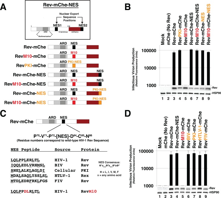
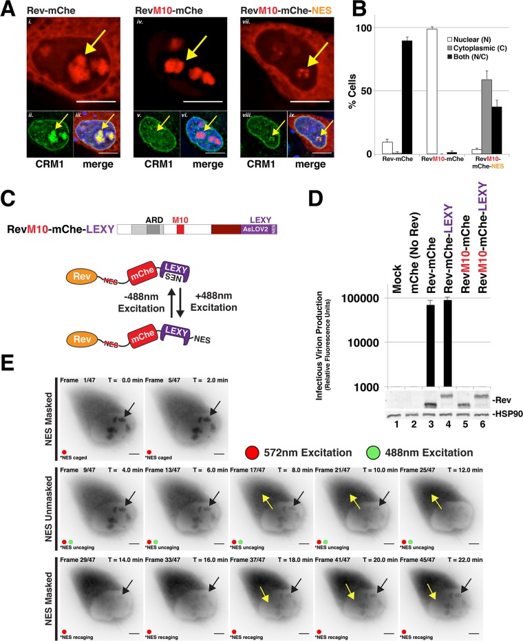
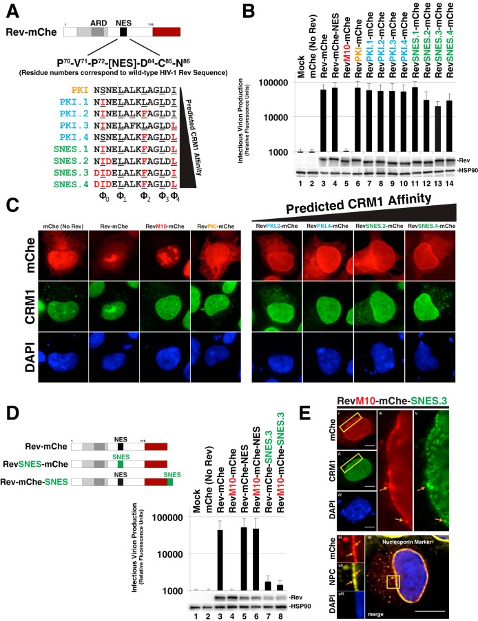
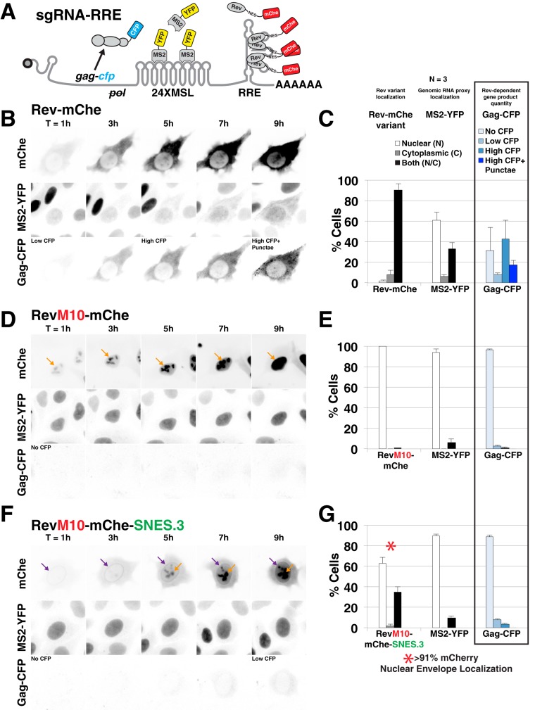
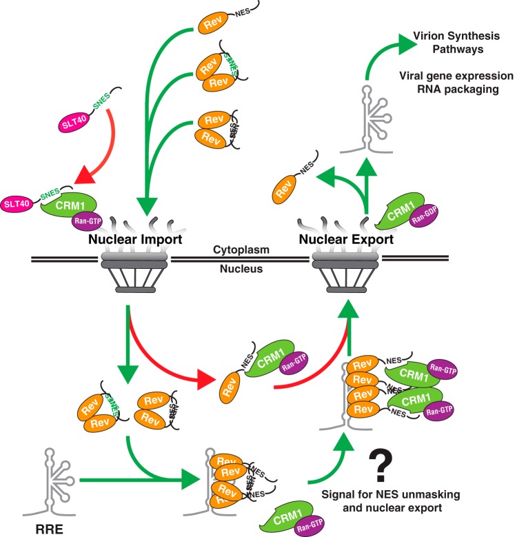
HIV-1's Rev protein forms a homo-oligomeric adaptor complex linking viral RNAs to the cellular CRM1/Ran-GTP nuclear export machinery through the activity of Rev's prototypical leucine-rich nuclear export signal (NES). In this study, we used a functional fluorescently tagged Rev fusion protein as a platform to study the effects of modulating Rev NES identity, number, position, or strength on Rev subcellular trafficking, viral RNA nuclear export, and infectious virion production. We found that Rev activity was remarkably tolerant of diverse NES sequences, including supraphysiological NES (SNES) peptides that otherwise arrest CRM1 transport complexes at nuclear pores. Rev's ability to tolerate a SNES was both position and multimerization dependent, an observation consistent with a model wherein Rev self-association acts to transiently mask the NES peptide(s), thereby biasing Rev's trafficking into the nucleus. Combined imaging and functional assays also indicated that NES masking underpins Rev's well-known tendency to accumulate at the nucleolus, as well as Rev's capacity to activate optimal levels of late viral gene expression. We propose that Rev multimerization and NES masking regulates Rev's trafficking to and retention within the nucleus even prior to RNA binding.

Importance: HIV-1 infects more than 34 million people worldwide causing >1 million deaths per year. Infectious virion production is activated by the essential viral Rev protein that mediates nuclear export of intron-bearing late-stage viral mRNAs. Rev's shuttling into and out of the nucleus is regulated by the antagonistic activities of both a peptide-encoded N-terminal nuclear localization signal and C-terminal nuclear export signal (NES). How Rev and related viral proteins balance strong import and export activities in order to achieve optimal levels of viral gene expression is incompletely understood. We provide evidence that multimerization provides a mechanism by which Rev transiently masks its NES peptide, thereby biasing its trafficking to and retention within the nucleus. Targeted pharmacological disruption of Rev-Rev interactions should perturb multiple Rev activities, both Rev-RNA binding and Rev's trafficking to the nucleus in the first place.

Keywords: CRM1; Gag; RNA trafficking; Rev; exportin-1; human immunodeficiency virus; nuclear export signal; nuclear pore complex; nucleolus; retroviruses.


Figures

FIG 1
FIG 1
Rev is robustly tolerant of changes to NES number, position, or identity. (A) Cartoon indicating Rev-mCherry (Rev-mChe) variants and relevant NES positions or modifications, native position (NES1), altered identity (PKI), inactivated (M10), and C-terminal position (NES2). Rev's arginine-rich domain (ARD, amino acids 34 to 50) encodes the nuclear localization signal (NLS) and RNA-binding activities. Rev's native NES (NES1 position; amino acids 73 to 83) is located within the disordered C-terminal domain (B) Capacity of Rev variants depicted in 1A to trans-complement Rev-minus HIV-1 YFP reporter viruses. 293T cells were transfected with plasmids encoding full-length, NL4-3-derived E-R-Rev-/YFP reporter proviruses, VSV-G, and either mCherry alone (No Rev control) or the indicated Rev-mChe variant. The lane 1 control lacks proviral DNA. Cell lysates and supernatants were harvested at 48 h posttransfection and processed for immunoblotting, and equivalent amounts of supernatant were used to infect target HeLa cells in order to gauge infectious virion production based on YFP fluorescence at 48 h postinfection (viral infectivity assay). Error bars represent standard deviations from the mean for three independent experiments. Rev and HSP90 (loading control) were detected using anti-Rev and anti-HSP90 antisera. (C) Depiction of additional Rev-mChe variants bearing alternative NES sequences. Predicted NES consensus-defining amino acids are underlined. BIV, bovine immunodeficiency virus; PKI, protein kinase A inhibitor; HTLV-1, human T-lymphotropic virus type 1; FIV, feline immunodeficiency virus. (D) Activities of the Rev variants shown in panel C were determined by viral infectivity assay as described for panel B.
FIG 2
FIG 2
Evidence for Rev exhibiting position-dependent NES masking. (A) Context-specific NES effects on Rev's subcellular localization. HeLa cells transfected to express E-R-Rev-/Luc and the indicated Rev-mChe variants were fixed, permeabilized, and DAPI stained 24 h posttransfection. Endogenous CRM1 was detected by indirect immunofluorescence using anti-CRM1 antisera. Yellow arrows highlight nucleolar accumulation of Rev and/or CRM1. Scale bars, 10 μm. (B) RevM10-mChe-NES exhibits less accumulation at or near the nucleolus. Rev-mChe subcellular distribution was quantified in individual cells as primarily nuclear (N), cytoplasmic (C), or equivalent in both compartments (N/C). Error bars represent the standard deviations from the mean for three independent transfections. (C) Depictions of RevM10-mChe-LEXY construct and blue light-regulated NES unmasking using the LEXY regulatory module (75). (D) Control experiment demonstrating that the activity of Rev-mChe-LEXY and RevM10-mChe-LEXY variants is equivalent to Rev-mChe constructs lacking LEXY. Viral infectivity was measured as for Fig. 1B. Error bars represent the standard deviations from the mean for three independent experiments. (E) Image panel shows selected frames from a representative time-lapse fluorescence microscopy experiment capturing mCherry fluorescence from RevM10-mChe-LEXY in HeLa cells. Red circles indicate exposure to 572-nm wavelength light (mCherry acquisition wavelength), and green circles indicate exposure to 488-nm wavelength light (LEXY activation wavelength). Black arrows indicate nucleolar Rev accumulation sites, and yellow arrows indicate direction of Rev transitions over time. Scale bars, 10 μm.
FIG 3
FIG 3
Rev tolerates supraphysiological NES domains in a position-dependent manner. (A) Panel of NES domains predicted to exhibit increasing CRM1 binding strength in the context of Rev-mChe. Wild-type PKI NES is labeled orange (same variant from Fig. 1). PKI NES-derived sequences with predicted increases in CRM1 affinity are labeled blue. PKI NES-derived sequences with predicted supraphysiological CRM1 binding affinity (i.e., bind to CRM1 even in the absence of Ran-GTP; SNESs) are labeled green. Amino acids shown red predicted to confer the increase of CRM1 affinity. (B) Even supraphysiological NESs had only modest effects (∼2-fold decreases) on Rev function in our trans-complementation infectivity assay described for Fig. 1B. Error bars represent the standard deviations from the mean for five independent experiments. (C) Nucleolar localization was decreased when Rev encoded an NES with increased CRM1 affinity. HeLa cells were transfected and prepared as for Fig. 2A. (D) Combining increases to NES strength with changes to NES position potently inhibits viral infectivity. Diagram of relevant Rev NES strength/context variants with our infectivity assay demonstrating >10-fold losses to infectious virion production. Error bars represent the standard deviations from the mean for five independent experiments. (E) Representative indirect immunofluorescence images showing Rev-mChe-SNES colocalizing with CRM1 and nucleoporins at the nuclear membrane. Yellow rectangles indicate regions of interest and orange arrows highlight colocalization. Scale bars, 10 μm.
FIG 4
FIG 4
SNES-arrested Rev/CRM1 complexes block Rev's ability to export vRNAs from the nucleus. (A) Diagram of subgenomic, intron-retaining HIV-1 gag-pol mRNA for live-cell imaging. This mRNA encodes 24 copies of MS2 coat protein binding loop (24XMSL) and a CFP-labeled Gag protein (Gag-CFP) for tracking Rev-dependent viral mRNA nuclear export and late viral gene expression (See Materials and Methods and reference for additional information). (B to G) Live cell imaging of Rev-mChe variants, viral mRNA trafficking, and Gag-CFP expression in HeLa cells stably producing MS2-YFP (HeLa.MS2-YFP) over a 9-h interval. Image capture was initiated <1 h posttransfection and fixed at ∼24 h posttransfection for endpoint analysis. (B) Wild-type Rev-mChe supports MS2-YFP translocation from the nucleus to the cytoplasm and Gag-CFP expression consistent with Rev-dependent viral mRNA nuclear export and translation. (C) Endpoint analysis of Rev, viral mRNA labeled by MS2-YFP proxy, and Gag-CFP. Rev and MS2-YFP distribution was quantified in individual cells for fluorescence signal as primarily nuclear (N), cytoplasmic (C), or readily detectable in both compartments (N/C). Gag-CFP was quantified based upon CFP expression level (no CFP, low CFP, or high CFP) and distribution of signal (diffuse or diffuse with punctate). Error bars represent the standard deviations from three independent transfections. A total of >300 cells were scored per condition for all transfection replicates combined. (D and E) RevM10-mChe is restricted to the nucleus (orange arrows) and does not activate viral mRNA export or Gag-CFP expression. (F and G) RevM10-mChe-SNES first accumulates at the nuclear envelope (purple arrows) and at later time points in the nucleolus (orange arrows) but does not trigger detectable viral mRNA export and supports only low levels of Gag-CFP expression. The red asterisk in panel G indicates that RevM10-mChe-SNES localization typically included signal at the nuclear envelope.
FIG 5
FIG 5
A C-terminal SNES blocks Rev-dependent viral mRNA export. Direct visualization of unspliced HIV-1 mRNA using fluorescence in situ hybridization (FISH). HeLa cells transfected to express the E-R-Rev-/Luc construct and the indicated Rev variant were processed as for Fig. 2A at 24 h posttransfection. FISH probes targeting the gag-pol reading frame of the E-R-Rev-/Luc construct were used to detect unspliced viral RNA (shown in magenta). Scale bars correspond to 10 μm. (A, C, and E) Representative images of Rev (red), mRNA (FISH, magenta), and nuclear DNA (DAPI, blue) for wild-type Rev-mApple (A), RevM10-mApple (C), and RevM10-mApple-SNES3 (E). (B, D, and F) RNA distribution was quantified in individual cells for RNA localization as primarily nuclear (N), cytoplasmic (C), or readily detectable in both compartments (N/C). A total of 100 cells were scored per condition.
FIG 6
FIG 6
Rev's capacity to mask a SNES is both context and multimerization dependent. (A) Diagram depicting shuttle YFP (S-YFP) reporter assay. This assay employs NLS- and NES-directed nucleocytoplasmic trafficking of S-YFP reporter to determine the disruptive effect of Rev variants encoding different NES configurations. (B) Inhibition of CRM1-dependent export promotes S-YFP nuclear accumulation. The image panel depicts individual transfected HeLa cells producing S-YFP and the indicated Rev variant (the example shown is Rev-mChe) in the absence (panels i to iv) or presence (panels v to viii) of 5 nM leptomycin B (LMB) as a control for CRM1-dependent nuclear export inhibition. Transfected HeLa cells were fixed at 24 h posttransfection and DAPI stained to demarcate nuclear and cytoplasmic compartments (nuclear border labeled by white dotted line). Scale bars, 10 μm. (C) Multimerization-deficient Rev encoding SNES in native context phenocopies C-terminal SNES and LMB treatment. The nuclear-to-cytoplasmic ratio (N/C ratio) of YFP fluorescence was measured from individual cells producing the indicated Rev variants in an S-YFP assay. Cells were transfected to express the indicated Rev variant and viral RNA and processed as for panel B. Nuclear and cytoplasmic YFP fluorescence was measured from YFP- and mCherry-positive HeLa cells and assigned using DAPI as a marker for nuclear-specific fluorescence signal for reference (see Fig. 6B, compare panels i to ii to panels v to vi). Error bars represent the standard deviations from the mean for four independent experiments (>250 cells measured per condition). Automated cell segmentation and fluorescence quantification were performed using KNIME/FIJI. (D) Wild-type HIV-1 Rev sequence (amino acids 34 to 83) indicating functional activities and amino acid substitutions conferring loss of RNA binding (M5), Rev multimerization (SLT40), and CRM1 binding (M10). (E) Model for regulation of multimerization-dependent NES masking. Physical masking of SNES in native NES1 context decreases interaction potential with CRM1, thus promoting the cytoplasmic accumulation of S-YFP, as measured in panel C.
FIG 7
FIG 7
NES context regulates optimal infectious virion production. Titration of Rev-mChe and RevM10-mChe-NES plasmids reveals functional differences between variants in our trans-complementation viral infectivity assay, as described for in Fig. 1B. Error bars represent the standard deviations from the mean for three independent experiments.
FIG 8
FIG 8
Working model for multimerization-dependent NES masking in Rev's nucleocytoplasmic trafficking scheme. Our data suggest Rev-Rev interactions promote Rev's nucleolar accumulation by inhibiting CRM1 from accessing Rev's NES prior to and during nuclear import (left NPC, green arrows). An unmasked NES disrupts Rev's capacity to accumulate in the nucleolus by blocking nuclear import (red arrow in cytoplasm, SNES) or driving immediate export of Rev from the nucleus (red arrow in nucleus, NES). In the nucleus, one or more Rev NES peptides are likely exposed through an unknown mechanism during formation of Rev/RNA complexes, thus serving as a signal for the recruitment of CRM1 (right NPC, green arrows). Nuclear export and dissolution of the complex promotes recycling of Rev and downstream events in the HIV-1 life cycle.

Similar articles

Cited by

References

    1. Kutay U, Güttinger S. 2005. Leucine-rich nuclear-export signals: born to be weak. Trends Cell Biol 15:121–124. doi:10.1016/j.tcb.2005.01.005. - DOI - PubMed
    1. Wente SR, Rout MP. 2010. The nuclear pore complex and nuclear transport. Cold Spring Harb Perspect Biol 2:a000562. doi:10.1101/cshperspect.a000562. - DOI - PMC - PubMed
    1. Güttler T, Görlich D. 2011. Ran-dependent nuclear export mediators: a structural perspective. EMBO J 30:3457–3474. doi:10.1038/emboj.2011.287. - DOI - PMC - PubMed
    1. Görlich D, Kutay U. 1999. Transport between the cell nucleus and the cytoplasm. Annu Rev Cell Dev Biol 15:607–660. doi:10.1146/annurev.cellbio.15.1.607. - DOI - PubMed
    1. Siddiqui N, Borden KLB. 2012. mRNA export and cancer. Wiley Interdiscip Rev RNA 3:13–25. doi:10.1002/wrna.101. - DOI - PubMed

Substances